X-Ray is a form of electromagnetic radiation with very high frequency and energy. X-rays lie between ultraviolet radiation and gamma radiation on the electromagnetic spectrum. A bone x-ray uses a very small dose of ionizing radiation to produce pictures of any bone in the body. It is commonly used to diagnose fractured bones or joint dislocation. Bone x-rays are the fastest and easiest way for your doctor to view and assess bone fractures, injuries, and joint abnormalities.
Chest x-ray (frontal view)
-
Lateral CXR rarely indicated and should be discussed with a consultant
-
Pre-operative CXR NOT to be done routinely at any age.
Respiratory indications
-
Infection – to exclude pneumonia
-
Inhaled foreign body >most lodge in the intrathoracic tracheobronchial tree.
- Need films in full inspiration and expiration to demonstrate air trapping or collapse.
-
for an air leak, haemothorax or wide mediastinum.
-
Rib views rarely indicated.
-
Pneumothorax – full inspiratory films adequate
-
diagnosis unclear
-
SEVERE attack – not responding to standard therapy
possible air leak. -
NB. Focal signs +/- fever is most likely due to mucus plug and viral illness rather than pneumonia.
Cardiac indications
-
Large thymic shadow is normal under the age of 2 years.
-
Normal cardio-thoracic ratio 0.5 ( infants up to 0.6 )
-
Heart murmurs – If careful examination suggests innocent murmur, no need for urgent CXR – but arrange appropriate follow up.
-
Hypertension – CXR is seldom useful.
Neonates (<6wks)
-
Septic screen – CXR indicated unless clear focus elsewhere
-
Respiratory distress – to exclude congestive cardiac failure or cardiomegaly
Limb x-rays & other imaging modalities
Comparative and Stress Views – rarely necessary and should not be routinely taken. However may be useful for complex fractures (after consultation) if initial x-rays unclear (eg. elbow)
Specific Indications/Contraindications:
Trauma
-
Follow up films after reduction of a displaced # should be done to assess the position.
-
Additional views are sometimes useful (eg. radial head views) and other fractures (eg. stress # or toddler #) might need Bone Scan or CT (these requests should be discussed with ED consultant & orthopedics and appropriate follow-up arranged).
Nonaccidental injury (to be seen by registrar or consultant)
- If child > 2yr x-rays should be limited to sites of clinically suspected injury.
-
Complete Skeletal Survey if child < 3 years (not available after-hours unless urgent)
- Bone Scan (if < 3yr) – can complement skeletal survey
-
metaphyseal
-
marked or unusual epiphyseal separation
-
of spine or ribs
-
unexplained skull intracranial- injury
Acutely painful hip
-
Plain xrays (AP and frog-leg lateral) will demonstrate slipped upper femoral epiphyses, Perthe’s and fractures.
-
USS/ bone scan may be indicated depending on clinical findings (discuss with specialty team or treating consultant).
Acutely swollen joint
Osteomyelitis
-
Early XR often shows no bony abnormality but may have deep soft tissue swelling.
-
Bone scan/MRI will demonstrate an abnormality earlier than XR (needs orthopedic team input)
Septic Arthritis
-
Normal XR or Bone scan does not exclude septic arthritis.
-
Ultrasound may be useful to demonstrate a joint effusion and soft tissue abnormality (discuss with orthopedic team or treating consultant)
Metabolic disorders
-
Rickets – XR of one wrist +/- one knee is most useful.
-
Osteogenesis Imperfecta – the very low threshold for an x-ray.
Pulled Elbow
-
If the injury mechanism and examination suggest radial head subluxation, an x-ray is unnecessary.
Abdominal x- rays
Suspected bowel obstruction/ perforation
-
A plain AXR will demonstrate most obstruction (dilated loops).
-
An erect AXR is indicated to exclude perforation
Suspected intussusception
-
A normal AXR does not exclude intussusception but is useful to exclude perforation or bowel obstruction in suspected intussusception.
Foreign Bodies
-
Ingested opaque FB requires a single survey AP film (mouth to anus).
-
Routine follow-up films are NOT indicated unless clinical symptoms develop.
Suspected Abdominal Mass
-
Initial investigation – plain AXR and ultrasound, then further as indicated
Blunt abdominal trauma
-
Needs early assessment by General Surgery.
-
CT scan is the best modality for diagnosing the intra-abdominal injury.
Unnecessary AXRs
If unsure whether AXR would be helpful – ask consultant or registrar for advice
AXR not indicated for:
-
Vague central abdominal pain.
-
Gastroenteritis.
-
Haematemesis.
-
Pyloric stenosis.
-
Uncomplicated appendicitis.
-
Chronic constipation, encopresis or enuresis (in the Emerg. Dept setting )
Abdominal & pelvic ultrasounds
If an urgent ultrasound is necessary, the patient should be discussed with the surgeon &/or the treating consultant.
Specific indications
Suspected intussusception
-
Ultrasound by experienced operators is the diagnostic modality of choice for intussusception.
-
However these patients are potentially unstable and should only be sent for an ultrasound after appropriate resuscitation including an IV, and treatment as well as notifying the surgeons and the treating consultants
Suspected pyloric stenosis
-
Ultrasound is a very sensitive test for pyloric stenosis
Abdominal pain
-
or iliac or pelvic pain in the pubertal female with possible ovarian pathology (requires full bladder), or if potential renal tract obstruction, early ultrasound recommended.
-
Abdominal ultrasound is a useful tool for many other abdominal pain presentations however the urgency of the request should be proportional to the symptoms.
Urinary tract imaging
-
Bacteriologically proved first UTI usually requires renal tract US (particularly <4 years old) but only occasionally MCU
Intracranial and skull imaging
Specific Indications for Skull X-rays
Only indicated in well-appearing children
NAI
-
as part of the skeletal survey (more sensitive than Bone Scan for skull fracture)
Plagiocephaly
-
Craniosynostosis (prematurely fused sutures) accounts for the minority of abnormal skull shapes. An SXR is useful to evaluate sutures but is ideally done via outpatient follow-up (Craniofacial or neurosurgical unit- RCH Deformational head clinic)
There are no other routine indications for skull XRay and any such requests should be discussed with the treating consultant.
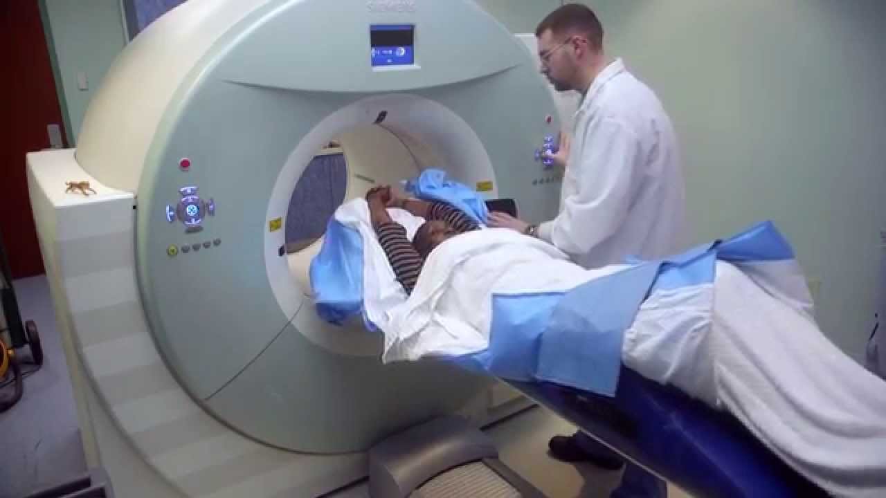
Specific Indications for CT Brain
-
The treating consultant should discuss the need for all CT scans.
-
The Neurosurgical team should be involved before CT for the unwell or potentially unstable patient who may need urgent interventions.
Head Trauma
-
Useful for rapid diagnosis of suspected intracranial injuries and is the preferred investigation if clinical evidence of intracranial injury.
-
Clinical deterioration is usually an indication for repeat CT examination.
Depressed conscious level of unknown cause
-
CT scan is indicated after appropriate stabilizing treatment.
Headaches
Clinical evaluation is the most important factor in determining the need for imaging.
CT scan indications
-
Abnormal neurological signs.
-
Unexplained decrease in visual acuity.
-
Headaches with seizures.
-
Marked change in behavior.
-
Enlarging head
-
Symptoms of raised intracranial pressure.
-
The increasing frequency of unexplained headaches or new onset of a severe or persistent headache
Seizures
-
Persistent abnormal neurological signs/impaired conscious state.
-
Focal neurological signs or EEG findings.
-
Failure to respond to anticonvulsant therapy.
-
Neurocutaneous lesions.
Abnormal Size / Shape Of Skull
Clinical examination is usually sufficient to diagnose an abnormality of the skull.
-
Large head – rapidly enlarging head needs imaging-US or CT scan.
-
Small head – nearly always pathological secondary to abnormal brain growth. Evaluate with CT or MRI scan, which is usually best organized via the managing outpatient physician
Specific Indications for cranial ultrasound
Large head
-
Rapidly enlarging head with open fontanelle.
Neurological concerns in neonates/ infants
-
Clinical usefulness will vary depending on the size of fontanelle and indications and should be discussed with the radiologist.
Spinal imaging
Any investigations other than plain x- rays should be ordered in consultation with the treating consultant &/or the appropriate specialty team.
NB. Down syndrome children have increased the risk of C1-2 instability.
Specific indications in Trauma:
Cervical spine
A normal Spinal Xray series or CT scan will not allow clearance of the neck in the unconscious or uncooperative patient
Thoraco-Lumbar Spine
-
Children poorly localize the level of the injury, therefore imaging the full length of thora columbar spine may be necessary (discuss with treating consultant).
-
If neurological signs present do a CT or MRI scan after consultation with Neurosurgery.
Specific Non-trauma indications:
Scoliosis
-
Plain films should include the entire spine
Potential cord compression
-
Needs discussion with the treating consultant and neurosurgical team.
Suspected focal vertebral pathology
-
Choice of imaging modality needs discussion with the treating consultant.
References

![]()
People Also Reading
 Ultrasound; Types, Uses,Procedures, Adverse Effects
 CT Scan; Types, Indications, Procedures, Side Effects, Results
 EMG; Types, Indications/Uses, Procedures, Results
 Electrocardiography; Types, Indications/Uses, Procedures
 ECG / EKG; Types, Indications/Uses, Procedures, Results
 Cosmetic Surgery, Types, Procedures, Indications
 Synovial Fluid – Types. Indications, Procedures
 Skin Grafting, Types, Indications, Procedures,
 Chemotherapy; Types, Procedures, Side Effects, Stage
 Lumbar Puncture / Spinal Tap; Types, Uses, Procedures, Results
 Barbiturate; Types, Indications/Uses, Contra Indications, Side Effects, Interactions
 Echocardiogram; Indications, Preparetion, Procedures
About the author