Swallowing problems for liquids and solids
Swallowing problems for liquids and solids/Achalasia is a rare neurodegenerative [...]
Swallowing problems for liquids and solids/Achalasia is a rare neurodegenerative [...]
Lower Esophageal Sphincter Spasm/Achalasia is a rare neurodegenerative motor smooth [...]
Altitude-Related Disorders/Altitude sickness occurs when you cannot get enough oxygen [...]
Vulvovaginitis/Vulvitis is inflammation of the vulva, the external female mammalian genitalia that [...]
What does pantothenic acid do for your body?/Pantothenic Acid is [...]
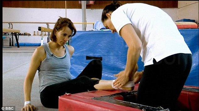
What causes swimmer's shoulder?/Swimmer’s shoulder is an umbrella term covering [...]
Fingernails Infection or Paronychia is an infection of the proximal [...]
Arthrocutaneouveal granulomatosis/Blau syndrome (BS) is a rare autosomal dominant, autoinflammatory [...]
What are the first signs of gangrene?/Wet Gangrene is characterized [...]
How does allopurinol prevent tumor lysis syndrome?/Tumor lysis syndrome (TLS) [...]
Diet, Exercise of Clubfoot I Can Do Not Going To [...]
Child Foot Deformities I Can Know Not Going To Doctor/Foot [...]
We Inspire with our business services
through the agency’s dream to strive for
the excellence.
© Avada Studio • All rights reserved.
Powered by WordPress