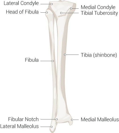
Tibia fractures are common injuries. The subcutaneous nature of the tibia makes it more prone to open injury. The musculature about the lower leg divides into four compartments separated by fascial tissue. Radiographs are essential in the initial evaluation of the fractures. In the case of injury or fracture of the lower extremity, the fascial tissue may have to be released by fasciotomies to prevent the sequelae of compartment syndrome. Treatment methods can be non-operative for minimally displaced fractures although operative fixation for displaced and open fractures is preferred.
Types of Tibia Fracture
Classifications
Some classifications help with treatment decisions.
Western and Tscherne
This is a classification of closed fracture soft tissue injury and is as follows:
-
Grade 0: Injuries from indirect forces with minimal soft tissue damage
-
Grade 1: Superficial contusion/ abrasion, simple fractures
-
Grade II: Deep abrasions, muscle/skin contusion, direct trauma, impending compartment syndrome
-
Grade III: Excessive skin contusion, crushed skin or muscle destruction, subcutaneous degloving, acute compartment syndrome, and rupture of a major blood vessel or nerve
The Gustilo-Anderson
This classification is used to assess open tibia fractures.
-
Type I is limited periosteal stripping, clean wound less than 1 cm
-
Type II mild to moderate periosteal stripping; wound greater than 1 cm in length
-
Type IIIA significant soft tissue injury, significant periosteal stripping with a wound that is usually greater than 1 cm in length with no flap required
-
Type IIIB is significant periosteal stripping and soft tissue injury with a flap required due to inadequate soft tissue coverage
-
Type IIIC these are significant soft tissue injury with a vascular injury requiring repair
Treatment of Tibia Fracture
Non-Operative Treatment
Closed-reduction and nonoperative treatment in a long leg cast is acceptable for fractures in less than 5 degrees of varus-valgus angulation, less than 10 degrees in anterior-posterior angulation, greater than 50% cortical apposition, less than 1-cm shortening and less than 10 to 20 degrees of flexion and less than 10 degrees of rotational malalignment after reduction.
External Fixation
Treatment of choice when significant soft tissue compromise is present or in polytrauma cases where damage-control orthopedics is needed.
Intramedullary Nailing (IMN)
This is the treatment of choice for operative fixation.
When comparing outcomes of IMN with external fixation, IMN is associated with decreased malalignment and compared to closed treatment, IMN is associated with decreased union time and time to weight bearing.
Percutaneous Plating-Shaft
This method is often used in the distal tibia or proximal-third fractures that are too proximal or distal for intramedullary nailing.
Amputation
This is another treatment method but can be difficult to get the patient to buy into this treatment. The mangled extremity severity score (MESS) can help predict when an amputation is necessary. A score of 7 or greater is highly predictive of amputation. MESS has a high specificity but low sensitivity in predicting amputations. Relative indications include significant soft tissue trauma, warm ischemia greater than 6 hours, and severe ipsilateral foot trauma. It is important to note that loss of plantar sensation is not an absolute indication for amputation.
Management of Tibial Fractures
Tibial plateau fracture: These fractures present with knee pain and effusion. They classically occur after a car hits a pedestrian’s fixed knee, which is known as a “bumper fracture.” They are classified using the Schatzker classification and managed by using nonsurgical or surgical methods to achieve stable alignment. Operative strategies include external fixation and open reduction internal fixation.[10]
-
Schatzker Classification
-
Type 1: lateral split fracture
-
Type 2: lateral split-depressed fracture
-
Type 3: lateral pure depression fracture
-
Type 4: medial fracture
-
Type 5: bicondylar fracture
-
Type 6: metaphyseal-diaphyseal disassociation
-
Tibial shaft fracture: Compared to most long bone fractures, tibial shaft fractures are more likely to be open because the medial surface is adjacent to the subcutaneous tissue. The fracture can have a low or high energy pattern. The low energy patterns are a result of torsional injury resulting in a spiral fracture. The high energy pattern is from a direct force that causes a wedge or oblique fracture. Nonoperative treatment is chosen for low-energy fractures that are minimally displaced while operative treatment is indicated for high-energy fractures including external fixation, intramedullary nailing, and percutaneous locking plate. These fractures can lead to extensive soft tissue injury, compartment syndrome, malunion, and bone loss. [rx]
Ankle fractures involving the distal tibia: These injuries generally present with ankle pain and swelling and an inability to bear weight. They are usually the result of severe inversion or eversion of the ankle joint. The Lauge-Hansen and Danis-Weber classifications are commonly used to determine the type of fracture. There are also several specific distal tibial fractures that have their own name. The Pilon fracture involves the distal tibia and its articular surface with the ankle joint, and the Tillaux fracture involves the anterolateral distal tibial epiphysis. Distal tibial fractures are most commonly treated with open reduction and internal fixation.[rx],[rx]
-
Lauge-Hansen Classification
-
Supination-adduction
-
Supination-external rotation
-
Pronation-abduction
-
Pronation-external rotation
-
-
Danis-Weber classification
-
Type A: fracture of lateral malleolus distal to the syndesmosis
-
Type B: fracture of the fibula at the level of syndesmosis
-
Type C: fracture of the fibula proximal to syndesmosis
-
References
![]()
People Also Reading
 Wrist Fracture: Causes, Symptoms, Diagnosis, Treatment
Le Fort Fracture – Causes, Symptoms, Treatment
Wrist Fracture: Causes, Symptoms, Diagnosis, Treatment
Le Fort Fracture – Causes, Symptoms, Treatment
 Penile Fracture; Causes, Symptoms, Diagnosis, Treatment
Ankle Fracture – Causes, Symptoms, Diagnosis, Treatment
Phalanx Fracture – Causes, Symptoms, Diagnosis, Treatment
Penile Fracture; Causes, Symptoms, Diagnosis, Treatment
Ankle Fracture – Causes, Symptoms, Diagnosis, Treatment
Phalanx Fracture – Causes, Symptoms, Diagnosis, Treatment
 Barton’s Fracture – Causes, Symptoms, Diagnosis, Treatment
What Is Odontoid Fracture? – Symptoms, Diagnosis, Treatment
Hangman’s Fracture – Symptoms, Diagnosis, Treatment
Barton’s Fracture – Causes, Symptoms, Diagnosis, Treatment
What Is Odontoid Fracture? – Symptoms, Diagnosis, Treatment
Hangman’s Fracture – Symptoms, Diagnosis, Treatment
 C2 C3 Vertebrae Fracture – Symptoms, Diagnosis, Treatment
Odontoid Fracture – Causes, Symptoms, Diagnosis, Treatment
Scaphoid Wrist Fracture: Causes, Symptoms, Diagnosis, Treatment
C2 C3 Vertebrae Fracture – Symptoms, Diagnosis, Treatment
Odontoid Fracture – Causes, Symptoms, Diagnosis, Treatment
Scaphoid Wrist Fracture: Causes, Symptoms, Diagnosis, Treatment
 Scapula Fracture; Causes, Symptoms, Diagnosis, Treatment
About the author