
Color Vision Deficiency/Color blindness is the inability to perceive color differences under normal lighting conditions. It is most commonly inherited from mutations on X chromosome and thus, more common in men than women. Prevalence of deficiency in European Caucasians is about 8% in men and about 0.4% in women and between 4% and 6.5% in men of Chinese and Japanese ethnicity.[rx] However, the male: female prevalence ratio is markedly different in Europeans and Asians.[rx]
Color blindness, also known as color vision deficiency, is the decreased ability to see color or differences in color.[rx] Simple tasks such as selecting ripe fruit, choosing clothing, and reading traffic lights can be more challenging.[2] Color blindness may also make some educational activities more difficult.[rx] However, problems are generally minor, and most people find that they can adapt.[rx]People with total color blindness (achromatopsia) may also have decreased visual acuity and be uncomfortable in bright environments.[rx]
Types of Color Vision Deficiency
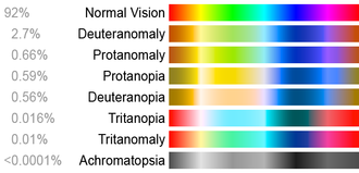
These color charts show how different colorblind people see compared to a person with normal color vision.
Types of color blindness and the terms used
| Cone system |
Red | Green | Blue | |||||||
| N=normal A=anomalous |
N | A | N | A | N | A | ||||
| 1 | Normal vision | Trichromat | Normal | |||||||
| 2 | Protanomaly | Anomalous Trichromat | Partially color blind | Red-green | ||||||
| 3 | Protanopia | Dichromat | Partially color blind | Red-green | ||||||
| 4 | Deuteranomaly | Anomalous Trichromat | Partially color blind | Red-green | ||||||
| 5 | Deuteranopia | Dichromat | Partially color blind | Red-green | ||||||
| 6 | Tritanomaly | Anomalous Trichromat | Partially color blind | Blue-yellow | ||||||
| 7 | Tritanopia | Dichromat | Partially color blind | Blue-yellow | ||||||
| 8 | Achromatopsia | Monochromat | Totally color blind | |||||||
| 9 | Tetrachromat | |||||||||
| 10 | ||||||||||
Based on clinical appearance, color blindness may be described as total or partial. Total color blindness is much less common than partial color blindness.[rx] There are two major types of color blindness: difficulty distinguishing between red and green, and difficulty distinguishing between blue and yellow.[rx][rx]
Red-Green Color Blindness
The most common types of hereditary color blindness are due to the loss or limited function of red cone (known as protan) or green cone (deutran) photopigments. This kind of color blindness is commonly referred to as red-green color blindness.
- Protanomaly – In males with protanomaly, the red cone photopigment is abnormal. Red, orange, and yellow appear greener and colors are not as bright. This condition is mild and doesn’t usually interfere with daily living. Protanomaly is an X-linked disorder estimated to affect 1 percent of males.
- Protanopia – In males with protanopia, there are no working red cone cells. Red appears as black. Certain shades of orange, yellow, and green all appear as yellow. Protanopia is an X-linked disorder that is estimated to affect 1 percent of males.
- Deuteranomaly – In males with deuteranomaly, the green cone photopigment is abnormal. Yellow and green appear redder and it is difficult to tell violet from blue. This condition is mild and doesn’t interfere with daily living. Deuteranomaly is the most common form of color blindness and is an X-linked disorder affecting 5 percent of males.
- Deuteranopia – In males with deuteranopia, there are no working green cone cells. They tend to see reds as brownish-yellow and greens as beige. Deuteranopia is an X-linked disorder that affects about 1 percent of males.
Blue-Yellow Color Blindness
Blue-yellow color blindness is rarer than red-green color blindness. Blue-cone (tritan) photopigments are either missing or have limited function.
- Tritanomaly – People with tritanomaly have functionally limited blue cone cells. Blue appears greener and it can be difficult to tell yellow and red from pink. Tritanomaly is extremely rare. It is an autosomal dominant disorder affecting males and females equally.
- Tritanopia – People with tritanopia, also known as blue-yellow color blindness, lack blue cone cells. Blue appears green and yellow appears violet or light grey. Tritanopia is an extremely rare autosomal recessive disorder affecting males and females equally.
Complete Color Blindness
People with complete color blindness (monochromacy) don’t experience color at all and the clearness of their vision (visual acuity) may also be affected. There are two types of monochromacy:
- Cone monochromacy – This rare form of color blindness results from a failure of two of the three cone cell photopigments to work. There is red cone monochromacy, green cone monochromacy, and blue cone monochromacy. People with cone monochromacy have trouble distinguishing colors because the brain needs to compare the signals from different types of cones in order to see color. When only one type of cone works, this comparison isn’t possible. People with blue cone monochromacy, may also have reduced visual acuity, near-sightedness, and uncontrollable eye movements, a condition known as nystagmus. Cone monochromacy is an autosomal recessive disorder.
- Rod monochromacy or achromatopsia – This type of monochromacy is rare and is the most severe form of color blindness. It is present at birth. None of the cone cells have functional photopigments. Lacking all cone vision, people with rod monochromacy see the world in black, white, and gray. And since rods respond to dim light, people with rod monochromacy tend to be photophobic – very uncomfortable in bright environments. They also experience nystagmus. Rod monochromacy is an autosomal recessive disorder.
Trichromacy
- Normal colour vision uses all three types of light cones correctly and is known as trichromacy. People with normal colour vision are known as trichromats.
Anomalous Trichromacy
- People with ‘faulty’ trichromatic vision will be colour blind to some extent and are known as anomalous trichromats. In people with this condition all of their three cone types are used to perceive light colours but one type of cone perceives light slightly out of alignment, so that there are three different types of effect produced depending upon which cone type is ‘faulty’
- The different anomalous conditions are protanomaly, which is a reduced sensitivity to red light, deuteranomaly which is a reduced sensitivity to green light and is the most common form of colour blindness and tritanomaly which is a reduced sensitivity to blue light and is extremely rare.
Dichromacy


Deuteranopia
- People with dichromatic colour vision have only two types of cones which are able to perceive colour i.e. they have a total absence of function of one cone type. Lack of ability to see colour is the easiest way to explain this condition but in actual fact it is a specific section of the light spectrum which can’t be perceived. For convenience we call these areas of the light spectrum ‘red’, ‘green’ or ‘blue’
Protanopia
- Protanopes are more likely to confuse:-
- Black with many shades of red
- Dark brown with dark green, dark orange and dark red
- Some blues with some reds, purples and dark pinks
- Mid-greens with some oranges
Deuteranopes
- Deuteranopes are more likely to confuse
Mid-reds with mid-greens
- Blue-greens with grey and mid-pinks
- Bright greens with yellows
- Pale pinks with light grey
- Mid-reds with mid-brown
- Light blues with lilac
Tritanopes
- The most common colour confusions for tritanopes are light blues with greys, dark purples with black, mid-greens with blues and oranges with reds. The images show how the beautiful colours of the pigments are lost to people suffering with each type of dichromatic vision.
Monochromacy (achromatopsia)

Normal Vision

Monochromacy
People with monochromatic vision can see no colour at all and their world consists of different shades of grey ranging from black to white, rather like only seeing the world on an old black and white television set. Achromatopsia is extremely rare, occuring only in approximately 1 person in 33,000 and its symptoms can make life very difficult. Usually someone with achromatopsia will need to wear dark glasses inside in normal light conditions.
Causes of Color Vision Deficiency
Color vision deficiencies can be classified as acquired or inherited.
- Acquired – Diseases, drugs (e.g., Plaquenil), and chemicals such as styrene[rx] or organic solvents[rx] may cause color blindness.[rx][rx]
- Inherited – There are three types of inherited or congenital color vision deficiencies: monochromacy, dichromacy, and anomalous trichromacy.
- Monochromacy – also known as “total color blindness”, is the lack of ability to distinguish colors (and thus the person views everything as if it were on a black and white television); caused by cone defect or absence. Monochromacy occurs when two or all three of the cone pigments are missing and color and lightness vision is reduced to one dimension.
- Rod monochromacy (achromatopsia) – is an exceedingly rare, nonprogressive inability to distinguish any colors as a result of absent or nonfunctioning retinal cones. It is associated with light sensitivity (photophobia), involuntary eye oscillations (nystagmus), and poor vision.
- Cone monochromacy – is a rare total color blindness that is accompanied by relatively normal vision, electroretinogram, and electrooculogram. Cone monochromacy can also be a result of having more than one type of dichromatic color blindness. People who have, for instance, both protanopia and tritanopia are considered to have cone monochromacy. Since cone monochromacy is the lack of/damage of more than one cone in retinal environment, having two types of dichromacy would be an equivalent.
- Dichromacy is hereditary – Protanopia and deuteranopia are hereditary and sex-linked, affecting predominantly males.
- Protanopia – is caused by the complete absence of red retinal photoreceptors. Protans have difficulties distinguishing between blue and green colors and also between red and green colors. It is a form of dichromatism in which the subject can only perceive light wavelengths from 400 nm to 650 nm, instead of the usual 700 nm. Pure reds cannot be seen, instead appearing black; purple colors cannot be distinguished from blues; more orange-tinted reds may appear as dim yellows, and all orange-yellow-green shades of too long a wavelength to stimulate the blue receptors appear as a similar yellow hue. It is present in 1% of males.
- Deuteranopia – affects hue discrimination in a similar way to protanopia, but without the dimming effect. Again, it is found in about 1% of the male population.[rx]
- Tritanopia – is a very rare color vision disturbance in which only the red and the green cone pigments are present, with a total absence of blue retinal receptors. Blues appear greenish, yellows and oranges appear pinkish, and purple colors appear deep red. It is related to chromosome 7; thus unlike protanopia and deuteranopia, tritanopia and tritanomaly are not sex-linked traits and can be acquired rather than inherited and can be reversed in some cases.
- Anomalous trichromacy – is a common type of inherited color vision deficiency, occurring when one of the three cone pigments is altered in its spectral sensitivity.
- Protanomaly – is a mild color vision defect in which an altered spectral sensitivity of red retinal receptors (closer to green receptor response) results in poor red-green hue discrimination. It is hereditary, sex-linked, and present in 1% of males. In contrast to other defects, in this case the L-cone is present but malfunctioning, whereas in protanopia the L-cone is completely missing.[rx]
- Deuteranomaly – caused by a similar shift in the green retinal receptors, is by far the most common type of color vision deficiency, mildly affecting red-green hue discrimination in 5% of European males. It is hereditary and sex-linked. In contrast to deuteranopia, the green-sensitive cones are not missing but malfunctioning.[rx]
- Tritanomaly – is a rare, hereditary color vision deficiency affecting blue-green and yellow-red/pink hue discrimination. It is related to chromosome 7.[rx] In contrast to tritanopia, the S-cone is malfunctioning but not missing.[rx]
Genetics
- Color blindness is typically an inherited genetic disorder. It is most commonly inherited from mutations on the X chromosome, but the mapping of the human genome has shown there are many causative mutations—mutations capable of causing color blindness originate from at least 19 different chromosomes and 56 different genes (as shown online at the Online Mendelian Inheritance in Man(OMIM)).
Other Causes
- Other causes – of color blindness include brain or retinal damage caused by accidents and other traumas which produce swelling of the brain in the occipital lobe, and damage to the retina caused by exposure to ultraviolet light (wavelengths 10 to 300 nm). Damage often presents itself later in life.
- Color blindness – may also present itself in the range of degenerative diseases of the eye, such as age-related macular degeneration, and as part of the retinal damage caused by diabetes. Vitamin A deficiency may also cause color blindness.[rx]
- Some subtle forms– of color blindness may be associated with chronic solvent-induced encephalopathy (CSE), caused by long-time exposure to solvent vapors.[rx]
- Red–green color blindness can be caused by ethambutol,[rx] a drug used in the treatment of tuberculosis.
- Parkinson’s disease (PD) – Because Parkinson’s disease is a neurological disorder, light-sensitive nerve cells in the retina where vision processing occurs may be damaged and cannot function properly.
- Cataracts – Clouding of the eye’s natural lens that occurs with cataracts can “wash out” color vision, making it much less bright. Fortunately, cataract surgery can restore bright color vision when the cloudy natural lens is removed and replaced with an artificial intraocular lens.
- Certain medications –For example, an anti-seizure drug called tiagabine has been shown to reduce color vision in about 41 percent of those taking the drug, although effects do not appear to be permanent.
- Leber’s hereditary optic neuropathy (LHON) – This type of inherited optic neuropathy can affect even carriers who don’t have other symptoms but do have a degree of color blindness. Red-green color vision defects primarily are noted with this condition.
- Kallman’s syndrome. This inherited condition involves failure of the pituitary gland, which can lead to incomplete or unusual gender-related development such as of sexual organs. Color blindness can be one symptom of this condition.
Symptoms of Color Vision Deficiency
The symptoms include
- Trouble seeing colors and the brightness of colors in the usual way;
- Inability to tell the difference between shades of the same or similar colors. This happens most with red and green, or blue and yellow.
- Difficulties recognising and identifying different colours beyond the age of around four years
- The inability to separate things by their colour.
Except in the most severe form, color blindness does not affect the sharpness of vision. The inability to see any color at all and to see everything only in shades of gray is called achromatopsia. This rare condition is often associated with:
- amblyopia
- nystagmus
- light sensitivity, and
- poor vision
Diagnosis of Color Vision Deficiency
Eye care professionals use a variety of tests to diagnose color blindness. These tests can quickly diagnose specific types of color blindness.
- The Ishihara Color Test – is the most common test for red-green color blindness. The test consists of a series of colored circles, called Ishihara plates, each of which contains a collection of dots in different colors and sizes. Within the circle are dots that form a shape clearly visible to those with normal color vision, but invisible or difficult to see for those with red-green color blindness.
- The newer Cambridge Color Test – uses a visual array similar to the Ishihara plates, except displayed on a computer monitor. The goal is to identify a C shape that is different in color from the background. The “C” is presented randomly in one of four orientations. When test-takers see the “C,” they are asked to press one of four keys that correspond to the orientation.
- The anomaloscope – uses a test in which two different light sources have to be matched in color. Looking through the eyepiece, the viewer sees a circle. The upper half is a yellow light that can be adjusted in brightness. The lower half is a combination of red and green lights that can be mixed in variable proportions. The viewer uses one knob to adjust the brightness of the top half, and another to adjust the color of the lower half. The goal is to make the upper and lower halves the same brightness and color. The HRR Pseudoisochromatic Color Test is another red-green color blindness test that uses color plates to test for color blindness.
- Ishihara color test. This checks for red-green color blindness. The doctor will ask you to look at a series of circles (also called plates) with dots of different colors and sizes. Some of the dots form shapes or one- or two-digit numbers. If you have trouble seeing red and green, those shapes will be hard to see, or you may not see them at all.
- Cambridge color test – This is a lot like the Ishihara test, except that you look at a computer screen. You’ll try to find a “C” shape that’s a different color than the background. It pops up randomly. When you see it, you press one of four keys.
- Anomaloscope – You look through an eyepiece and see a circle. The top half of the circle is a yellow light. The bottom half is made up of red and green lights. You turn knobs until both halves are the same color and brightness. Doctors use this test to check for trouble seeing red and green.
- Farnsworth-Munsell 100 hue test – This uses blocks or pegs that are different shades of the same color. Your task is to line them up a certain way. This test checks to see if you can pick up slight color changes. Companies that need workers to see colors correctly sometimes use it.
- Farnsworth lantern test – The U.S. military uses this to see if recruits have a mild or severe form of color blindness. You can serve in the armed forces if your condition is mild.
Treatment of Color Vision Deficiency
There is generally no treatment to cure color deficiencies. ″The American Optometric Association reports a contact lens on one eye can increase the ability to differentiate between colors, though nothing can make you truly see the deficient color.″[rx]
- Lenses – Optometrists can supply colored spectacle lenses or a single red-tint contact lens to wear on the non-dominant eye, but although this may improve discrimination of some colors, it can make other colors more difficult to distinguish. A 1981 review of various studies to evaluate the effect of the X-chrom contact lens concluded that, while the lens may allow the wearer to achieve a better score on certain color vision tests, it did not correct color vision in the natural environment.[rx] A case history using the X-Chrom lens for a rod monochromat is reported[rx] and an X-Chrom manual is online.[rx]
- Lenses that filter certain wavelengths – of light can allow people with a cone anomaly, but not dichromacy, to see better separation of colors, especially those with classic “red/green” color blindness. They work by notching out wavelengths that strongly stimulate both red and green cones in a deuter- or protanomalous person, improving the distinction between the two cones’ signals. As of 2012, sunglasses that notch out color wavelengths are available commercially.[rx]
- Apps –Many mobile applications have been developed to help colorblind people to view colors in a better way. Many applications launch a simulation of colorblindness to allow people with normal vision to understand how people with color blindness see the world
How Does Color Blindness Affect Daily Life?
- Color blindness can make it difficult to read color-coded information such as bar graphs and pie charts. This can be particularly troubling for children who aren’t yet diagnosed with color blindness, since educational materials are often color-coded. Children with red-green color blindness may also have difficulty reading a green chalkboard when yellow chalk is used. Art classes, which require selecting appropriate colors of paint or crayons, may be challenging.
- Color blindness can go undetected for some time since children will often try to hide their disorder. It’s important to have children tested, particularly boys, if there is a family history of color blindness. Many school systems offer vision screening tests that include color blindness testing. Once a child is diagnosed, he or she can learn to ask for help with tasks that require color recognition.
- Simple everyday tasks like cooking meat to the desired color or selecting ripe produce can be a challenge for adults. Children might find food without bright color as less appetizing. Traffic lights pose challenges, since they have to be read by the position of the light. Since most lights are vertical, with green on bottom and red on top, if a light is positioned horizontally, a color blind person has to do a quick mental rotation to read it. Reading maps or buying clothes that match colors can also be difficult. However, these are relatively minor inconveniences and most people with color blindness learn to adapt.
References

![]()
People Also Reading
 Color Vision Deficiency Symptoms, Treatment
 Color Vision Deficiency Treatment, Symptoms, Life Style
 Color Vision Deficiency Prevention, Symptoms, Surgery
 Color Vision Deficiency Test Diagnosis, Prevention
 Color Blindness Causes, Symptoms, Treatment
 Color Blindness Types, Causes, Symptoms, Treatment
 Color Blindness Symptoms, Diagnosis, Treatment
 Color Blindness, Causes, Symptoms, Diagnosis, Treatment
 Color Blindness Treatment, Types, Complications
 Color Blindness Diagnosis, Treatment, Prevention
 Black Color Under Eye, Causes, Symptoms, Treatment
 Black Color Under Eye Causes, Symptoms, Treatment
About the author