Spinal Cord Injury; Causes, Symptoms, Diagnosis is damage to the spinal cord that causes temporary or permanent changes in its function. Symptoms may include loss of muscle function, sensation, or autonomic function in the parts of the body served by the spinal cord below the level of the injury. Injury can occur at any level of the spinal cord and can be complete injury, with a total loss of sensation and muscle function, or incomplete, meaning some nervous signals are able to travel past the injured area of the cord. Depending on the location and severity of damage, the symptoms vary, from numbness to paralysis to incontinence. Long-term outcomes also range widely, from full recovery to permanent tetraplegia (also called quadriplegia) or paraplegia. Complications can include muscle atrophy, pressure sores, infections, and breathing problems.
Spinal Cord Injury; Causes, Symptoms, Diagnosis
Defined as spinal cord injury with some preserved motor or sensory function below the injury level including
voluntary anal contraction (sacral sparing)
sacral sparing critical to separate complete vs. incomplete injury
OR palpable or visible muscle contraction below the injury level
OR perianal sensation present
Epidemiology
11,000 new cases/year in the US
34% incomplete tetraplegia
central cord syndrome most common
17% incomplete paraplegia
remaining 47% are complete
Prognosis most important prognostic variable relating to neurologic recovery is the completeness of the lesion (severity of neurologic deficit)
Nervous system
Central nervous system
includes the brain & spinal cord the spinal cord ends at L3 at birth and L1 at maturity
Peripheral nervous system’ contains the
cranial nerves
peripheral nerves
Autonomic nervous system > sympathetic system
A total of 22 ganglia (3 cervical, 11 thoracics, 4 lumbar, 4 sacral) > cervical ganglia
the three cervical include the stellate, middle, and superior
the middle ganglion is most at risk at the level of C6 where it lies close to the medial border of the longus colli muscles
injury to the middle ganglion/sympathetic chain will lead to Horner’s syndrome
Parasympathetic nervous system
hypogastric plexus formed by S2, S3, S4 parasympathetic fibers and lumbar sympathetic fibers (splanchnic nerves)
The spinal cord extends from brainstem to inferior border of L1
conus medullaris is termination of the spinal cord
filum terminal is a residual fragment of the spinal cord that extends from conus medullaris to sacrum.
thecal sac the dural surrounded sac that extends from the spinal cord and contains CSF, nerve roots and the cauda equina
cauda equina nerve roots and filum terminal surrounded by dura that extend from the spinal cord
Embryology of the spinal cord
Neural Tube
becomes spinal cord
formed from the primitive Streak, which turns into the primitive (midsagittal) groove > which turns into the Neural Tube
failure of the neural tube to close leads to
anencephaly when it fails to close cranially
spinal Bifida occulta, meningocele, myelomeningocele when it fails to close distally
Neural crest
Forms dorsal to neural tube becomes the
peripheral nervous system
pia mater
spinal ganglia
sympathetic trunk
Notochord
Forms ventral to neural tube >becomes
vertebral bodies
intervertebral discs
nucleus pulposus from cells of notochord
annulus from sclerotomal cells associated with segmentation
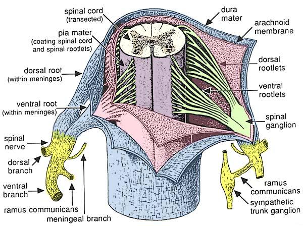
Layers of the spinal cord
Layers of the spinal cord include the
dura mater (outside)
arachnoid
pia mater (inside)
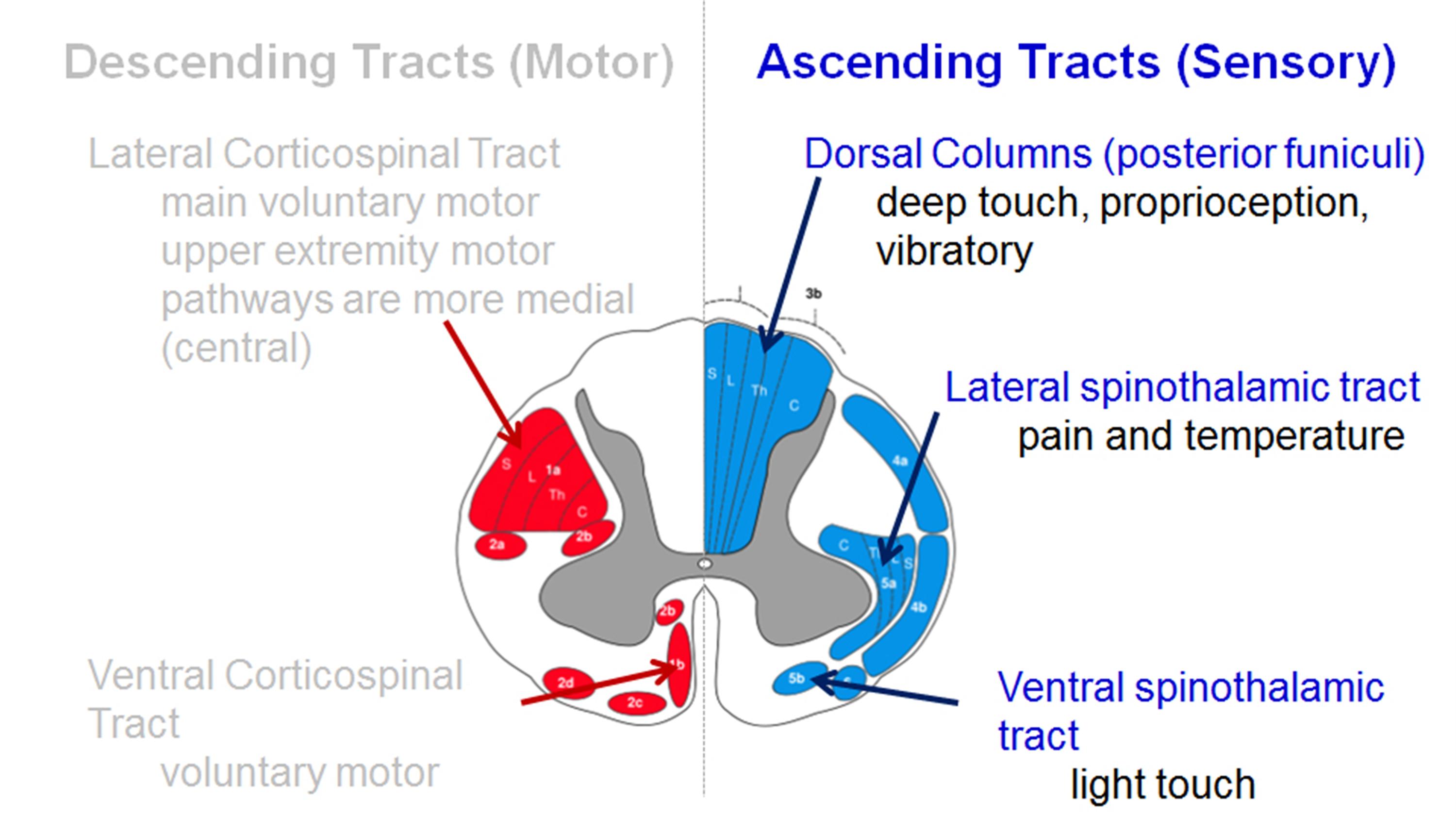
Spinal Cord Functional Tracts
Ascending Tracts (Sensory)
dorsal columns (posterior funiculi)
deep touch, proprioception, vibratory
lateral spinothalamic tract
pain and temperature
a site of chordotomy to alleviate intractable pain
upper extremity motor pathways are more medial(central) which explains why a central cord injury affects the upper extremities more than the lower extremities
ventral corticospinal tract
voluntary motor
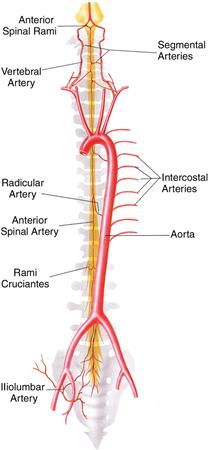
Blood Supply of the spinal cord
spinal cord blood supply provided by
anterior spinal artery
primary blood supply of anterior 2/3 of the spinal cord, including both the lateral corticospinal tract and ventral corticospinal tract
posterior spinal artery (right and left)
primary blood supply to the dorsal sensory columns
Artery of Adamkiewicz
the largest anterior segmental artery
typically arises from a left posterior intercostal artery, which branches from the aorta, and supplies the lower two-thirds of the spinal cord via the anterior spinal artery
significant variation exists
in 75% it originates on the left side between the T8 and L1 vertebral segments
Cerebral Spinal Fluid
unction
a colorless fluid that occupies the subarachnoid space surrounding the brain, spinal cord, and ventricular system
the subarachnoid space is between the arachnoid mater and pia mater
provides mechanical and immunological protection for the brain, spinal cord, and thecal sac
Production
location
most human cerebrospinal fluid (CSF) is produced by the choroid plexus in the third, fourth, and lateral ventricles of the brain.
CSF is an ultrafiltrate of blood plasma through the permeable capillaries of the choroid plexus
volume
total CSF volume between brain, spinal cord, and thecal sac is ~150 mL
CSF formation occurs at a rate of ~500mL per day
thus the total amount of CSF is turned over 3-4 times per day
Cauda equina syndrome, which involves damage to nerve roots at the caudal end of the cord, is not a spinal cord syndrome. However, it mimics conus medullaris syndrome, causing distal leg paresis and sensory loss in and around the perineum and anus (saddle anesthesia), as well as bladder, bowel, and pudendal dysfunction (eg, urinary retention, urinary frequency, urinary or fecal incontinence, erectile dysfunction, loss of rectal tone, abnormal bulbocavernosus and anal wink reflexes). In cauda equina syndrome (unlike in spinal cord injury), muscle tone and deep tendon reflexes are decreased in the legs.
Spinal Contusions: The most common type of spinal cord injury. The spinal cord is bruised but not severed. Inflammation and bleeding occur near the injury as a result of the injury.
Injuries to Individual Nerve Cells: Loss of sensory and motor functions in the area of the body to which the injured nerve root corresponds.
Flexion Fracture Pattern
Complete and Incomplete Spinal Cord Injury
The terms, ‘Complete,’ and, ‘Incomplete,’ in reference to a spinal cord injury is associated with the type of lesion in the person’s spine.
A person who is completely paralyzed below the lesion has a, ‘Complete,’ SCI.
A person who experiences partial paralysis below the lesion on their spine has an, ‘Incomplete,’ SCI.
Persons with incomplete SCI might have some sensation below the lesion, yet have no movement. There are a number of types of incomplete spinal cord injuries. Every person with an incomplete spinal cord injury is unique in regards to their injury.
Compression fracture: While the front (anterior) of the vertebra breaks and loses height, the back (posterior) part of it does not. This type of fracture is usually stable and rarely associated with neurologic problems.
Axial burst fracture: The vertebra loses height on both the front and back sides. It is often caused by a fall from a height and landing on the feet.
Extension Fracture Pattern
Flexion/distraction (Chance) fracture: The vertebra is literally pulled apart (distraction). This can happen in accidents such as a head-on car crash, in which the upper body is thrown forward while the pelvis is stabilized by a lap seat belt.
Rotation Fracture Pattern
Transverse process fracture: This fracture is uncommon and results from rotation or extreme sideways (lateral) bending, and usually does not affect stability.
Fracture-dislocation: This is an unstable injury involving bone and/or soft tissue in which a vertebra may move off an adjacent vertebra (displaced). These injuries frequently cause serious spinal cord compression.
method to scale ASIA classification
Grading Scales
There are two well-known scales used to grade and prognosticate SCI. The Frankel scale was developed during World War I, but is less commonly used today. It is a basic scale that grades the SCI based on level and is used to evaluate functional recovery. There are five grades used in the Frankel scale, which essentially divide completely versus incomplete spinal injuries as follows:
A — complete paralysis (no motor/sensory below level of injury);
B — sensory present below the level of injury;
C — incomplete injury with motor and sensory function below the level of injury;
D — fair to good motor function below the level of injury; and
E — normal function (no motor of the sensory deficit).3
The American Spinal Injury Association (ASIA) Impairment Scale (AIS) is a more widely used and more refined scale. Based on the Frankel scale’s five grading levels, the AIS was originally developed in 1982 and has undergone six revisions, with the most recent occurring in 2002. The AIS differs from the Frankel scale in that it more clearly defines complete and incomplete injury by determining sacral sparing (presence of rectal motor function or sensory function at S4-S5 dermatome), determining the presence of neurologic level of injury using sensory and motor evaluation in bilateral extremities, and by determining, in incomplete injuries, where partial zones of sensory or motor preservation exist.
Central Cord Syndrome
The most common of all partial cord syndromes is central cord syndrome, which is distinguished from the other cord syndromes by the fact that the upper extremities are significantly more affected from the motor perspective than the lower extremities are. The most common mechanism of injury is a hyperextension injury, and it is usually seen after a fall in an older population with preexisting spinal stenosis or arthritis. The injury to the spinal cord affects the central portions of the corticospinal and spinothalamic tracts, resulting in the disproportionate pattern of symptoms between the upper and lower extremities. Patients typically have greater weakness in the proximal muscles than in the distal ones. Sensory symptoms are also appreciable, with some patients presenting with dysesthesias of their upper extremities as their predominant symptom.
Epidemiology
incidence
most common incomplete cord injury
demographics
often in elderly with minor extension injury mechanisms
due to anterior osteophytes and posterior infolded ligament flavum
Pathophysiology
believed to be caused by spinal cord compression and central cord edema with selective destruction of lateral corticospinal tract white matter
anatomy of spinal cord explains why upper extremities and hand preferentially affected
hands and upper extremities are located “centrally” in the corticospinal tract
Symptoms
the weakness with hand dexterity most affected
hyperpathia
burning in distal upper extremity
physical exam
loss
motor deficit worse in UE than LE (some preserved motor function)
hands have a more pronounced motor deficit than arms
good prognosis although full functional recovery rare
usually ambulatory at final follow up
usually, regain bladder control
upper extremity and hand recovery is unpredictable and patients often have permanent clumsy hands
Recovery occurs in a typical pattern
lower extremity recovers first
bowel and bladder function next
proximal upper extremity next
hand function last to recover
Anterior Cord Syndrome
Anterior cord syndrome is usually sustained due to a hyperflexion injury to the cervical cord but can occur anywhere in the spinal column. Hyperflexion of the cord causes direct contusion to the cord or can result in the protrusion of disc contents, bony fragments that have fractured, or, rarely, can cause direct laceration or thrombosis to the anterior spinal artery. Since the injury to the cord is bilateral, the pattern of symptoms that accompany this injury includes bilateral motor paralysis and loss of pinprick, temperature, and pain sensation below the level of injury. Since the posterior aspect of the cord is preserved, so is proprioception and vibratory sensation.
The overall prognosis for anterior cord syndrome is poor. Improvement in motor function can be seen within the first 24 hours following injury, but usually does not occur after the first day. After 30 days following injury, there is little to no additional recovery of function.
A condition characterized by
motor dysfunction
dissociated sensory deficit below the level of SCI
Pathophysiology >Injury to anterior spinal cord caused by
direct compression (osseous) of the anterior spinal cord
anterior spinal artery injury
anterior 2/3 spinal cord supplied by the anterior spinal artery
Mechanism
usually, result of flexion/ compression injury
lower extremity affected more than upper extremity
loss
LCT (motor)
LST (pain, temperature)
preserved
DC (proprioception, vibratory sense)
Prognosis
worst prognosis of incomplete SCI
most likely to mimic complete cord syndrome
10-20% chance of motor recovery
Brown-Séquard Syndrome
Brown-Séquard syndrome is an anatomic or functional hemisection of the cord, which has several potential causes. From a trauma perspective, Brown-Séquard is commonly the result of penetrating trauma to the spinal cord. However, more commonly it is due to inherent spinal or compressive lesions such as tumors or epidural hematomas. Classic Brown-Séquard syndrome, in its purest form, is described as a loss of ipsilateral motor function, proprioception, vibratory and pressure sensation, and contralateral loss of temperature and pain sensation below the level of injury. Although the pure form of Brown-Séquard syndrome is rarely seen, a partial form of Brown-Séquard is more common. Interestingly, because the fibers of the lateral spinothalamic tract decussate one or two levels above or below where the injury may occur, it is possible to see ipsilateral pain and temperature sensory loss above the level of injury.
Caused by complete cord hemitransection
usually seen with penetrating trauma
Exam
ipsilateral deficit
LCS tract
motor function
dorsal columns
proprioception
vibratory sense
contralateral deficit
LST
pain
temperature
spinothalamic tracts cross at spinal cord level (classically 2-levels below)
Prognosis
excellent prognosis
99% ambulatory at final follow up
best prognosis for function motor activity
Posterior Cord Syndrome
Introduction
very rare
Exam
loss
proprioception
preserved
motor, pain, light touch
Neurological History and Examination
Taking a detailed history and performing a careful examination can help the doctor to determine the site of a specific neurological lesion and reach a diagnosis, or at least differential diagnoses. A systematic approach is required.
This is a general article, attempting to cover all aspects of neurological history and examination. You are referred to other related articles were relevant for more detail.
Mental state examination may also be an important consideration and this is covered in the separate Mini-Mental State Examination (MMSE) article.
Observation of the patient
Gait
Look at the patient’s gait as they enter the room.
Note if there evidence of, for example, hemiparesis, foot drop, ataxic gait, a typical Parkinsonian gait.
See separate Abnormal Gait and Gait Abnormalities in Children articles.
Speech
Note any problem with articulation (dysarthria). Here comprehension is retained and speech construction is normal. There is usually weakness or incoordination of the orolingual muscles. Ask the patient to say ‘West Register Street’ if you are uncertain.
Note any problem with phonation (dysphonia). This is usually due to laryngeal problems which can cause voice hoarseness. There may be reduced speech volume.
Note any problem with language function (dysphasia). This is due to a lesion in the language areas of the dominant hemisphere.
Involuntary movements
Establish whether there is evidence of involuntary movements – for example, tremor, tics, chorea, hemiballismus, or orofacial dyskinesias.
History
Specific emphasis should be placed on the following:
Presenting complaint / Ask about the symptoms
What are they?
Which part of the body do they affect? Are they localized or more widespread?
When did they start?
How long do they last for?
Were they sudden, rapid or gradual in onset? Is there a history of trauma?
Are the symptoms static or deteriorating, or are there exacerbations and remissions? For example, worsening of symptoms with hot environments – eg, sauna, hot bath or hot weather in demyelinating disorders (called Uhthoff’s sign).
Does anything trigger the symptoms – eg, exercise, sleep, posture or external stimuli such as light or smell?
Ask about any associated symptoms (other features of neurological disease):
Some neurological problems can present years after a causative event.
Enquire about other medical problems, past and present. These may give clues to the diagnosis. For example:
A person in atrial fibrillation may be producing multiple tiny emboli.
There may be vascular problems or recurrent miscarriage to suggest antiphospholipid syndrome.
There may be diabetes mellitus.
Ask about pregnancy, delivery, and neonatal health.
Ask about any infections, convulsions or injuries in infancy, childhood or adult life. Particularly ask about the head or spinal injury, meningitis or encephalitis.
The systematic inquiry is very important here. For example:
Loss of weight and appetite may suggest malignancy and this may be a paraneoplastic syndrome.
The gain in weight may have precipitated diabetes mellitus.
Polyuria may suggest diabetes mellitus. The difficulty with micturition or constipation may be part of the neurological problem but was not volunteered in the general history. In men, enquire about erectile dysfunction.
Social history
Note smoking and drinking habits. Alcohol is a significant neurotoxin, both centrally and peripherally.
Ask about drugs, including prescribed, over-the-counter and illicit (such as cocaine usage that can be linked to cardiovascular problems). This includes complementary and alternative medicines.
Ask about occupation and what it involves. There may be exposed to toxins. Is repetitive strain injury likely? Is there prolonged visual work which may predispose to a tension-type headache or a migraine? The job may involve driving but the patient has admitted to convulsions. He/she may work at heights or in a dangerous environment.
Ask about marital status. Has there been recent bereavement or divorce which may have affected symptoms?
Ask about sexual orientation and consider the likelihood of sexually transmitted infection – eg, syphilis, HIV.
Family history
Consider if there may be a genetic basis or predisposition. For example:
A cousin with Duchenne muscular dystrophy or Becker’s muscular dystrophy would be very important for a boy who cannot run like his peers.
Huntington’s chorea is unusual in that it is a familial disease that does not present until well into adult life.
A family history of, for example, type 2 diabetes mellitus, cerebral aneurysm, neuropathies, epilepsy, migraine or vascular disease may be important.
Examination
Examination of speech
Look for spontaneous speech, fluency and use of appropriate words during conversation.
Ask the patient to name objects.
Ask the patient to carry out some commands to assess their comprehension.
Ask the patient to read aloud. This can show evidence of any dyslexia.
Ask the patient to repeat a simple sentence. Inability to do this suggests a conduction dysphasia.
Look at the patient’s handwriting. There may be problems with the form, grammar or syntax, which may suggest a more global language problem and not just a speech disorder.
Examination of the neck
Examine the neck movements:
Is there evidence of degenerative disease which may be producing radicular symptoms in the upper limbs? Examine flexion, extension, and rotation.
Look for Lhermitte’s sign: neck flexion causes an electric shock-like feeling on the limbs. It is due to disease in cervical spinal cord sensory tracts (seen in, for example, multiple sclerosis, syringomyelia, tumors) .
Is there any neck stiffness? This can be a sign of meningeal irritation. The chin can normally touch the chest when the neck is flexed but this is not possible if neck stiffness is present. This may be a sign of meningitis or subarachnoid hemorrhage.
Palpate the supraclavicular fossae:
Look for enlarged lymph nodes or cervical ribs.
Listen for any bruits:
Listen at the carotid bifurcation at the angle of the jaw for carotid bruits.
Listen over the supraclavicular fossa for vertebral or subclavian bruits.
A common carotid bruit may be heard by listening between these two sites.
Listen with the bell of the stethoscope over a closed eyelid for bruits due to cerebral arteriovenous malformations.
Listen for cardiac murmurs to ensure that any bruit heard is not just due to the transmission of these.
Note that just because a bruit is not heard, it does not mean that there is no significant stenosis present.
Cranial nerves
Examination of the cranial nerves takes practice. For their function and examination, see separate Examination of the Cranial Nerves article. This should include testing of the olfactory, optic, oculomotor, trochlear, abducent, trigeminal, facial, vestibulocochlear, glossopharyngeal, vagus, accessory and hypoglossal nerves.
Examination of the sensory system
See separate Neurological Examination of the Upper Limbs and Neurological Examination of the Lower Limbs articles. Both the upper and lower limbs should be examined. Work in a methodical way. A logical progression is required when examining each sensory modality. The following sensory modalities should be tested:


About the author