Foot Bones is a complex structure comprised of over 26 bones, 30 joints, numerous tendons, ligaments, and muscles responsible for our ability to stand upright, supporting the weight of the entire body and provides the base for the mechanism for bipedal gait. The foot corresponds to the portion of the lower extremity distal to the ankle and divides into hind, mid and forefoot. The articular surfaces of each bone have a covering of hyaline cartilage, and each joint is invested by a capsule and supported by ligamentous structures. The numerous muscles of the foot are attached to the bones by tendons, which allows the contraction of the muscles to exert force on the osseous structures. This complex anatomy allows the foot to adapt to uneven terrain during heel strike and become a stiff lever for better propulsion during step off. The importance of these structures to activities of daily living not surprisingly results in injury or pain to the feet as a common cause for presentation to the emergency department or primary care clinics. While the ligamentous and soft tissues are essential to foot function, this paper will primarily discuss the osseous structures.[rx][rx][rx][rx]

Structure and Function of Foot Bones
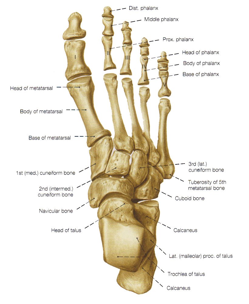
As previously stated, the foot is comprised of 26 bones distal to the ankle, which can subdivide into the hindfoot, midfoot, and forefoot.[rx] The osseous structures can also be broken down into groups termed tarsal, metatarsal, and phalanges.[rx][rx][rx] The talus and calcaneus, two of the seven tarsal bones, comprise the hindfoot. The hindfoot joins to the more proximal lower extremity at the ankle joint, which is comprised of the tibia, fibula, and talus. The distal tibia (medial malleolus, posterior malleolus, and tibial plafond) and distal fibula (lateral malleolus) form a recess termed the mortis, which articulates with the body of the talus termed the talar dome.[rx][rx][rx][rx]
The talus is the second largest tarsal bone. It slopes inferomedially terminating as the convex cartilage covered head that articulates with the navicular. The body of the talus articulates with the calcaneus at the subtalar joint, comprised of multiple facets, posterolateral (posterior facet), middle facet at the sustentaculum tali, located medially, and anterior facet, anteromedially. The superior talar body forms the talar dome and is the articular surface of the tibiotalar joint. The talar neck lies between the body and head of the talus and forms the roof of the tarsal canal that opens laterally as the sinus tarsi. The posterior process of the talus consists of the medial and lateral tubercles with the flexor hallucis longus tendon lying within the groove between them. An elongated lateral tubercle is a normal anatomic variant called a Stieda process. An osseous ridge is ordinarily present at the distal aspect of the talar neck and represents the site of attachment of the joint capsule and ligamentous structures.[rx][rx][rx]
The talus is approximately 60% covered with hyaline cartilage and is devoid of muscular or tendinous attachments. This unique anatomy renders it susceptible to avascular necrosis despite arterial contributions from 3 arteries. The posterior tibial artery supplies the body, and the anterior tibial artery supplies the superior half of the head and neck. Approximately one cm proximal to the division into the medial and lateral plantar arteries, the tarsal canal artery arises from the posterior tibial artery.[rx][rx][rx] The tarsal sinus artery provides the central and lateral two-thirds of the talar body and anastomoses with the artery of the tarsal sinus and gives off the deltoid branches that supply the remaining talus.[rx]
Inferior to the talus is the largest tarsal bone, the calcaneus. The calcaneus articulates with the talus superiorly at the anterior, middle, and posterior facets. Each facet is an encapsulated synovial joint with the middle and anterior facets within the same joint capsule. Between the posterior and middle facets, the calcaneal sulcus runs posteromedially and forms the floor of the sinus tarsi. Anteriorly, the calcaneus articulates with the cuboid.[rx] The calcaneus is meant to withstand the stresses of weight-bearing, and approximately 50% of our body weight is borne on the subtalar joints and calcaneus, generally split equally between the two feet in the absence of altered weight-bearing/gait. On radiographs, the traction trabeculae radiate from the inferior cortex, and compression trabeculae radiate posteriorly from the anterior and posterior subtalar facets with a small area with relatively few trabeculations called the neutral triangle.[rx][rx]
The midfoot consists of the cuboid, the navicular, the cuneiforms (medial, middle, lateral), and five of the seven tarsal bones. The navicular is curved concave proximally and convex distally. The concave posterior portion of the navicular receives the ellipsoid shaped talar head forming a ball and socket joint similar to the hip and named coxa pedis.[rx] There are three facets along the distal convexity that form articulations with the cuneiforms and one inferolateral facet forming the cuboid articulation. Medially, the posterior tibial tendon attaches to the navicular tuberosity.[rx]
The cuboid is a roughly cube-shaped bone that ossifies between the 9th fetal month and six months postnatally. Proximally, it articulates with the calcaneus and distally with the lateral cuneiform and 4 and 5 metatarsals. Rarely, it will articulate with the head of the talus. At the inferolateral margin of the cuboid, there is a sulcus containing the peroneus longus tendon.[rx][rx][rx]
The talonavicular and calcaneocuboid articulations link the hindfoot to the midfoot, collectively called the Chopart joint, named for the famous French surgeon Francois Chopart.[rx]
The Cuneiforms articulate proximally with the navicular and distally with the first-third metatarsals and form an arch with a keystone like configuration. The medial cuneiform is the largest and has articulating facets with the first metatarsal distally, a vertical square-shaped facet for the dorsomedial 2nd metatarsal base, laterally with the intermediate or middle cuneiform and proximally with the navicular. The medial cuneiform usually has two ossification centers and ossifies in the 2nd year of life. Importantly, the Lisfranc ligament complex attaches to the distal/lateral aspect of the medial cuneiform running obliquely to the base of the second metatarsal and transmits a large axial load into the second metatarsal during ambulation.[rx][rx][rx]
The middle (intermediate cuneiform) is the smallest of the three cuneiforms. It articulates with the navicular and the second metatarsal, proximally and distally, respectively, as well as the other cuneiforms medially and laterally. Since it is shorter than the adjacent cuneiforms, it forms a mortise and holds the second metatarsal base. The medial cuneiform has one ossification center and ossifies in the third year of life.[rx][rx][rx]
The lateral (third cuneiform) articulates with the cuboid, navicular, third metatarsal, and the middle cuneiform. The plantar surface receives slips from the posterior tibial tendon, and sometimes the flexor hallucis brevis.[rx]
The forefoot is composed of the five metatarsals and the 14 phalanges. Each metatarsal can subdivide into three contiguous regions: base, shaft (diaphysis), and head.
The junction of the midfoot to the forefoot is at the tarsometatarsal articulations collectively termed the LisFranc joint, named for the French surgeon Jacques LisFranc de Saint-Martin. The first metatarsal articulates with the medial cuneiform, the second metatarsal with the middle cuneiform, the third with the lateral, and the fourth/fifth with the cuboid joining the midfoot to the forefoot.[rx]
The first (greater) metatarsal deserves special attention. At the plantar aspect of the head, there are medial and lateral grooves to accommodate the sesamoid bones. The intersesamoidal ridge separates these grooves. The intersesamoidal ligament links the medial and lateral sesamoid bones. These structures form a portion of the plantar plate complex.[rx][rx]
The 14 phalanges are analogous to the fingers with two phalanges in the great toe and three in each of the remaining digits. Segmental anomalies are not infrequent.
Functionally, the osseous structures of the foot form three arches. The calcaneus, cuboid, fourth and fifth rays form the rigid lateral arch. The medial arch can dynamically vary in shape, allowing for uneven terrain and has the medial three rays, cuneiforms, navicular, talus, and calcaneus. Finally, the transverse arch runs obliquely along the tarsometatarsal joints.[rx]
Seven tarsal bones
-
Cuboid bone
-
Calcaneus or heel bone
-
Three cuneiforms (medial, middle and lateral)
-
Navicular bone
-
The talus bone which is just below the ankle joint
Five metatarsal bones: number 1 to 5 medial to lateral
Fourteen Phalanges
-
The first digit has two phalanges.
-
The second through fifth digits each have three phalanges
Sesamoid bones
-
The foot also has sesamoid bones that help improve stability and function. The two sesamoid bones are near the first metatarsal bone, where it connects to the big toe. Both sesamoids are within the tendon of flexor hallucis brevis. One sesamoid is usually located on the lateral aspect of the first metatarsal, whereas the other one is often on the medial side. In some individuals, only a single sesamoid may be present near the first metatarsal phalangeal joint.
Foot compartments
-
The forefoot contains the phalanges and metatarsals.
-
The midfoot consists of the five tarsal bones, three cuneiforms, the navicular, and the cuboid.
-
The hindfoot is composed of two tarsal bones, the calcaneus and the talus.
Foot joints (major)
-
Subtalar – articulation between the talus and calcaneus comprised of 6 facets, three on each bone, divided into two joints, anterior (anterior and middle facets) and posterior.[rx]
-
Chopart – Also known as the midtarsal joint. Joins the hindfoot to the midfoot. It is made up of the talonavicular and calcaneocuboid articulations. Named for the French surgeon Francois Chopart.[rx]
-
LisFranc – Junction of the mid and forefoot. It is comprised of the tarsometatarsal joints. Named for the French surgeon Jacques LisFranc de Saint-Martin.[rx]

Blood Supply and Lymphatics of Foot Bones
The anterior and posterior tibial arteries perfuse the foot. The anterior tibial artery bifurcates, becoming the dorsalis pedis along the dorsal/medial aspect of the foot and the lateral tarsal artery. Together these vessels supply perfusion to the dorsal foot. Distally these vessels converge, forming the transversely running arcuate artery. The posterior tibial artery is palpable at the level of the medial malleolus posteriorly in the region of the proximal tarsal tunnel. It courses anteriorly along the medial aspect of the ankle and plantar hindfoot giving off the artery of the tarsal canal before forming the medial and lateral plantar arteries. The medial and lateral plantar arteries anastomose distally as the transversely running deep plantar arch. Terminal branches of the arcuate and deep plantar arch perfuse the toes. The fibular or peroneal artery runs along the posterior/lateral ankle and hindfoot. There are communicating arteries and anastomosis between the branches of the anterior tibial, posterior tibial, and fibular arteries providing collateral circulation.[rx][rx][rx]
Nerves of Foot Bones
Branches of the saphenous, superficial fibular, deep fibular, medial plantar, lateral plantar, and calcaneal nerves are responsible for innervation of the foot.[rx]
Sensation to the medial ankle and foot derives from the saphenous nerve, a branch of the femoral nerve. This nerve can sometimes suffer an injury during Achilles tendon repair resulting in pain and burning sensation in the region of the medial hindfoot.[rx]
The common fibular nerve divides into the superficial and deep fibular nerves near the level of the knee. The superficial fibular nerve is a terminal branch of the common fibular nerve supplying sensation to the majority of the dorsal foot. The deep fibular nerve innervates the extensor digitorum brevis and extensor hallucis brevis muscles as well as providing sensation to the first web space (between first and second digits).[rx]
The tibial nerve branches into the medial and lateral plantar nerves at the level of the ankle on the medial side (tarsal tunnel). The medial plantar nerve supplies sensation to the plantar aspect of the first through third digits and medial aspect of the fourth digit. Additionally, it provides motor innervation to the flexor digitorum brevis, flexor hallucis brevis, abductor hallucis brevis, and 1st lumbrical.[rx][rx] The lateral plantar branch supplies sensation to the lateral plantar aspect of the 4th digit and the entire plantar aspect of the 5th digit. It also provides motor innervation to the quadratus plantae, abductor digiti minimi, and flexor digiti minimi.[rx]
The sural nerve is unique in that it arises from both the tibial and common fibular nerves responsible for sensation to the lateral hind and midfoot. Calcaneal branches of both the tibial and sural nerves supply innervation to the heel.[rx]
Muscles
The muscles responsible for motor/movement of the foot can categorize into extrinsic (originating outside the foot) and intrinsic (located entirely within the foot). The extrinsic muscles further subdivide into anterior, posterior, and lateral compartments. The intrinsic muscles fall into two categories; dorsal and plantar. The plantar muscles then further divide into four layers.[rx]
Extrinsic
Anterior Compartment
-
Anterior tibialis
-
Extensor hallucis longus
-
Extensor digitorum longus
-
Peroneus tertius
Posterior Compartment
Superficial
-
Gastrocnemius
-
Soleus
-
Plantaris
Deep (posterior/medial)
-
Posterior tibialis
-
Flexor hallucis longus
-
Flexor digitorum longus
Lateral
-
Peroneus longus
-
Peroneus brevis
Intrinsic
Dorsal
-
Extensor digitorum brevis
-
Extensor hallucis brevis
-
Dorsal interossei
Plantar
-
First Layer
-
Abductor hallucis
-
Flexor digitorum brevis
-
Abductor digiti minimi
-
-
Second Layer
-
Quadratus plantae
-
Lumbricals
-
-
Third Layer
-
Flexor hallucis brevis
-
Adductor hallucis
-
Flexor digiti minimi brevis
-
-
Fourth Layer
-
Plantar interossei
-
Muscles Attachment and Foot Bones
The fascia plays a key role in dividing and attaching muscles in the foot. The relationship of the fascia with the muscles can be further described by the compartment they are in.
-
Medial compartment: The medial plantar fascia overlies the abductor hallucis, flexor hallucis brevis, and the flexor hallucis longus tendon.
-
Central compartment: The central plantar fascia overlies the flexor digitorum brevis, the tendon of the flexor hallucis longus, the tendons and musculature of the flexor digitorum longus, the quadratus plantae, the lumbricals, and the adductor hallucis.
-
Lateral compartment: The lateral plantar fascia overlies the abductor and flexor digiti minimi brevis.
-
Interosseous compartment: The plantar and dorsal interosseous fascias border the interosseous muscles.
-
Dorsal compartment: The dorsal fascia overlies the extensors hallucis brevis and extensor digitorum brevis.
To simplify the organization of the muscles, the following will break them up into those that act upon the foot and ankle and those classified as intrinsic.[rx][rx][rx]
Foot And Ankle
Peroneus Longus
-
The peroneus longus is one of the three muscles that span the lateral leg – peroneus may also be interchanged with fibular, referring to the lateral bone of the lower leg running deep to the peroneal muscles
-
Origin: The peroneus longus muscle originates on the head of the fibula and the upper half of the fibular shaft – this muscle crosses the ankle joint and courses deep into the foot and passes into a groove of the cuboid bone.
-
Insertion: the posterolateral aspect of the medial cuneiform bone and the lateral portion of the base of the first metatarsal
-
Action: The peroneus longus acts to evert the foot, plantarflex the ankle and adds support to the transverse arch of the foot
-
Blood Supply: Anterior tibial artery
-
Innervation: Superficial peroneal nerve
Peroneus Brevis
-
The peroneus brevis is another of the three muscles spanning the lateral leg and may also be called fibularis brevis, referring to the fibula
-
Origin: The peroneus brevis originates on the inferior two-thirds of the lateral fibula and courses posteriorly to the lateral malleolus of the fibula ultimately
-
Insertion: The styloid process of the fifth metatarsal
-
Action: The primary action of the peroneus brevis is to evert the foot and plantar flex the ankle
-
Blood Supply: Peroneal artery
-
Innervation: The superficial peroneal nerve innervates the peroneus brevis muscle
Peroneus Tertius
-
The peroneus tertius is the third and final muscle of the lateral peroneus or fibular muscles
-
Origin: The peroneus tertius originates from the middle fibular shaft
-
Insertion: The dorsal surface of the fifth metatarsal
-
Action: Dorsiflex, evert, and abduct the foot
-
Blood Supply: The peroneus tertius primarily receives its blood supply from the anterior tibial artery
-
Innervation: Peroneus tertius innervation comes from the deep peroneal nerve, an innervation different than its similarly named peroneal counterparts
Anterior Tibialis
-
The anterior tibialis is the most prominent muscle in the anterior leg and is often visible during dorsiflexion of the foot
-
Origin: The lateral condyle of the tibia and the proximal half to two-thirds of the tibial shaft.
-
Insertion: Occurs after passing under the extensor retinaculum and is on the medial and plantar surfaces of the medial cuneiform and base of the 1st metatarsal.
-
Action: Dorsiflex the ankle and invert the hindfoot
-
Blood Supply: Anterior tibial artery
-
Innervation: Comes from the deep peroneal nerve
Posterior Tibialis
-
Origin: The superior two-thirds of the medial posterior surface of the tibia
-
Insertion: The tendon courses distally, splitting into two at the calcaneonavicular ligament, to insert on the tuberosity of the navicular bone (superficial slip) and the plantar surfaces of the metatarsals two to four (deep slip)
-
Action: The posterior tibialis is the primary inverter of the foot but also adducts, plantar flexes, and aides in supination of the foot
-
Blood Supply: Sural, peroneal, and posterior tibial arteries
-
Innervation: Tibial nerve
Extensor Digitorum Longus
-
Origin: Lateral tibial condyle and continues distally to split into four tendons after the level of the extensor retinaculum
-
Insertion: Dorsum of the middle and distal phalanges
-
Action: Extend the second through fifth digits and dorsiflex the ankle
-
Blood Supply: anterior tibial artery
-
Innervation: deep peroneal nerve
Flexor Digitorum Longus
-
Origin: Posterior surface of the tibia distal to the popliteal line
-
Insertion: Continues distally to split into four individual tendons which insert on the plantar surfaces of the bases of the second through fifth distal phalanges
-
Action: Flex the digits two through five and may aid in plantar flexion of the ankle
-
Blood Supply: Posterior tibial artery
-
Innervation: Tibial nerve
Flexor Hallucis Longus
-
Origin: inferior two-thirds of the posterior fibula
-
Insertion: The plantar surface of the base of the distal phalanx of the great toe
-
Action: Flex the great toe but may minimally supinate and plantar flex the ankle
-
Blood Supply: Peroneal and posterior tibial artery
-
Innervation: Tibial nerve
Gastrocnemius
-
The gastrocnemius is the most superficial calf muscle
-
Origin: femoral condyles
-
Insertion: thick Achilles tendon inserting on the calcaneus.
-
Action: Plantarflex the ankle.
-
Blood Supply: Sural branch of the popliteal artery
-
Innervation: Tibial nerve
Soleus
-
The soleus is the deep muscle of the posterior leg and makes up most of the bulk of the calf
-
Origin: Upper quarter of the posterior fibula and the middle third of the posterior tibial shaft
-
Insertion: The soleus eventually joins the gastrocnemius to for the Achilles tendon to insert on the calcaneus
-
Action: The action is to plantarflex the ankle
-
Blood Supply: Posterior tibial, peroneal, and sural arteries
-
Innervation: Tibial nerve
Intrinsic
Dorsal
Extensor Digitorum Brevis
-
Origin: Dorsal surface of the calcaneus
-
Insertion: The base of the proximal phalanx of digits two through four
-
Action: Extend the toes
-
Blood Supply: Dorsalis pedis
-
Innervation: Deep peroneal nerve
Dorsal Interosseus
-
The dorsal interossei muscles (3) exist between digits two through five – the two adjacent muscles form a central tendon and act to abduct the metatarsal-phalangeal joints and innervation comes from the lateral plantar nerve
Extensor Hallucis Brevis
-
Origin: Dorsal surface of the calcaneus
-
Insertion: The base of the proximal phalanx of the great toe
-
Action: Extend the great toe
-
Blood Supply: Dorsalis pedis.
-
Innervation: Deep peroneal nerve
Plantar
1st Layer
Abductor Hallucis
-
Origin: Calcaneal tuberosity
-
Insertion: Base of the great toe and the proximal phalanx.
-
Action: Abduct the great toe
-
Blood Supply: Medial plantar artery
-
Innervation: Medial plantar nerve
Flexor Digitorum Brevis
-
Origin: Calcaneal tuberosity
-
Insertion: The middle phalanx of digits two thorugh five
-
Action: Flex the digits two through five
-
Blood Supply: Medial plantar artery
-
Innervation: Medial plantar nerve
Abductor Digiti Minimi
-
Origin: Calcaneal tuberosity
-
Insertion: Base of the fifth metatarsal
-
Action: Abduct the 5th digit
-
Blood Supply: Lateral plantar artery
-
Innervation: Lateral plantar nerve lateral plantar artery
2nd Layer
Quadratus Plantae
-
Origin: Plantar surface of the calcaneus
-
Insertion: Flexor digitorum longus tendon
-
Action: Help flex the distal phalanges
-
Blood Supply: Lateral plantar artery
-
Innervation: Llateral plantar nerve
Lumbricals
-
There are four muscles referred to as lumbricals in the foot
-
Origin: Flexor digitorum longus tendon
-
Insertion: Extensor digitorum longus tendon
-
Action: Flex the metatarsophalangeal joints and extend the interphalangeal joints
-
Blood Supply: Medial and lateral plantar arteries
-
Innervation: Medial and lateral plantar nerve
3rd Layer
Flexor Hallucis Brevis
-
Origin: The cuboid and the lateral cuneiform
-
Insertion: Proximal phalanx of the great toe
-
Action: Flex the great toe
-
Blood Supply: Medial plantar artery
-
Innervation: Medial plantar nerve
Oblique and Transverse Head of Adductor Hallucis
-
The adductor hallucis has two heads, an oblique head, and a transverse head
-
Origin: The oblique head originates at the proximal ends of the metatarsals two thourgh four, and the transverse head originates via MTP ligaments of digits three through five
-
Insertion: inserts at the proximal phalanx of the great toe
-
Action: The primary action is to adduct the great toe
-
Blood Supply: First plantar metatarsal artery
-
Innervation: Deep branch of lateral plantar
Flexor Digiti Minimi Brevis
-
Origin: Base of the fifth metatarsal
-
Insertion: Proximal phalanx of the fifth metatarsal
-
Action: The primary action is to flex the fifth digit
-
Blood Supply: Lateral Plantar artery
-
Innervation: Lateral plantar nerve
4th Layer
Plantar Interosseous
-
The plantar interossei (3)
-
Origin: medial aspect of the individual metatarsals of digits three through five
-
Insertion: The proximal phalanges
-
Action: Adduct the digits
-
Blood Supply: Plantar metatarsal artery
-
Innervation: Lateral plantar nerve
References
![]()
People Also Reading
 Flexor Digiti Minimi Brevis Muscle – Anatomy, Nerve Supply
Flexor Digiti Minimi Brevis – Anatomy, Nerve Supply, Function
Flexor Hallucis Brevis – Anatomy, Nerve Supply, Functions
Abductor Hallucis – Anatomy, Nerve Supply, Function
Abductor Hallucis Muscle – Anatomy, Nerve Supply, Function
Flexor Hallucis Brevis Muscle – Anatomy, Nerve Supply, Function
Adductor Hallucis – Anatomy, Nerve Supply, Function
Foot Muscles – Anatomy, Nerve Supply, Functions
Adductor Hallucis Muscle – Anatomy, Nerve Supply, Function
Flexor Digitorum Brevis Muscle – Nerve Supply, Functions
How Many Bones Are Contain in Foot? Nerve Supply
Flexor Digitorum Brevis – Anatomy, Nerve Supply, Function
Flexor Digiti Minimi Brevis Muscle – Anatomy, Nerve Supply
Flexor Digiti Minimi Brevis – Anatomy, Nerve Supply, Function
Flexor Hallucis Brevis – Anatomy, Nerve Supply, Functions
Abductor Hallucis – Anatomy, Nerve Supply, Function
Abductor Hallucis Muscle – Anatomy, Nerve Supply, Function
Flexor Hallucis Brevis Muscle – Anatomy, Nerve Supply, Function
Adductor Hallucis – Anatomy, Nerve Supply, Function
Foot Muscles – Anatomy, Nerve Supply, Functions
Adductor Hallucis Muscle – Anatomy, Nerve Supply, Function
Flexor Digitorum Brevis Muscle – Nerve Supply, Functions
How Many Bones Are Contain in Foot? Nerve Supply
Flexor Digitorum Brevis – Anatomy, Nerve Supply, Function
About the author