Autonomic nervous system (ANS), formerly the vegetative nervous system, is a division of the peripheral nervous system that supplies smooth muscle and glands, and thus influences the function of internal organs. The autonomic nervous system is a control system that acts largely unconsciously and regulates bodily functions such as the heart rate, digestion, respiratory rate, pupillary response, urination, and sexual arousal. This system is the primary mechanism in control of the fight-or-flight response.
Within the brain, the autonomic nervous system is regulated by the hypothalamus. Autonomic functions include control of respiration, cardiac regulation (the cardiac control center), vasomotor activity (the vasomotor center), and certain reflex actions such as coughing, sneezing, swallowing and vomiting. Those are then subdivided into other areas and are also linked to ANS subsystems and nervous systems external to the brain. The hypothalamus, just above the brain stem, acts as an integrator for autonomic functions, receiving ANS regulatory input from the limbic system to do so.
Specific learning objectives for the discussion of the autonomic nervous system include the following:
-
Explain how various regions of the central nervous system regulate autonomic nervous system function;
-
Explain how autonomic reflexes contribute to homeostasis;
-
Describe how the neuroeffector junction in the autonomic nervous system differs from that of a neuron-to-neuron synapse;
-
Compare and contrast the anatomical features of the sympathetic and parasympathetic systems;
-
For each neurotransmitter in the autonomic nervous system, list the neurons that release them and the type and location of receptors that bind with them;
-
Describe the mechanism by which neurotransmitters are removed;
-
Distinguish between cholinergic and adrenergic receptors;
-
Describe the overall and specific functions of the sympathetic system;
-
Describe the overall and specific functions of the parasympathetic system; and
-
Explain how the effects of the catecholamines differ from those of direct sympathetic stimulation.
Autonomic Nervous System
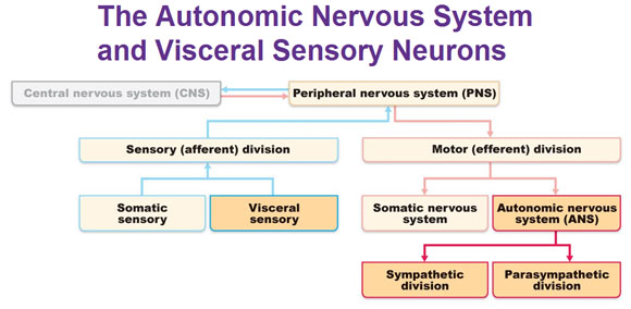
Up until now we’ve covered everything in this organizational chart below except for the ANS and visceral sensory system, which are highlighted in yellow.

The autonomic nervous system has two divisions:
Sympathetic and Parasympathetic. They mostly innervate the same structures but cause opposite effects. The sympathetic division mobilizes the body during extreme situations such as exercise, excitement and emergencies. Colloquially known as “fight or flight.” The parasympathetic division controls routine maintenance functions such as to conserve body energy and is colloquially called as “rest and digest.”
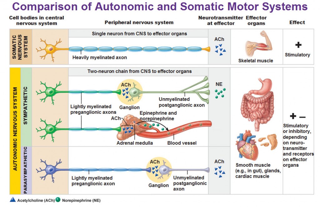
To make sense of the picture above, note the following…
A neuron found in the parasympathetic nervous system has:
LONG myelinated axon –> ganglion –> SHORT unmyelinated axon.
A neuron found in the sympathetic nervous system has:
SHORT myelinated axon –> ganglion –> LONG unmyelinated axon.
Now is a good time to make sure we differentiate that the ganglion we are talking about is not the same as the dorsal root ganglia. The “ganglia” in the ANS are motor ganglia made up of cell bodies of motor neurons. The “dorsal root ganglia” is in the sensory-somatic part of the peripheral nervous system and are sensory ganglia made up of cell bodies of sensory neurons. Review the location of the dorsal root ganglia here: CNS:
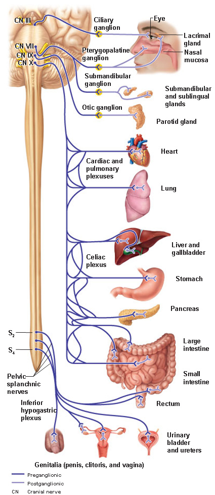
The Parasympathetic Division

The Sympathetic Division (T1 – L2)
Adrenal Medulla
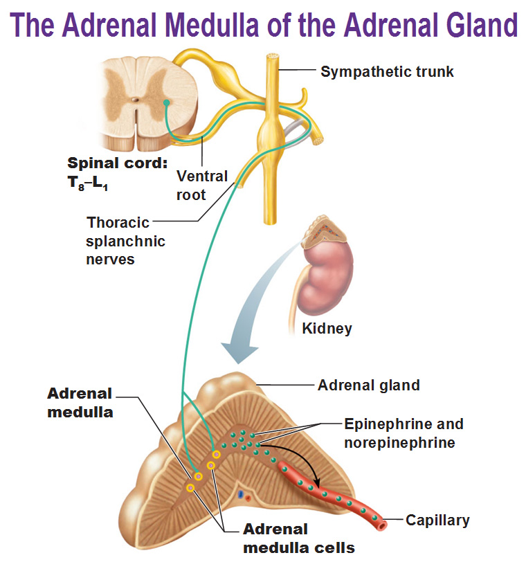
The adrenal medulla is GIGANTIC. It’s the largest sympathetic ganglia. The cells are made of modified neurons that have short axons and no nerve processes. The adrenal cortex is an endocrine organ and the outer layer is the adrenal cortex while the inner layer is the medulla. When stimulated by preganglionic sympathetic fibers from T8-L1, they secrete large quantities of the excitatory hormones norepinephrine and epinephrine (adrenaline) into nearby capillaries. When these two hormones are released in the blood they amplify all of this fight or flight stuff to give you more energy.
courtesy by Rx
Function
Sympathetic and parasympathetic divisions typically function in opposition to each other. But this opposition is better termed complementary in nature rather than antagonistic. For an analogy, one may think of the sympathetic division as the accelerator and the parasympathetic division as the brake. The sympathetic division typically functions in actions requiring quick responses. The parasympathetic division functions with actions that do not require immediate reaction. The sympathetic system is often considered the “fight or flight” system, while the parasympathetic system is often considered the “rest and digest” or “feed and breed” system.
However, many instances of sympathetic and parasympathetic activity cannot be ascribed to “fight” or “rest” situations. For example, standing up from a reclining or sitting position would entail an unsustainable drop in blood pressure if not for a compensatory increase in the arterial sympathetic tonus. Another example is the constant, second-to-second, modulation of heart rate by sympathetic and parasympathetic influences, as a function of the respiratory cycles. In general, these two systems should be seen as permanently modulating vital functions, in usually antagonistic fashion, to achieve homeostasis. Some typical actions of the sympathetic and parasympathetic systems are listed below.
Sympathetic nervous system
Promotes a fight-or-flight response, corresponds with arousal and energy generation, and inhibits digestion
- Diverts blood flow away from the gastro-intestinal (GI) tract and skin via vasoconstriction
- Blood flow to skeletal muscles and the lungs is enhanced (by as much as 1200% in the case of skeletal muscles)
- Dilates bronchioles of the lung through circulating epinephrine, which allows for greater alveolar oxygen exchange
- Increases heart rate and the contractility of cardiac cells (myocytes), thereby providing a mechanism for enhanced blood flow to skeletal muscles
- Dilates pupils and relaxes the ciliary muscle to the lens, allowing more light to enter the eye and enhances far vision
- Provides vasodilation for the coronary vessels of the heart
- Constricts all the intestinal sphincters and the urinary sphincter
- Inhibits peristalsis
- Stimulates orgasm
Parasympathetic nervous system
The parasympathetic nervous system has been said to promote a “rest and digest” response, promotes calming of the nerves return to regular function, and enhancing digestion. Functions of nerves within the parasympathetic nervous system include
- Dilating blood vessels leading to the GI tract, increasing the blood flow.
- Constricting the bronchiolar diameter when the need for oxygen has diminished
- Dedicated cardiac branches of the vagus and thoracic spinal accessory nerves impart parasympathetic control of the heart (myocardium)
- Constriction of the pupil and contraction of the ciliary muscles, facilitating accommodation and allowing for closer vision
- Stimulating salivary gland secretion, and accelerates peristalsis, mediating digestion of food and, indirectly, the absorption of nutrients
- Sexual. Nerves of the peripheral nervous system are involved in the erection of genital tissues via the pelvic splanchnic nerves 2–4. They are also responsible for stimulating sexual arousal.
Enteric nervous system
The enteric nervous system is the intrinsic nervous system of the gastrointestinal system. It has been described as “the Second Brain of the Human Body”. Its functions include:
- Sensing chemical and mechanical changes in the gut
- Regulating secretions in the gut
- Controlling peristalsis and some other movements
Neurotransmitters
At the effector organs, sympathetic ganglionic neurons release noradrenaline (norepinephrine), along with other cotransmitters such as ATP, to act on adrenergic receptors, with the exception of the sweat glands and the adrenal medulla:
- Acetylcholine is the preganglionic neurotransmitter for both divisions of the ANS, as well as the postganglionic neurotransmitter of parasympathetic neurons. Nerves that release acetylcholine are said to be cholinergic. In the parasympathetic system, ganglionic neurons use acetylcholine as a neurotransmitter to stimulate muscarinic receptors.
- At the adrenal medulla, there is no postsynaptic neuron. Instead the presynaptic neuron releases acetylcholine to act on nicotinic receptors. Stimulation of the adrenal medulla releases adrenaline (epinephrine) into the bloodstream, which acts on adrenoceptors, thereby indirectly mediating or mimicking sympathetic activity.
References


