Category Archive Neurology

Ablative Nerve Block – Indications, Contraindications

Ablative Nerve Block/Ablation is the destruction (also called ablation) of nerves is a method that may be used to reduce certain kinds of chronic pain by preventing transmission of pain signals. It is a safe procedure in which a portion of nerve tissue is destroyed or removed to cause an interruption in pain signals and reduce pain in that area.

Radiofrequency ablation, also called rhizotomy, is a non-surgical, minimally invasive procedure that uses heat to reduce or stop the transmission of pain. Radiofrequency waves ablate, or “burn,” the nerve that is causing the pain, essentially eliminating the transmission of pain signals to the brain.

This procedure is most commonly used to treat chronic pain and conditions such as arthritis of the spine (spondylosis) and sacroiliitis. It is also used to treat neck, back, knee, pelvic and peripheral nerve pain. The benefits of radiofrequency ablation include: avoiding surgery, immediate pain relief, little to no recovery time, decreased need for pain medication, improved function, and a quicker return to work and other activities.

Types of Ablation

Three types or variations of RFA may be used to produce heat lesions on tissues and are discussed below.

  • Conventional continuous radiofrequency (CRF) ablation – uses a needle that delivers continuous high-voltage current to produce a heat lesion. The tip of the needle is heated between 140 to 176 degrees Fahrenheit.
  • Pulsed radiofrequency (PRF) ablation – uses a needle that delivers short bursts of high-voltage current with silent phases in between when no current is passed. The needle is heated to approximately 107 degrees Fahrenheit in PRF.
  • Water-cooled radiofrequency (WCRF) ablation – uses a specialized needle that is heated up to 140 degrees Fahrenheit but also cooled by a continuous flow of water. The water allows a regulated flow of current and also prevents the needle tip from being over-heated.
  • Pulsed radiofrequency – Applies heat in a pulsatile manner, but uses lower temperatures in a than conventional radiofrequency to avoid neurolysis.
  • Cooled radiofrequency – Utilizes specialized electrodes that are cooled with water-flowing along its shaft, but not at the active tip. This allows higher temperatures and more spherical lesion sizes to be achieved at the target site and less risk of tissue damage superficial to the target.
  • Cryoablation – causes the destruction of axons by disrupting the vasa nervorum. It is increasing in popularity due to a decreased incidence of post-procedural hyperalgesia and neuroma formation compared to conventional radiofrequency.

Anatomy and Physiology

Facet-mediated pain is due to facet arthropathy or facet arthritis which arise during the degenerative process of the spinal column. A single facet joint is composed of the inferior articulating process of one vertebra and the superior articulating process of the vertebra directly inferior.

Medial branches from the dorsal rami of spinal nerve roots at the same level and one level above provide sensory innervation to the facet (for example, the L4-L5 facet joint is innervated by the medial branches of L3 and L4). The medial branches typically course over the lateral border of the superior articulating process.

An insulated electrode with a non-insulated tip is advanced toward the concavity that is formed between the superior articulating process and the adjacent transverse process, which is in proximity to the nerve that is suspected of causing the symptoms. It is in this region where the use of high-frequency or radiofrequency energy is generated to produce a lesion via coagulative necrosis, thereby disrupting afferent pain signals. In Pain Medicine, the use of radiofrequency ablation most commonly involves targeting the specific medial branches of the dorsal rami that innervate pain-producing facet joints under fluoroscopic guidance. However, further investigations regarding the efficacy of ablation in other common pain syndromes continue to diversify its use.

Indications of Ablation

Nerve ablation has typically been used in the treatment of facet-mediated axial back pain of the cervical and lumbar spine that has failed conservative therapy.

  • Additionally, it has other uses such as relief of chronic neck pain after whiplash and chronic headache syndromes due to occipital and trigeminal neuralgia.
  • Ablation is performed after successful analgesia of the suspected trouble-causing nerve with a local anesthetic. There continue to study investigating the role of ablation in peripheral nerve-mediated pain outside of the spinal column, particularly in knee osteoarthritis and plantar fasciitis.
  • Ablative nerve blocks, with cooled radiofrequency ablation of the genicular nerve, have been shown to be an effective pain reliever for refractory osteoarthritis of the knee. Furthermore, it has been shown to improve knee function for up to six months following the procedure.

Contraindications of Ablation

Absolute contraindications for ablative nerve blocks are few, but these include active local infection at the site of needle insertion and elevated intracranial pressure. The use of anti-coagulants provides a unique challenge for practitioners, as the clinical judgment must be used following accepted guidelines. Current guidelines put forth by ASRA (American Society of Regional Anesthesia and Pain Medicine) recommend that:

  • Aspirin to be stopped 6 days before the procedure
  • Clopidogrel to be stopped 7 days before the procedure
  • Apixaban to be stopped 3 to 5 days before the procedure
  • Rivaroxaban to be stopped 3 days before the procedure
  • Warfarin to be stopped 5 days before the procedure
  • Intravenous heparin to be stopped 4 hours before the procedure

Other relative contraindications that the clinician must consider before the procedure include:

  • Neurologic abnormalities
  • Concerning clinical or imaging finding
  • Definitive causes of low back pain (for example, disc herniation, spondylolisthesis, spondylosis, spinal stenosis, malignancy, infection or trauma)
  • Lack of pain relief from prior diagnostic nerve blocks

Equipment of Ablative Nerve Block

The procedure is routinely done in a sterile procedure suite, with the patient lying prone on a procedure table.

Key components of the procedure include:

  • C-arm mobile fluoroscopic unit
  • Local anesthetic
  • continuous high-frequency generator with built-in thermocouple, impedance, voltage and amperage monitor
  • Introducer needles (50 to 150 mm in length, 18 to 22 gauge in diameter)
  • Active and ground electrodes

The necessary equipment includes the following:

  • Chlorhexidine gluconate or povidone iodine
  • Ultrasound probe with a sterile probe cover and gel (if applicable)
  • Fluoroscopy equipment (if applicable)
  • Nerve stimulator (if applicable)
  • Local anesthetic, typically 1% lidocaine, for superficial layer local anesthesia
  • Regional block local anesthetic test solution (2% lidocaine or 1.5% mepivacaine)
  • A 10- to 20-mL syringe with extension tubing
  • Block needle (length-variable depending on depth of targeted peripheral nerve) or spinal/epidural needle if neuraxial approach (needle characteristics based on body habitus and provider preference)
  • Chemical neurolytic agent (alcohol, phenol), if applicable
  • Radiofrequency probe, if applicable
  • Cryo machine and cryoprobe with cooling agents (i.e. nitrous oxide, carbon dioxide), if applicable

Agents used for chemical neurolysis primarily include 50-100% alcohol and 5% to 15% phenol, although the use of other agents including hypertonic saline, glycerol, ammonium salt solutions, and chlorocresol has also been reported. The mechanism of action of alcohol neurolysis is axonal and Schwann cell destruction from phospholipid extraction in the cell membrane and lipoprotein precipitation. Phenol infiltration causes damage from protein coagulation and degeneration.

Personnel

As with other interventional spinal procedures, only physicians specifically trained in fluoroscopically guidance procedures should perform radiofrequency ablation. Qualified physicians typically undergo residency training in the fields of Anesthesiology, Physiatry, Neurology, Psychiatry, or Neurosurgery. This is followed by an interventional pain or spine fellowship that allows adequate training under an experienced interventionalist prior to performing the procedure independently. Support staff for the procedure can include an assistant to draw up medications and operate the radiofrequency generator and radiology technician to operate the C-arm, under the guidance of the practicing physician.

Technique of Ablative Nerve Block

The destruction of tissue via radiofrequency must occur after successful diagnostic anesthetic nerve blocks have located the target nerve. During the procedure, the patient should receive little to no sedation, as they must define what they are experiencing during stimulation and lesioning of the nerve.

  • The patient is placed in a comfortable position with adequate exposure of the region overlying the target nerve. The skin is cleaned and prepped, and the target is found using fluoroscopy. A small amount of local anesthetic is injected subcutaneously at the point of needle insertion.
  • The introducer needle is then inserted through the skin, subcutaneous tissue, and muscle toward the target using fluoroscopy to guide the trajectory.
  • Once the tip of the needle is placed satisfactorily close to the target, the active electrode is inserted through the needle.
  • Sensory stimulation is used at first, to recreate the painful symptoms that the patient experiences, thus locating the target.
  • This is followed by motor stimulation to ensure that the active tip is not close to the motor nerves.
  • Once confirmed, the generator produces a continuous output of voltage at the tip of the electrode, where it will be kept at 80 degrees Celsius for approximately one minute, creating a zone of thermally induced coagulation.

Complications of Ablative Nerve Block

Nerve ablation is a minimally invasive, relatively low-risk procedure. There is a limited side effect profile to ablative nerve blocks. However, adverse events may occur during the placement of the introducer needle or during the ablative process. Advancement of the introducer needle has the potential to cause vascular or neural insult along the trajectory that it is traveling, while the process of thermal ablation may lead to burns (due to errors in ground pad placement), worsened pain, sensory loss or new-onset neuropathic pain.

Complications are most common after intracranial ablation of the trigeminal ganglion, which may manifest as facial numbness, dysesthesia, anesthesia dolorosa, corneal anesthesia, keratitis, and trigeminal motor dysfunction.

Adverse events from ablation of lumbar medial branches are far and few between with transient postoperative pain dominating as the premier adverse event.

As with any invasive technique, the risk of allergy to materials or anesthetic, hematoma formation, and infection must be considered.

References

Loading

If the article is helpful, please Click to Star Icon and Rate This Post!
[Total: 0 Average: 0]

Water-cooled Radiofrequency (WCRF) Ablation

Water-cooled radiofrequency (WCRF) ablation uses a specialized needle that is heated up to 140 degrees Fahrenheit but also cooled by a continuous flow of water. The water allows a regulated flow of current and also prevents the needle tip from being over-heated

Pulsed Radiofrequency (PRF) Ablation /Ablation is the destruction (also called ablation) of nerves is a method that may be used to reduce certain kinds of chronic pain by preventing transmission of pain signals. It is a safe procedure in which a portion of nerve tissue is destroyed or removed to cause an interruption in pain signals and reduce pain in that area.

Radiofrequency ablation, also called rhizotomy, is a non-surgical, minimally invasive procedure that uses heat to reduce or stop the transmission of pain. Radiofrequency waves ablate, or “burn,” the nerve that is causing the pain, essentially eliminating the transmission of pain signals to the brain.

This procedure is most commonly used to treat chronic pain and conditions such as arthritis of the spine (spondylosis) and sacroiliitis. It is also used to treat neck, back, knee, pelvic and peripheral nerve pain. The benefits of radiofrequency ablation include: avoiding surgery, immediate pain relief, little to no recovery time, decreased need for pain medication, improved function, and a quicker return to work and other activities.

Types of Ablation

Three types or variations of RFA may be used to produce heat lesions on tissues and are discussed below.

  • Conventional continuous radiofrequency (CRF) ablation – uses a needle that delivers continuous high-voltage current to produce a heat lesion. The tip of the needle is heated between 140 to 176 degrees Fahrenheit.
  • Pulsed radiofrequency (PRF) ablation – uses a needle that delivers short bursts of high-voltage current with silent phases in between when no current is passed. The needle is heated to approximately 107 degrees Fahrenheit in PRF.
  • Water-cooled radiofrequency (WCRF) ablation – uses a specialized needle that is heated up to 140 degrees Fahrenheit but also cooled by a continuous flow of water. The water allows a regulated flow of current and also prevents the needle tip from being over-heated.
  • Pulsed radiofrequency – Applies heat in a pulsatile manner, but uses lower temperatures in a than conventional radiofrequency to avoid neurolysis.
  • Cooled radiofrequency – Utilizes specialized electrodes that are cooled with water-flowing along its shaft, but not at the active tip. This allows higher temperatures and more spherical lesion sizes to be achieved at the target site and less risk of tissue damage superficial to the target.
  • Cryoablation – causes the destruction of axons by disrupting the vasa nervorum. It is increasing in popularity due to a decreased incidence of post-procedural hyperalgesia and neuroma formation compared to conventional radiofrequency.

Anatomy and Physiology

Facet-mediated pain is due to facet arthropathy or facet arthritis which arise during the degenerative process of the spinal column. A single facet joint is composed of the inferior articulating process of one vertebra and the superior articulating process of the vertebra directly inferior.

Medial branches from the dorsal rami of spinal nerve roots at the same level and one level above provide sensory innervation to the facet (for example, the L4-L5 facet joint is innervated by the medial branches of L3 and L4). The medial branches typically course over the lateral border of the superior articulating process.

An insulated electrode with a non-insulated tip is advanced toward the concavity that is formed between the superior articulating process and the adjacent transverse process, which is in proximity to the nerve that is suspected of causing the symptoms. It is in this region where the use of high-frequency or radiofrequency energy is generated to produce a lesion via coagulative necrosis, thereby disrupting afferent pain signals. In Pain Medicine, the use of radiofrequency ablation most commonly involves targeting the specific medial branches of the dorsal rami that innervate pain-producing facet joints under fluoroscopic guidance. However, further investigations regarding the efficacy of ablation in other common pain syndromes continue to diversify its use.

Indications of Ablation

Nerve ablation has typically been used in the treatment of facet-mediated axial back pain of the cervical and lumbar spine that has failed conservative therapy.

  • Additionally, it has other uses such as relief of chronic neck pain after whiplash and chronic headache syndromes due to occipital and trigeminal neuralgia.
  • Ablation is performed after successful analgesia of the suspected trouble-causing nerve with a local anesthetic. There continue to study investigating the role of ablation in peripheral nerve-mediated pain outside of the spinal column, particularly in knee osteoarthritis and plantar fasciitis.
  • Ablative nerve blocks, with cooled radiofrequency ablation of the genicular nerve, have been shown to be an effective pain reliever for refractory osteoarthritis of the knee. Furthermore, it has been shown to improve knee function for up to six months following the procedure.

Contraindications of Ablation

Absolute contraindications for ablative nerve blocks are few, but these include active local infection at the site of needle insertion and elevated intracranial pressure. The use of anti-coagulants provides a unique challenge for practitioners, as the clinical judgment must be used following accepted guidelines. Current guidelines put forth by ASRA (American Society of Regional Anesthesia and Pain Medicine) recommend that:

  • Aspirin to be stopped 6 days before the procedure
  • Clopidogrel to be stopped 7 days before the procedure
  • Apixaban to be stopped 3 to 5 days before the procedure
  • Rivaroxaban to be stopped 3 days before the procedure
  • Warfarin to be stopped 5 days before the procedure
  • Intravenous heparin to be stopped 4 hours before the procedure

Other relative contraindications that the clinician must consider before the procedure include:

  • Neurologic abnormalities
  • Concerning clinical or imaging finding
  • Definitive causes of low back pain (for example, disc herniation, spondylolisthesis, spondylosis, spinal stenosis, malignancy, infection or trauma)
  • Lack of pain relief from prior diagnostic nerve blocks

Equipment

The procedure is routinely done in a sterile procedure suite, with the patient lying prone on a procedure table.

Key components of the procedure include:

  • C-arm mobile fluoroscopic unit
  • Local anesthetic
  • continuous high-frequency generator with built-in thermocouple, impedance, voltage and amperage monitor
  • Introducer needles (50 to 150 mm in length, 18 to 22 gauge in diameter)
  • Active and ground electrodes

The necessary equipment includes the following:

  • Chlorhexidine gluconate or povidone iodine
  • Ultrasound probe with a sterile probe cover and gel (if applicable)
  • Fluoroscopy equipment (if applicable)
  • Nerve stimulator (if applicable)
  • Local anesthetic, typically 1% lidocaine, for superficial layer local anesthesia
  • Regional block local anesthetic test solution (2% lidocaine or 1.5% mepivacaine)
  • A 10- to 20-mL syringe with extension tubing
  • Block needle (length-variable depending on depth of targeted peripheral nerve) or spinal/epidural needle if neuraxial approach (needle characteristics based on body habitus and provider preference)
  • Chemical neurolytic agent (alcohol, phenol), if applicable
  • Radiofrequency probe, if applicable
  • Cryo machine and cryoprobe with cooling agents (i.e. nitrous oxide, carbon dioxide), if applicable

Agents used for chemical neurolysis primarily include 50-100% alcohol and 5% to 15% phenol, although the use of other agents including hypertonic saline, glycerol, ammonium salt solutions, and chlorocresol has also been reported. The mechanism of action of alcohol neurolysis is axonal and Schwann cell destruction from phospholipid extraction in the cell membrane and lipoprotein precipitation. Phenol infiltration causes damage from protein coagulation and degeneration.

Personnel

As with other interventional spinal procedures, only physicians specifically trained in fluoroscopically guidance procedures should perform radiofrequency ablation. Qualified physicians typically undergo residency training in the fields of Anesthesiology, Physiatry, Neurology, Psychiatry, or Neurosurgery. This is followed by an interventional pain or spine fellowship that allows adequate training under an experienced interventionalist prior to performing the procedure independently. Support staff for the procedure can include an assistant to draw up medications and operate the radiofrequency generator and radiology technician to operate the C-arm, under the guidance of the practicing physician.

Technique

The destruction of tissue via radiofrequency must occur after successful diagnostic anesthetic nerve blocks have located the target nerve. During the procedure, the patient should receive little to no sedation, as they must define what they are experiencing during stimulation and lesioning of the nerve.

  • The patient is placed in a comfortable position with adequate exposure of the region overlying the target nerve. The skin is cleaned and prepped, and the target is found using fluoroscopy. A small amount of local anesthetic is injected subcutaneously at the point of needle insertion.
  • The introducer needle is then inserted through the skin, subcutaneous tissue, and muscle toward the target using fluoroscopy to guide the trajectory.
  • Once the tip of the needle is placed satisfactorily close to the target, the active electrode is inserted through the needle.
  • Sensory stimulation is used at first, to recreate the painful symptoms that the patient experiences, thus locating the target.
  • This is followed by motor stimulation to ensure that the active tip is not close to the motor nerves.
  • Once confirmed, the generator produces a continuous output of voltage at the tip of the electrode, where it will be kept at 80 degrees Celsius for approximately one minute, creating a zone of thermally induced coagulation.

Complications

Nerve ablation is a minimally invasive, relatively low-risk procedure. There is a limited side effect profile to ablative nerve blocks. However, adverse events may occur during the placement of the introducer needle or during the ablative process. Advancement of the introducer needle has the potential to cause vascular or neural insult along the trajectory that it is traveling, while the process of thermal ablation may lead to burns (due to errors in ground pad placement), worsened pain, sensory loss or new-onset neuropathic pain.

Complications are most common after intracranial ablation of the trigeminal ganglion, which may manifest as facial numbness, dysesthesia, anesthesia dolorosa, corneal anesthesia, keratitis, and trigeminal motor dysfunction.

Adverse events from ablation of lumbar medial branches are far and few between with transient postoperative pain dominating as the premier adverse event.

As with any invasive technique, the risk of allergy to materials or anesthetic, hematoma formation, and infection must be considered.

References

Loading

If the article is helpful, please Click to Star Icon and Rate This Post!
[Total: 0 Average: 0]

Pulsed Radiofrequency (PRF) Ablation

Pulsed Radiofrequency (PRF) Ablation /Ablation is the destruction (also called ablation) of nerves is a method that may be used to reduce certain kinds of chronic pain by preventing transmission of pain signals. It is a safe procedure in which a portion of nerve tissue is destroyed or removed to cause an interruption in pain signals and reduce pain in that area.

Radiofrequency ablation, also called rhizotomy, is a non-surgical, minimally invasive procedure that uses heat to reduce or stop the transmission of pain. Radiofrequency waves ablate, or “burn,” the nerve that is causing the pain, essentially eliminating the transmission of pain signals to the brain.

This procedure is most commonly used to treat chronic pain and conditions such as arthritis of the spine (spondylosis) and sacroiliitis. It is also used to treat neck, back, knee, pelvic and peripheral nerve pain. The benefits of radiofrequency ablation include: avoiding surgery, immediate pain relief, little to no recovery time, decreased need for pain medication, improved function, and a quicker return to work and other activities.

Types of Ablation

Three types or variations of RFA may be used to produce heat lesions on tissues and are discussed below.

  • Conventional continuous radiofrequency (CRF) ablation – uses a needle that delivers continuous high-voltage current to produce a heat lesion. The tip of the needle is heated between 140 to 176 degrees Fahrenheit.
  • Pulsed radiofrequency (PRF) ablation – uses a needle that delivers short bursts of high-voltage current with silent phases in between when no current is passed. The needle is heated to approximately 107 degrees Fahrenheit in PRF.
  • Water-cooled radiofrequency (WCRF) ablation – uses a specialized needle that is heated up to 140 degrees Fahrenheit but also cooled by a continuous flow of water. The water allows a regulated flow of current and also prevents the needle tip from being over-heated.
  • Pulsed radiofrequency – Applies heat in a pulsatile manner, but uses lower temperatures in a than conventional radiofrequency to avoid neurolysis.
  • Cooled radiofrequency – Utilizes specialized electrodes that are cooled with water-flowing along its shaft, but not at the active tip. This allows higher temperatures and more spherical lesion sizes to be achieved at the target site and less risk of tissue damage superficial to the target.
  • Cryoablation – causes the destruction of axons by disrupting the vasa nervorum. It is increasing in popularity due to a decreased incidence of post-procedural hyperalgesia and neuroma formation compared to conventional radiofrequency.

Anatomy and Physiology

Facet-mediated pain is due to facet arthropathy or facet arthritis which arise during the degenerative process of the spinal column. A single facet joint is composed of the inferior articulating process of one vertebra and the superior articulating process of the vertebra directly inferior.

Medial branches from the dorsal rami of spinal nerve roots at the same level and one level above provide sensory innervation to the facet (for example, the L4-L5 facet joint is innervated by the medial branches of L3 and L4). The medial branches typically course over the lateral border of the superior articulating process.

An insulated electrode with a non-insulated tip is advanced toward the concavity that is formed between the superior articulating process and the adjacent transverse process, which is in proximity to the nerve that is suspected of causing the symptoms. It is in this region where the use of high-frequency or radiofrequency energy is generated to produce a lesion via coagulative necrosis, thereby disrupting afferent pain signals. In Pain Medicine, the use of radiofrequency ablation most commonly involves targeting the specific medial branches of the dorsal rami that innervate pain-producing facet joints under fluoroscopic guidance. However, further investigations regarding the efficacy of ablation in other common pain syndromes continue to diversify its use.

Indications of Ablation

Nerve ablation has typically been used in the treatment of facet-mediated axial back pain of the cervical and lumbar spine that has failed conservative therapy.

  • Additionally, it has other uses such as relief of chronic neck pain after whiplash and chronic headache syndromes due to occipital and trigeminal neuralgia.
  • Ablation is performed after successful analgesia of the suspected trouble-causing nerve with a local anesthetic. There continue to study investigating the role of ablation in peripheral nerve-mediated pain outside of the spinal column, particularly in knee osteoarthritis and plantar fasciitis.
  • Ablative nerve blocks, with cooled radiofrequency ablation of the genicular nerve, have been shown to be an effective pain reliever for refractory osteoarthritis of the knee. Furthermore, it has been shown to improve knee function for up to six months following the procedure.

Contraindications of Ablation

Absolute contraindications for ablative nerve blocks are few, but these include active local infection at the site of needle insertion and elevated intracranial pressure. The use of anti-coagulants provides a unique challenge for practitioners, as the clinical judgment must be used following accepted guidelines. Current guidelines put forth by ASRA (American Society of Regional Anesthesia and Pain Medicine) recommend that:

  • Aspirin to be stopped 6 days before the procedure
  • Clopidogrel to be stopped 7 days before the procedure
  • Apixaban to be stopped 3 to 5 days before the procedure
  • Rivaroxaban to be stopped 3 days before the procedure
  • Warfarin to be stopped 5 days before the procedure
  • Intravenous heparin to be stopped 4 hours before the procedure

Other relative contraindications that the clinician must consider before the procedure include:

  • Neurologic abnormalities
  • Concerning clinical or imaging finding
  • Definitive causes of low back pain (for example, disc herniation, spondylolisthesis, spondylosis, spinal stenosis, malignancy, infection or trauma)
  • Lack of pain relief from prior diagnostic nerve blocks

Equipment

The procedure is routinely done in a sterile procedure suite, with the patient lying prone on a procedure table.

Key components of the procedure include:

  • C-arm mobile fluoroscopic unit
  • Local anesthetic
  • continuous high-frequency generator with built-in thermocouple, impedance, voltage and amperage monitor
  • Introducer needles (50 to 150 mm in length, 18 to 22 gauge in diameter)
  • Active and ground electrodes

The necessary equipment includes the following:

  • Chlorhexidine gluconate or povidone iodine
  • Ultrasound probe with a sterile probe cover and gel (if applicable)
  • Fluoroscopy equipment (if applicable)
  • Nerve stimulator (if applicable)
  • Local anesthetic, typically 1% lidocaine, for superficial layer local anesthesia
  • Regional block local anesthetic test solution (2% lidocaine or 1.5% mepivacaine)
  • A 10- to 20-mL syringe with extension tubing
  • Block needle (length-variable depending on depth of targeted peripheral nerve) or spinal/epidural needle if neuraxial approach (needle characteristics based on body habitus and provider preference)
  • Chemical neurolytic agent (alcohol, phenol), if applicable
  • Radiofrequency probe, if applicable
  • Cryo machine and cryoprobe with cooling agents (i.e. nitrous oxide, carbon dioxide), if applicable

Agents used for chemical neurolysis primarily include 50-100% alcohol and 5% to 15% phenol, although the use of other agents including hypertonic saline, glycerol, ammonium salt solutions, and chlorocresol has also been reported. The mechanism of action of alcohol neurolysis is axonal and Schwann cell destruction from phospholipid extraction in the cell membrane and lipoprotein precipitation. Phenol infiltration causes damage from protein coagulation and degeneration.

Personnel

As with other interventional spinal procedures, only physicians specifically trained in fluoroscopically guidance procedures should perform radiofrequency ablation. Qualified physicians typically undergo residency training in the fields of Anesthesiology, Physiatry, Neurology, Psychiatry, or Neurosurgery. This is followed by an interventional pain or spine fellowship that allows adequate training under an experienced interventionalist prior to performing the procedure independently. Support staff for the procedure can include an assistant to draw up medications and operate the radiofrequency generator and radiology technician to operate the C-arm, under the guidance of the practicing physician.

Technique

The destruction of tissue via radiofrequency must occur after successful diagnostic anesthetic nerve blocks have located the target nerve. During the procedure, the patient should receive little to no sedation, as they must define what they are experiencing during stimulation and lesioning of the nerve.

  • The patient is placed in a comfortable position with adequate exposure of the region overlying the target nerve. The skin is cleaned and prepped, and the target is found using fluoroscopy. A small amount of local anesthetic is injected subcutaneously at the point of needle insertion.
  • The introducer needle is then inserted through the skin, subcutaneous tissue, and muscle toward the target using fluoroscopy to guide the trajectory.
  • Once the tip of the needle is placed satisfactorily close to the target, the active electrode is inserted through the needle.
  • Sensory stimulation is used at first, to recreate the painful symptoms that the patient experiences, thus locating the target.
  • This is followed by motor stimulation to ensure that the active tip is not close to the motor nerves.
  • Once confirmed, the generator produces a continuous output of voltage at the tip of the electrode, where it will be kept at 80 degrees Celsius for approximately one minute, creating a zone of thermally induced coagulation.

Complications

Nerve ablation is a minimally invasive, relatively low-risk procedure. There is a limited side effect profile to ablative nerve blocks. However, adverse events may occur during the placement of the introducer needle or during the ablative process. Advancement of the introducer needle has the potential to cause vascular or neural insult along the trajectory that it is traveling, while the process of thermal ablation may lead to burns (due to errors in ground pad placement), worsened pain, sensory loss or new-onset neuropathic pain.

Complications are most common after intracranial ablation of the trigeminal ganglion, which may manifest as facial numbness, dysesthesia, anesthesia dolorosa, corneal anesthesia, keratitis, and trigeminal motor dysfunction.

Adverse events from ablation of lumbar medial branches are far and few between with transient postoperative pain dominating as the premier adverse event.

As with any invasive technique, the risk of allergy to materials or anesthetic, hematoma formation, and infection must be considered.

References

Loading

If the article is helpful, please Click to Star Icon and Rate This Post!
[Total: 0 Average: 0]

Conventional Continuous Radiofrequency (CRF) Ablation

Conventional Continuous Radiofrequency (CRF) Ablation/Ablation is the destruction (also called ablation) of nerves is a method that may be used to reduce certain kinds of chronic pain by preventing transmission of pain signals. It is a safe procedure in which a portion of nerve tissue is destroyed or removed to cause an interruption in pain signals and reduce pain in that area.

Radiofrequency ablation, also called rhizotomy, is a non-surgical, minimally invasive procedure that uses heat to reduce or stop the transmission of pain. Radiofrequency waves ablate, or “burn,” the nerve that is causing the pain, essentially eliminating the transmission of pain signals to the brain.

This procedure is most commonly used to treat chronic pain and conditions such as arthritis of the spine (spondylosis) and sacroiliitis. It is also used to treat neck, back, knee, pelvic and peripheral nerve pain. The benefits of radiofrequency ablation include: avoiding surgery, immediate pain relief, little to no recovery time, decreased need for pain medication, improved function, and a quicker return to work and other activities.

Types of Ablation

Three types or variations of RFA may be used to produce heat lesions on tissues and are discussed below.

  • Conventional continuous radiofrequency (CRF) ablation – uses a needle that delivers continuous high-voltage current to produce a heat lesion. The tip of the needle is heated between 140 to 176 degrees Fahrenheit.
  • Pulsed radiofrequency (PRF) ablation – uses a needle that delivers short bursts of high-voltage current with silent phases in between when no current is passed. The needle is heated to approximately 107 degrees Fahrenheit in PRF.
  • Water-cooled radiofrequency (WCRF) ablation – uses a specialized needle that is heated up to 140 degrees Fahrenheit but also cooled by a continuous flow of water. The water allows a regulated flow of current and also prevents the needle tip from being over-heated.
  • Pulsed radiofrequency – Applies heat in a pulsatile manner, but uses lower temperatures in a than conventional radiofrequency to avoid neurolysis.
  • Cooled radiofrequency – Utilizes specialized electrodes that are cooled with water-flowing along its shaft, but not at the active tip. This allows higher temperatures and more spherical lesion sizes to be achieved at the target site and less risk of tissue damage superficial to the target.
  • Cryoablation – causes the destruction of axons by disrupting the vasa nervorum. It is increasing in popularity due to a decreased incidence of post-procedural hyperalgesia and neuroma formation compared to conventional radiofrequency.

Anatomy and Physiology

Facet-mediated pain is due to facet arthropathy or facet arthritis which arise during the degenerative process of the spinal column. A single facet joint is composed of the inferior articulating process of one vertebra and the superior articulating process of the vertebra directly inferior.

Medial branches from the dorsal rami of spinal nerve roots at the same level and one level above provide sensory innervation to the facet (for example, the L4-L5 facet joint is innervated by the medial branches of L3 and L4). The medial branches typically course over the lateral border of the superior articulating process.

An insulated electrode with a non-insulated tip is advanced toward the concavity that is formed between the superior articulating process and the adjacent transverse process, which is in proximity to the nerve that is suspected of causing the symptoms. It is in this region where the use of high-frequency or radiofrequency energy is generated to produce a lesion via coagulative necrosis, thereby disrupting afferent pain signals. In Pain Medicine, the use of radiofrequency ablation most commonly involves targeting the specific medial branches of the dorsal rami that innervate pain-producing facet joints under fluoroscopic guidance. However, further investigations regarding the efficacy of ablation in other common pain syndromes continue to diversify its use.

Indications of Ablation

Nerve ablation has typically been used in the treatment of facet-mediated axial back pain of the cervical and lumbar spine that has failed conservative therapy.

  • Additionally, it has other uses such as relief of chronic neck pain after whiplash and chronic headache syndromes due to occipital and trigeminal neuralgia.
  • Ablation is performed after successful analgesia of the suspected trouble-causing nerve with a local anesthetic. There continue to study investigating the role of ablation in peripheral nerve-mediated pain outside of the spinal column, particularly in knee osteoarthritis and plantar fasciitis.
  • Ablative nerve blocks, with cooled radiofrequency ablation of the genicular nerve, have been shown to be an effective pain reliever for refractory osteoarthritis of the knee. Furthermore, it has been shown to improve knee function for up to six months following the procedure.

Contraindications of Ablation

Absolute contraindications for ablative nerve blocks are few, but these include active local infection at the site of needle insertion and elevated intracranial pressure. The use of anti-coagulants provides a unique challenge for practitioners, as the clinical judgment must be used following accepted guidelines. Current guidelines put forth by ASRA (American Society of Regional Anesthesia and Pain Medicine) recommend that:

  • Aspirin to be stopped 6 days before the procedure
  • Clopidogrel to be stopped 7 days before the procedure
  • Apixaban to be stopped 3 to 5 days before the procedure
  • Rivaroxaban to be stopped 3 days before the procedure
  • Warfarin to be stopped 5 days before the procedure
  • Intravenous heparin to be stopped 4 hours before the procedure

Other relative contraindications that the clinician must consider before the procedure include:

  • Neurologic abnormalities
  • Concerning clinical or imaging finding
  • Definitive causes of low back pain (for example, disc herniation, spondylolisthesis, spondylosis, spinal stenosis, malignancy, infection or trauma)
  • Lack of pain relief from prior diagnostic nerve blocks

Equipment

The procedure is routinely done in a sterile procedure suite, with the patient lying prone on a procedure table.

Key components of the procedure include:

  • C-arm mobile fluoroscopic unit
  • Local anesthetic
  • continuous high-frequency generator with built-in thermocouple, impedance, voltage and amperage monitor
  • Introducer needles (50 to 150 mm in length, 18 to 22 gauge in diameter)
  • Active and ground electrodes

The necessary equipment includes the following:

  • Chlorhexidine gluconate or povidone iodine
  • Ultrasound probe with a sterile probe cover and gel (if applicable)
  • Fluoroscopy equipment (if applicable)
  • Nerve stimulator (if applicable)
  • Local anesthetic, typically 1% lidocaine, for superficial layer local anesthesia
  • Regional block local anesthetic test solution (2% lidocaine or 1.5% mepivacaine)
  • A 10- to 20-mL syringe with extension tubing
  • Block needle (length-variable depending on depth of targeted peripheral nerve) or spinal/epidural needle if neuraxial approach (needle characteristics based on body habitus and provider preference)
  • Chemical neurolytic agent (alcohol, phenol), if applicable
  • Radiofrequency probe, if applicable
  • Cryo machine and cryoprobe with cooling agents (i.e. nitrous oxide, carbon dioxide), if applicable

Agents used for chemical neurolysis primarily include 50-100% alcohol and 5% to 15% phenol, although the use of other agents including hypertonic saline, glycerol, ammonium salt solutions, and chlorocresol has also been reported. The mechanism of action of alcohol neurolysis is axonal and Schwann cell destruction from phospholipid extraction in the cell membrane and lipoprotein precipitation. Phenol infiltration causes damage from protein coagulation and degeneration.

Personnel

As with other interventional spinal procedures, only physicians specifically trained in fluoroscopically guidance procedures should perform radiofrequency ablation. Qualified physicians typically undergo residency training in the fields of Anesthesiology, Physiatry, Neurology, Psychiatry, or Neurosurgery. This is followed by an interventional pain or spine fellowship that allows adequate training under an experienced interventionalist prior to performing the procedure independently. Support staff for the procedure can include an assistant to draw up medications and operate the radiofrequency generator and radiology technician to operate the C-arm, under the guidance of the practicing physician.

Technique

The destruction of tissue via radiofrequency must occur after successful diagnostic anesthetic nerve blocks have located the target nerve. During the procedure, the patient should receive little to no sedation, as they must define what they are experiencing during stimulation and lesioning of the nerve.

  • The patient is placed in a comfortable position with adequate exposure of the region overlying the target nerve. The skin is cleaned and prepped, and the target is found using fluoroscopy. A small amount of local anesthetic is injected subcutaneously at the point of needle insertion.
  • The introducer needle is then inserted through the skin, subcutaneous tissue, and muscle toward the target using fluoroscopy to guide the trajectory.
  • Once the tip of the needle is placed satisfactorily close to the target, the active electrode is inserted through the needle.
  • Sensory stimulation is used at first, to recreate the painful symptoms that the patient experiences, thus locating the target.
  • This is followed by motor stimulation to ensure that the active tip is not close to the motor nerves.
  • Once confirmed, the generator produces a continuous output of voltage at the tip of the electrode, where it will be kept at 80 degrees Celsius for approximately one minute, creating a zone of thermally induced coagulation.

Complications

Nerve ablation is a minimally invasive, relatively low-risk procedure. There is a limited side effect profile to ablative nerve blocks. However, adverse events may occur during the placement of the introducer needle or during the ablative process. Advancement of the introducer needle has the potential to cause vascular or neural insult along the trajectory that it is traveling, while the process of thermal ablation may lead to burns (due to errors in ground pad placement), worsened pain, sensory loss or new-onset neuropathic pain.

Complications are most common after intracranial ablation of the trigeminal ganglion, which may manifest as facial numbness, dysesthesia, anesthesia dolorosa, corneal anesthesia, keratitis, and trigeminal motor dysfunction.

Adverse events from ablation of lumbar medial branches are far and few between with transient postoperative pain dominating as the premier adverse event.

As with any invasive technique, the risk of allergy to materials or anesthetic, hematoma formation, and infection must be considered.

References

Loading

If the article is helpful, please Click to Star Icon and Rate This Post!
[Total: 0 Average: 0]

Ablation – Indications, Contraindications, Procedure

Ablation is the destruction (also called ablation) of nerves is a method that may be used to reduce certain kinds of chronic pain by preventing transmission of pain signals. It is a safe procedure in which a portion of nerve tissue is destroyed or removed to cause an interruption in pain signals and reduce pain in that area.

Radiofrequency ablation, also called rhizotomy, is a non-surgical, minimally invasive procedure that uses heat to reduce or stop the transmission of pain. Radiofrequency waves ablate, or “burn,” the nerve that is causing the pain, essentially eliminating the transmission of pain signals to the brain.

This procedure is most commonly used to treat chronic pain and conditions such as arthritis of the spine (spondylosis) and sacroiliitis. It is also used to treat neck, back, knee, pelvic and peripheral nerve pain. The benefits of radiofrequency ablation include: avoiding surgery, immediate pain relief, little to no recovery time, decreased need for pain medication, improved function, and a quicker return to work and other activities.

Types of Ablation

Three types or variations of RFA may be used to produce heat lesions on tissues and are discussed below.

  • Conventional continuous radiofrequency (CRF) ablation – uses a needle that delivers continuous high-voltage current to produce a heat lesion. The tip of the needle is heated between 140 to 176 degrees Fahrenheit.3
  • Pulsed radiofrequency (PRF) ablation – uses a needle that delivers short bursts of high-voltage current with silent phases in between when no current is passed. The needle is heated to approximately 107 degrees Fahrenheit in PRF.
  • Water-cooled radiofrequency (WCRF) ablation – uses a specialized needle that is heated up to 140 degrees Fahrenheit but also cooled by a continuous flow of water. The water allows a regulated flow of current and also prevents the needle tip from being over-heated.
  • Pulsed radiofrequency – Applies heat in a pulsatile manner, but uses lower temperatures in a than conventional radiofrequency to avoid neurolysis.
  • Cooled radiofrequency – Utilizes specialized electrodes that are cooled with water-flowing along its shaft, but not at the active tip. This allows higher temperatures and more spherical lesion sizes to be achieved at the target site and less risk of tissue damage superficial to the target.
  • Cryoablation – causes the destruction of axons by disrupting the vasa nervorum. It is increasing in popularity due to a decreased incidence of post-procedural hyperalgesia and neuroma formation compared to conventional radiofrequency.

Anatomy and Physiology

Facet-mediated pain is due to facet arthropathy or facet arthritis which arise during the degenerative process of the spinal column. A single facet joint is composed of the inferior articulating process of one vertebra and the superior articulating process of the vertebra directly inferior.

Medial branches from the dorsal rami of spinal nerve roots at the same level and one level above provide sensory innervation to the facet (for example, the L4-L5 facet joint is innervated by the medial branches of L3 and L4). The medial branches typically course over the lateral border of the superior articulating process.

An insulated electrode with a non-insulated tip is advanced toward the concavity that is formed between the superior articulating process and the adjacent transverse process, which is in proximity to the nerve that is suspected of causing the symptoms. It is in this region where the use of high-frequency or radiofrequency energy is generated to produce a lesion via coagulative necrosis, thereby disrupting afferent pain signals. In Pain Medicine, the use of radiofrequency ablation most commonly involves targeting the specific medial branches of the dorsal rami that innervate pain-producing facet joints under fluoroscopic guidance. However, further investigations regarding the efficacy of ablation in other common pain syndromes continue to diversify its use.

Indications of Ablation

Nerve ablation has typically been used in the treatment of facet-mediated axial back pain of the cervical and lumbar spine that has failed conservative therapy.

  • Additionally, it has other uses such as relief of chronic neck pain after whiplash and chronic headache syndromes due to occipital and trigeminal neuralgia.
  • Ablation is performed after successful analgesia of the suspected trouble-causing nerve with a local anesthetic. There continue to study investigating the role of ablation in peripheral nerve-mediated pain outside of the spinal column, particularly in knee osteoarthritis and plantar fasciitis.
  • Ablative nerve blocks, with cooled radiofrequency ablation of the genicular nerve, have been shown to be an effective pain reliever for refractory osteoarthritis of the knee. Furthermore, it has been shown to improve knee function for up to six months following the procedure.

Contraindications of Ablation

Absolute contraindications for ablative nerve blocks are few, but these include active local infection at the site of needle insertion and elevated intracranial pressure. The use of anti-coagulants provides a unique challenge for practitioners, as the clinical judgment must be used following accepted guidelines. Current guidelines put forth by ASRA (American Society of Regional Anesthesia and Pain Medicine) recommend that:

  • Aspirin to be stopped 6 days before the procedure
  • Clopidogrel to be stopped 7 days before the procedure
  • Apixaban to be stopped 3 to 5 days before the procedure
  • Rivaroxaban to be stopped 3 days before the procedure
  • Warfarin to be stopped 5 days before the procedure
  • Intravenous heparin to be stopped 4 hours before the procedure

Other relative contraindications that the clinician must consider before the procedure include:

  • Neurologic abnormalities
  • Concerning clinical or imaging finding
  • Definitive causes of low back pain (for example, disc herniation, spondylolisthesis, spondylosis, spinal stenosis, malignancy, infection or trauma)
  • Lack of pain relief from prior diagnostic nerve blocks

Equipment

The procedure is routinely done in a sterile procedure suite, with the patient lying prone on a procedure table.

Key components of the procedure include:

  • C-arm mobile fluoroscopic unit
  • Local anesthetic
  • continuous high-frequency generator with built-in thermocouple, impedance, voltage and amperage monitor
  • Introducer needles (50 to 150 mm in length, 18 to 22 gauge in diameter)
  • Active and ground electrodes

The necessary equipment includes the following:

  • Chlorhexidine gluconate or povidone iodine
  • Ultrasound probe with a sterile probe cover and gel (if applicable)
  • Fluoroscopy equipment (if applicable)
  • Nerve stimulator (if applicable)
  • Local anesthetic, typically 1% lidocaine, for superficial layer local anesthesia
  • Regional block local anesthetic test solution (2% lidocaine or 1.5% mepivacaine)
  • A 10- to 20-mL syringe with extension tubing
  • Block needle (length-variable depending on depth of targeted peripheral nerve) or spinal/epidural needle if neuraxial approach (needle characteristics based on body habitus and provider preference)
  • Chemical neurolytic agent (alcohol, phenol), if applicable
  • Radiofrequency probe, if applicable
  • Cryo machine and cryoprobe with cooling agents (i.e. nitrous oxide, carbon dioxide), if applicable

Agents used for chemical neurolysis primarily include 50-100% alcohol and 5% to 15% phenol, although the use of other agents including hypertonic saline, glycerol, ammonium salt solutions, and chlorocresol has also been reported. The mechanism of action of alcohol neurolysis is axonal and Schwann cell destruction from phospholipid extraction in the cell membrane and lipoprotein precipitation. Phenol infiltration causes damage from protein coagulation and degeneration.

Personnel

As with other interventional spinal procedures, only physicians specifically trained in fluoroscopically guidance procedures should perform radiofrequency ablation. Qualified physicians typically undergo residency training in the fields of Anesthesiology, Physiatry, Neurology, Psychiatry, or Neurosurgery. This is followed by an interventional pain or spine fellowship that allows adequate training under an experienced interventionalist prior to performing the procedure independently. Support staff for the procedure can include an assistant to draw up medications and operate the radiofrequency generator and radiology technician to operate the C-arm, under the guidance of the practicing physician.

Technique

The destruction of tissue via radiofrequency must occur after successful diagnostic anesthetic nerve blocks have located the target nerve. During the procedure, the patient should receive little to no sedation, as they must define what they are experiencing during stimulation and lesioning of the nerve.

  • The patient is placed in a comfortable position with adequate exposure of the region overlying the target nerve. The skin is cleaned and prepped, and the target is found using fluoroscopy. A small amount of local anesthetic is injected subcutaneously at the point of needle insertion.
  • The introducer needle is then inserted through the skin, subcutaneous tissue, and muscle toward the target using fluoroscopy to guide the trajectory.
  • Once the tip of the needle is placed satisfactorily close to the target, the active electrode is inserted through the needle.
  • Sensory stimulation is used at first, to recreate the painful symptoms that the patient experiences, thus locating the target.
  • This is followed by motor stimulation to ensure that the active tip is not close to the motor nerves.
  • Once confirmed, the generator produces a continuous output of voltage at the tip of the electrode, where it will be kept at 80 degrees Celsius for approximately one minute, creating a zone of thermally induced coagulation.

Complications

Nerve ablation is a minimally invasive, relatively low-risk procedure. There is a limited side effect profile to ablative nerve blocks. However, adverse events may occur during the placement of the introducer needle or during the ablative process. Advancement of the introducer needle has the potential to cause vascular or neural insult along the trajectory that it is traveling, while the process of thermal ablation may lead to burns (due to errors in ground pad placement), worsened pain, sensory loss or new-onset neuropathic pain.

Complications are most common after intracranial ablation of the trigeminal ganglion, which may manifest as facial numbness, dysesthesia, anesthesia dolorosa, corneal anesthesia, keratitis, and trigeminal motor dysfunction.

Adverse events from ablation of lumbar medial branches are far and few between with transient postoperative pain dominating as the premier adverse event.

As with any invasive technique, the risk of allergy to materials or anesthetic, hematoma formation, and infection must be considered.

References

Loading

If the article is helpful, please Click to Star Icon and Rate This Post!
[Total: 0 Average: 0]

Charlie Horse; Causes, Symptoms, Diagnosis, Treatment

Charlie horse is a popular colloquial term used for painful involuntary spasms or cramps in the leg muscles, typically lasting anywhere from a few seconds to about a day. It is a sudden and involuntary contraction of one or more of your muscles. If you’ve ever been awakened in the night or stopped in your tracks by a sudden charley horse, you know that muscle cramps can cause severe pain.

Painful involuntary skeletal muscle contractions, or cramps, are common patient complaints and may be classified as examples of true cramp, tetany, contracture, or dystonia. The pathophysiologic and clinical features of each of these diagnoses are described. The approach to the patient with cramps should emphasize the history, physical examination, and, if the diagnosis is unclear, minimal routine laboratory data. Although many therapies have been proposed for ordinary cramps, the best evidence supports stretching exercises and quinine. Areas for future study of this common symptom are proposed.

Muscle cramps are a common problem characterized by a sudden, painful, involuntary contraction of the muscle. These true cramps, which originate from peripheral nerves, may be distinguished from other muscle pain or spasm. Medical history, physical examination, and a limited laboratory screen help to determine the various causes of muscle cramps. Despite the “benign” nature of cramps, many patients find the symptom very uncomfortable. Treatment options are guided both by experience and by a limited number of therapeutic trials. Quinine sulfate is an effective medication, but the side-effect profile is worrisome, and other membrane-stabilizing drugs are probably just as effective. Patients will benefit from further studies to better define the pathophysiology of muscle cramps and to find more effective medications with fewer side-effects.

A muscle  cramp is a sudden, involuntary muscle contraction or over-shortening; while generally temporary and non-damaging, they can cause mild-to-excruciating pain, and a paralysis-like immobility of the affected muscle(s). Onset is usually sudden, and it resolves on its own over a period of several seconds, minutes, or hours. Cramps may occur in a skeletal muscle or smooth muscle. Skeletal muscle cramps may be caused by muscle fatigue or a lack of electrolytes (e.g., low sodium, low potassium, or low magnesium, although the evidence has been mixed). Cramps of smooth muscle may be due to menstruation or gastroenteritis.

Types of Charley Horse

 Praphysiologic muscle cramps

Paraphysiologic muscle cramps are commonly experienced, but they have no underlying medical condition. This includes cramps associated with:

  • Pregnancy – Up to 30% of pregnant women will experience leg cramps during their pregnancy, most frequently during the last trimester;
  • Exercise-The cause of exercise-associated muscle cramps (EAMC) is still largely unknown. It is believed to be highly related to physiological stress such as

Most people at some time of their life will experience an idiopathic muscle cramp. These cramps usually occur at night in the leg and foot muscles and can be triggered by even the slightest shortening of skeletal muscle.

Charley horse

Idiopathic muscle cramps can be classified into the following

  • Nocturnal cramps – Cramps that occur at night when in bed;
  • Autosomal dominant cramping disease – An inherited disease involving recurrent muscle cramping;
  • Continuous muscle fiber activity syndromes – Including Isaac’s syndrome, Stiff-man syndrome and cramp fasciculation syndrome (the uncontrollable twitching of muscles beneath the skin);
  • Sathoyoshi’s syndrome – Syndrome of progressive muscle spasm which includes diarrhea and alopecia;
  • Myokymia – Spontaneous muscle quivering;
  • Myokymia-hyperhidrosis syndrome – Myokymia paired with excessive perspiration;
  • Familial insulin resistance with acanthocytosis (spiky red blood cells) and acral hypertrophy – A rare syndrome characterized by enlarged hands and feet, velvety skin and muscle cramps;
  • Cancer-induced cramps 
  • Natural muscle atrophy – (muscle wasting): that occurs in people over 65 predisposes them to muscle cramps. 

Symptomatic Charley Horse

Symptomatic muscle cramps constitute the majority of muscle cramps and are related to underlying disease in a number of different body systems.

Central and peripheral nervous system

  • Motor neuron disease including amyotrophic lateral scoliosis;
  • Parkinson’s disease;
  • Multiple sclerosis (MS);
  • Tetanus (lockjaw);
  • Radiculopathy; and
  • Peripheral neuropathies.

Muscular system

  • Some myopathies (diseases that affect skeletal muscles); and
  • Myotonia (temporary muscle stiffness or contraction).

Cardiovascular system

  • Venous disease (disease of the veins);
  • Peripheral arterial disease (arteriosclerosis); and
  • Hypertension (high blood pressure).

Diseases of the endocrine-metabolic system

  • Hypothyroidism (under-activity of the thyroid gland) and hyperthyroidism (over-activity of the thyroid gland);
  • Hyperparathyroidism (disease of the parathyroid gland); and
  • Deficiency of corticotropin (a hormone secreted by the pituitary gland).

Muscle cramps are also associated with

  • Liver cirrhosis and other liver dysfunction;
  • Bartter syndrome (disorder defect in the loop of Henle of the kidney);
  • Gitelman’s syndrome (an inherited defect in the distal convoluted tubule of the kidney);
  • Conn’s syndrome (disease of the adrenal gland);
  • Addison’s disease (disease of the adrenal gland);
  • Uraemia (an illness that accompanies kidney failure) and dialysis;
  • Heat cramps (cramps due to dehydration and electrolyte depletion);
  • Toxic.

Risk Factors for Charley Horse

The exact cause of muscle cramp is not known, but risk factors may include:

  • Tight, inflexible muscles
  • Poor physical condition
  • Poor muscle tone
  • Inadequate diet
  • Physical overexertion
  • The physical exertion of cold muscles
  • Muscle injury
  • Muscle fatigue
  • Excessive perspiration
  • Dehydration – caused by, for example, about of gastroenteritis
  • Reduced blood supply (ischemia)
  • Wearing high-heeled shoes for lengthy periods.

Causes  of Charley Horse

The cause of muscle cramps isn’t always known. Muscle cramps may be brought on by many conditions or activities, such as:

  • Exercising – injury, or overuse of muscles.
  • Pregnancy – Cramps may occur because of decreased amounts of minerals, such as calcium and magnesium, especially in the later months of pregnancy.
  • Exposure to cold temperatures – especially to cold water.
  • Other medical conditions – such as blood flow problems (peripheral arterial disease), kidney disease, thyroid disease, and multiple sclerosis.
  • Standing on a hard surface for a long time – sitting for a long time, or putting your legs in awkward positions while you sleep.
  • Not having enough potassium – calcium, and other minerals in your blood.
  • Being dehydrated – which means that your body has lost too much fluid.
  • Taking certain medicines – such as antipsychotics, birth control pills, diuretics, statins, and steroids.
  • Straining or overusing a muscle – This is the most common cause.
  • Compression of your nerves – from problems such as a spinal cord injury or a pinched nerve in the neck or back
  • Dehydration
  • Low levels of electrolytes such as magnesium, potassium, or calcium
  • Not enough blood getting to your muscles
  • Pregnancy
  • Certain medicines
  • Getting dialysis

Possible Causes of Charley Horse

Muscle cramps can have many possible causes. They include

  • Poor blood circulation in the legs
  • Overexertion of the calf muscles while exercising
  • Insufficient stretching before exercise
  • Exercising in the heat
  • Muscle fatigue
  • Dehydration
  • Magnesium and/or potassium deficiency
  • Calcium deficiency in pregnant women
  • Malfunctioning nerves, which could be caused by a problem such as a spinal cord injury or pinched nerve in the neck or back
  • Calcium deficiency (in expectant mothers)
  • Dehydration
  • Inadequate/insufficient stretching before starting your workout
  • Malfunctioning nerves as a result of such problems as pinched nerves in the back or the neck, or spinal cord injury
  • Muscle fatigue

Drugs causing Charley Horse

Always exclude a medicine-related cause. Implicated drugs include

  • Salbutamol and terbutaline
  • Raloxifene
  • Opiate withdrawal
  • Diuretics cause electrolyte loss
  • Nifedipine
  • Phenothiazines
  • Penicillamine
  • Nicotinic acid
  • Statin

Medications that can cause muscle cramps include

  • Lasix (furosemide), Microzide (hydrochlorothiazide), and other diuretics (“water pills”) used to remove fluid from the body
  • Aricept (donepezil), used to treat Alzheimer’s disease
  • Prostigmine (neostigmine), used for myasthenia gravis
  • Procardia (nifedipine), a treatment for angina and high blood pressure
  • Evista (raloxifene), an osteoporosis treatment
  • Brethine (terbutaline), Proventil and Ventolin (albuterol), asthma medications
  • Tasmar (tolcapone), a medication used to treat Parkinson’s disease
  • Statin medications for cholesterol such as Crestor (rosuvastatin), Lescol (fluvastatin), Lipitor (atorvastatin), Mevacor (lovastatin), Pravachol (pravastatin), or Zocor (simvastatin)

Cramps may also occur in association with metabolic disturbance, including:

  • Hyponatremia
  • Hypokalemia and hyperkalemia
  • Hypocalcemia
  • Hypomagnesaemia
  • Hypoglycemia

One or more of these may be the underlying etiology in many of the causes listed below. Blood tests measure the extracellular environment but do not reflect the intracellular fluid which is probably more important.

  • Arterial insufficiency.
  • Acute or chronic diarrhea.
  • Excessive heat and sweating causing Na+ depletion.
  • Hypothyroidism (associated with weakness, enlarged muscles, and painful muscle spasms).
  • Hyperthyroidism (associated with myopathy).
  • Lead poisoning.
  • Sarcoidosis.
  • Hyperparathyroidism (hypercalcemia).
  • Heavy alcohol ingestion and cirrhosis.
  • Hyperventilation-induced respiratory alkalosis.
  • Haemodialysis.
  • Parenteral nutrition.
  • Lower motor neuron disorders including amyotrophic lateral sclerosis, polyneuropathies, recovered poliomyelitis, peripheral nerve injury, and nerve root compression.

Many patients who complain of “cramps” actually have some other related phenomenon. What characteristics, then, can be helpful in making the correct diagnosis?

  • Quality – It is useful to find out about onset (abrupt or slow?), sensation (painful? stiff?), and appearance (if an abnormal posture is described, have the patient demonstrate it). With true cramps, the onset is sudden, the muscle feels taut and painful, and usually, it is visibly and palpably knotted (although this may not be readily apparent if only part of the muscle is involved).
  • Location – Determine if muscles are involved singly or in groups if certain muscles are consistently involved, and if the cramps remain limited or spread. While ordinary cramps often affect the leg (especially calf) muscles, other locations may point to a specific syndrome such as carpopedal spasms in tetany or unilateral facial involvement in hemifacial spasms.
  • Duration  Ordinary cramps last from seconds to several minutes if severe. Fleeting twitches or prolonged contractions suggest another type of disorder.
  • Precipitating or relieving maneuvers –  Attempt to uncover any relation to exercise; specifically, find out not only if the occurrence is at rest or during or after exercise but also the duration and intensity of exercise that may initiate cramps. Moreover, clarify if the cramps appear only sporadically or can be predictably expected at a certain level of exertion. One may also want to ask about other potential triggers such as movement, sensory or emotional stimuli, hyperventilation, cold, or fasting. Assess if anything alleviates the cramps.
  • The course of symptoms  A relatively recent onset suggests the possibility of acquired intercurrent illness, whereas affliction since youth may point to an inherited disorder.

Includes

  • Restless legs (Ekbom’s syndrome).
  • Intermittent claudication and ischaemic rest pain.
  • Muscle injury or strain.
  • Hypnagogic muscle jerking (when falling asleep).
  • Lumbar nerve root entrapment.
  • Ruptured Baker’s cyst.
  • Deep vein thrombosis or thrombophlebitis.
  • Peripheral neuropathy.
  • Occupational cramps – eg, writer’s cramp or musician’s cramp (focal dystonias, usually affecting the upper limb).
  • Causes of generalized muscle pain – eg, polymyositis, toxoplasmosis, alcohol-related myopathy, Guillain-Barré syndrome, polymyalgia rheumatic, Parkinsonism, fibromyalgia.

Potential investigations include:

  • U&Es
  • LFTs
  • TFTs
  • Serum calcium or magnesium
  • Creatine kinase
  • Lead levels


Limited evidence supports treating nocturnal leg cramps with exercise and stretching, or with medications such as magnesium, calcium-channel blockers, vitamin B or vitamin C. Quinine is no longer recommended to treat leg cramps.

General

  • In most cases the aetiology is benign and the patient needs to be reassured of this whilst steps are taken to help alleviate the problem. Exclude known causes of muscle cramps without excessive and unnecessary investigation.
  • Management depends upon the cause of the problem. Review drugs. Address any correctable problems – eg, use of diuretics and electrolyte imbalance.
  • The severity of symptoms and their impact on sleep, mood and quality of life will determine whether treatment is required. Asking patients to keep a sleep and cramp diary may be helpful to assess progress.
  • The evidence base for management of this common but usually benign condition is not strong.

Drug for Charley Horse

Types of prescription medications include

  • Analgesics – Prescription-strength drugs that relieve pain but not inflammation.
  • Anti-depressants – drugs that block pain messages from your brain and boost the effects of endorphins (your body’s natural painkillers).
  • Corticosteroids – Also known as oral steroids, these medications reduce inflammation.
  • Muscle Relaxants – These medications provide relief from spinal muscle spasms.
  • Neuropathic Agents – Drugs that address neuropathic—or nerve-related—pain. This includes burning, numbness, and tingling.
  • NSAIDs Prescription-strength drugs that reduce both pain and inflammation.
  • Opoid   Also known as narcotics, these medications are intense pain relievers that should only be used under a doctor’s careful supervision.
  • Topical Medications – These prescription-strength creams, gels, ointments, patches, and sprays help relieve pain and inflammation through the skin.

Non-drug

There is only limited evidence for the use of non-drug therapies for the treatment of lower-limb muscle cramps.

  • Passive stretching and massage of the affected muscle. This will help ease the pain of an acute attack – eg, for calf cramping, straighten the leg with dorsiflexion of the ankle or heel walk until the acute pain resolves.
  • Regular stretching of the calf muscles throughout the day. This may help to prevent acute attacks. Some people recommend stretching three times daily whilst others advocate stretching before going to bed.
  • Using a pillow to raise the feet through the night or raising the foot of the bed. This may help to prevent attacks in some people.
  • Note that whilst stretching exercises are unlikely to do harm, evidence for their efficacy is contradictory. In sport, stretching is widely advocated as likely to reduce injury and cramp but the quality of evidence tends to be poor, with failure to distinguish benefit from that due to improvement in physical fitness from training.
  • Avoiding over-training and risky conditions (eg, hot and humid environmental conditions) can be useful in preventing cramps

Quinine

  • some preliminary studies have found that a number of people benefit from taking quinine. There is no information yet about quinine’s safety and long-term effectiveness. Some doctors may recommend quinine if the stretching has not helped, attacks are frequent, and/or the patient’s quality of life is being undermined by the leg cramps. A course of treatment usually lasts from four to six weeks – the patient takes the medication just before going to bed.
  • Pregnant women should not take quinine. Individuals who had a previous reaction to quinine, those with previous hemolytic anemia, optic neuritis, and/or glucose 6-phosphate dehydrogenase deficiency should not take quinine.
  • As the quinine dosage is very low, side effects are rare. In rare cases the patient may develop a blood disorder. Some patients may develop cinchonism after long-term quinine therapy, which may cause vomiting, nausea, vision and/or hearing problems and dizziness.

How do I Avoid Muscle Cramp

In looking at the above risk factors, you can reduce your risk of cramping by trying the following tips:

  • Maintain your fitness.
  • Stretch regularly. 5 minutes of gentle stretching on the floor during a TV ad break each evening can be sufficient to drastically improve flexibility. Hold each stretch so that you are straining but not feeling pain, and release after 30 seconds.
  • Ensure you are getting the right minerals to maintain proper muscle function. Eating a varied diet is the best way to get a range of minerals. Cacao is a rich natural source of magnesium.
  • Try a supplement. Getting your RDI of some minerals is tough to do from food alone (adult women, for example, need 1300mg of calcium each day. That’s more than 4 glasses of milk). If you think you are struggling to get enough nutrients into your diet to stop cramping, you may benefit from taking a multivitamin that contains magnesium and nutrients to aid muscle relaxation or:
  •  Incorporate a Magnesium powder sourced from wholefoods (more readily absorbed by the body than tablets) OR Cacao Powder (cacao is rich in magnesium and potassium). It is possible to have too much magnesium, yet the side effects at the upper limit of recommended daily magneisum intake have not been shown to produce toxic effects when ingested as naturally occurring magnesium in food (according to nrv.gov.au) as contrasted against magnesium tablets.
  • Try a Calcium Supplement or Chia Seeds (chia seeds are incredibly rich in calcium, critical for muscles).
  • Ensure you are drinking enough fluids throughout the day, especially before, during and after periods of physical exertion. Drink a glass of water first thing when you wake up in the morning to top up any fluids lost during sleep.
  • Know your exercise boundaries. In hot/humid conditions, take it easy to avoid excessive perspiration.
  • If muscle cramps persist despite trying the above, see your doctor. Genetic factors, diseases such as atherosclerosis or sciatica or some prescription medications may be contributing.

Natural remedies for muscle cramps

  • Mix 1 part oil of wintergreen – (available from pharmacies or essential oils suppliers) with 4 parts vegetable oil and massage it into the cramp. Wintergreen contains methyl salicylate (related to aspirin), which relieves pain and stimulates blood flow. You can use this mixture several times a day, but not with a heating pad–it could burn your skin. (Caution: Note that wintergreen is highly toxic when swallowed.)
  • Cramp bark has long been used as a muscle relaxant – A medical herbalist can makeup both a tonic to be taken internally and a rubbing lotion from this herb.
  • Take a long, warm shower or soak in the bath – For added relief, pour in half a cup of Epsom salts. The magnesium in Epsom salts promotes muscle relaxation.
  • Place an electric heating pad or a hot face washer – on the troublesome muscle to relax the cramp and increase blood flow to the affected tissue. Set the pad on low, apply for 20 minutes, then remove it for at least 20 minutes before reapplying.
  • Find the central point of the cramp – Press this spot with your thumb, the heel of your hand or a loosely clenched fist. Hold the pressure for 10 seconds, ease off for 10 seconds, then press again. You should feel some discomfort but not excruciating pain. After repeating this action several times, the pain should start to diminish.
  • Muscle cramps from dehydration – Cramps are often caused by dehydration, so if you get cramps frequently, drink more water.

If you tend to get cramps during exercise, drink at least 2 cups of water 2 hours before each work-out. Then stop and drink 100–250ml every 10 to 20 minutes during your exercise sessions. If you sweat a lot, consider a sports drink, such as Lucozade Sport, that replaces lost sodium and other electrolytes.

Minerals that prevent muscle cramps

  • Low levels of minerals known as electrolytes – which include potassium, sodium, calcium and magnesium, can contribute to cramps. You probably don’t need more sodium (salt) in your diet, but you may need other minerals. Good food sources of magnesium are wholegrain breads and cereals, nuts and beans. Potassium is in most fruits and vegetables, especially bananas, oranges and apricot. And dairy foods supply calcium. If you change your diet and you still get cramps, take 500mg of calcium and 500mg of magnesium twice a day, adding up to 1000mg of each supplement, or as professionally prescribed. Some people who get leg cramps due to a magnesium deficiency obtain rapid relief from supplements. Don’t take magnesium without calcium; the 2 minerals work as a pair.
  • If you take diuretics for high blood pressure – your increased need to urinate may be robbing you of potassium. The result is a condition called hypokalemia, which can cause fatigue, muscle weakness and muscle cramps. Ask your doctor if you can switch to a blood-pressure medication that isn’t a diuretic.


Muscle cramps at night

  • Drink a glass of tonic water – which contains quinine, before bedtime. Research supports the use of quinine for nocturnal leg cramps, but don’t take it as tablets; they can have serious side effects, such as ringing in the ears and disturbed vision.
  • To prevent night-time calf cramps – try not to sleep with your toes pointed. And don’t tuck in your sheets too tightly as this tends to bend your toes downwards, causing cramp.
  • Take 250mg of vitamin E a day –  to improve arterial blood flow, thus potentially preventing night-time leg cramps. Muscle cramps are usually temporary and don’t cause permanent damage, but contact your doctor if the cramp or spasm lasts for more than a day, or if it continues to bother you despite trying these home remedies. And call immediately if the spasm occurs in the lower back or neck, accompanied by pain that radiates down your leg or into your arm. Finally, if abdominal cramps occur in the lower right-hand part of your belly, it could signal appendicitis.


References

Charlie horse

Loading

If the article is helpful, please Click to Star Icon and Rate This Post!
[Total: 0 Average: 0]

Meningitis Symptoms, Diagnosis, Treatment

Meningitis Symptoms/Meningitis is inflammation of the meninges covering the brain. It is a pathological definition. The cerebrospinal fluid (CSF) typically exhibits an elevated number of leucocytes (or a pleocytosis). In adults, >5 leucocytes/μL is defined as elevated. Bacterial or viral meningitis is confirmed by the detection of a pathogen in the CSF. Bacterial meningitis may also be suggested by symptoms of meningism and appropriate bacteria in the blood.

Meningitis is an inflammation of the membranes (meninges) surrounding your brain and spinal cord. The inflammation may be caused by infection with viruses, bacteria, or other microorganisms, and less commonly by certain drugs. Meningitis can be life-threatening because of the inflammation’s proximity to the brain and spinal cord; therefore, the condition is classified as a medical emergency.

Types of Meningitis

Bacterial

Meningitis caused by bacteria can be deadly and requires immediate medical attention. Vaccines are available to help protect against some kinds of bacterial meningitis. Streptococcus pneumoniae- A causative bacteria of meningitis.

The types of bacteria that cause bacterial meningitis vary according to the infected individual’s age group.

  • In premature babies and newborns up to three months old – common causes are group B streptococci (subtypes III which normally inhabit the vagina and are mainly a cause during the first week of life) and bacteria that normally inhabit the digestive tract such as Escherichia coli (carrying the K1 antigen). Listeria monocytogenes (serotype IVb) is transmitted by the mother before birth and may cause meningitis in the newborn.
  • Older children are more commonly affected byNeisseria meningitidis (meningococcus) and Streptococcus pneumoniae (serotypes 6, 9, 14, 18 and 23) and those under five by Haemophilus influenzae type B (in countries that do not offer vaccination).
  • In adults –  Neisseria meningitidis and Streptococcus pneumoniae together cause 80% of bacterial meningitis cases. Risk of infection with Listeria monocytogenes is increased in persons over 50 years old. The introduction of the pneumococcal vaccine has lowered rates of pneumococcal meningitis in both children and adults.
  • Recent skull trauma potentially – allows nasal cavity bacteria to enter the meningeal space. Similarly, devices in the brain and meninges, such as cerebral shunts, extraventricular drains or Ommaya reservoirs, carry an increased risk of meningitis. In these cases, the persons are more likely to be infected with Staphylococci, Pseudomonas, and other Gram-negative bacteria. These pathogens are also associated with meningitis in people with an impaired immune system.
  • Tuberculous meningitis – which is meningitis caused by Mycobacterium tuberculosis, is more common in people from countries in which tuberculosis is endemic, but is also encountered in persons with immune problems, such as AIDS.

Viral

  • Meningitis caused by viruses is serious but often is less severe than bacterial meningitis. People with normal immune systems who get viral meningitis usually get better on their own. There are vaccines to prevent some kinds of viral meningitis.
  • Viruses that cause meningitis include enteroviruses, herpes simplex virus (generally type 2, which produces most genital sores; less commonly type 1), varicella zoster virus (known for causing chickenpox and shingles), mumps virus, HIV, and LCMV. Mollaret’s meningitis is a chronic recurrent form of herpes meningitis; it is thought to be caused by herpes simplex virus type 2.

Fungal

  • Meningitis caused by fungi is rare, but people can get it by inhaling fungal spores from the environment. People with certain medical conditions, like diabetes, cancer, or HIV, are at higher risk of fungal meningitis.
  • There are a number of risk factors for fungal meningitis, including the use of immunosuppressants (such as after organ transplantation), HIV/AIDS, and the loss of immunity associated with aging. It is uncommon in those with a normal immune system but has occurred with medication contamination.
  • Symptom onset is typically more gradual, with headaches and fever being present for at least a couple of weeks before diagnosis. The most common fungal meningitis is cryptococcal meningitis due to Cryptococcus neoformans.
  • In Africa, cryptococcal meningitis is now the most common cause of meningitis in multiple studies, and it accounts for 20–25% of AIDS-related deaths in Africa. Other less common fungal pathogens which can cause meningitis include: Coccidioides immitisHistoplasma capsulatumBlastomyces dermatitidis, and Candidaspecies.

Parasitic

  • Various parasites can cause meningitis or can affect the brain or nervous system in other ways. Overall, parasitic meningitis is much less common than viral and bacterial meningitis.
  • A parasitic cause is often assumed when there is a predominance of eosinophils (a type of white blood cell) in the CSF. The most common parasites implicated are Angiostrongylus cantonensisGnathostoma spinigerumSchistosoma, as well as the conditions cysticercosis, toxocariasis, baylisascariasis, paragonimiasis, and a number of rarer infections and noninfective conditions.

Aseptic meningitis

Aseptic meningitis is a term referring to the broad category of meningitis that is not caused by bacteria. Approximately 50% of aseptic meningitis is due to viral infections. Other less common causes include

  • drug reactions or allergies, and
  • inflammatory diseases like lupus.

Non-infectious

  • Meningitis may occur as the result of several non-infectious causes: the spread of cancer to the meninges (malignant or neoplastic meningitis) and certain drugs (mainly non-steroidal anti-inflammatory drugs, antibiotics and intravenous immunoglobulins).
  • It may also be caused by several inflammatory conditions, such as sarcoidosis (which is then called neurosarcoidosis), connective tissue disorders such as systemic lupus erythematosus, and certain forms of vasculitis (inflammatory conditions of the blood vessel wall), such as Behçet’s disease. Epidermoid cysts and dermoid cysts may cause meningitis by releasing irritant matter into the subarachnoid space.

Amebic Meningitis

  • Primary amebic meningoencephalitis (PAM) is a rare and devastating infection of the brain caused by Naegleria fowleriNaegleria fowleri is a free-living microscopic ameba that lives in warm water and soil.

Pathophysiology

Meningitis typically occurs through two routes of inoculation:

Hematogenous Seeding

  • Bacterial droplets colonize the nasopharynx and enter the bloodstream after the mucosal invasion. Upon making their way to the subarachnoid space, the bacteria cross the blood-brain barrier, causing a direct inflammatory and immune-mediated reaction.

Direct Contiguous Spread

  • Organisms can enter the cerebrospinal fluid (CSF) via neighboring anatomic structures (otitis media, sinusitis), foreign objects (medical devices, penetrating trauma) or during operative procedures.

Viruses can penetrate the central nervous system (CNS) via retrograde transmission along neuronal pathways or by hematogenous seeding.

Causes of Meningitis

Causative organisms.

Organism Comment
Streptococcus pneumoniae Commonest organism
Affects healthy children
Additional risk factors: basilar skull or cribriform fracture, asplenism, HIV, and cochlear implants
Neisseria meningitidis Can cause epidemic, endemic, or sporadic infections
Haemophilus influenza type B Reduced incidence after introduction of the vaccination program
Group B streptococcus The less common pathogens
Group B streptococcus, E. Coli and L. monocytogenes more common in neonates
Escherichia coli
Non typeable H. influenzae
Other gram-negative bacilli
Listeria monocytogenes
Group A streptococci
Staphylococcal species Penetrating head trauma and neurosurgery
Streptococci
Aerobic gram-negative bacilli

Several strains of bacteria can cause acute bacterial meningitis, most commonly

  • Streptococcus pneumoniae (pneumococcus) – This bacterium is the most common cause of bacterial meningitis in infants, young children, and adults in the United States. It more commonly causes pneumonia or ear or sinus infections. A vaccine can help prevent this infection.
  • Neisseria meningitidis (meningococcus) – This bacterium is another leading cause of bacterial meningitis. These bacteria commonly cause an upper respiratory infection but can cause meningococcal meningitis when they enter the bloodstream. This is a highly contagious infection that affects mainly teenagers and young adults. It may cause local epidemics in college dormitories, boarding schools, and military bases. A vaccine can help prevent infection.
  • Haemophilus influenzae (Haemophilus) – Haemophilus influenza type b (Hib) bacterium was once the leading cause of bacterial meningitis in children. But new Hib vaccines have greatly reduced the number of cases of this type of meningitis.
  • Listeria monocytogenes (listeria) – These bacteria can be found in unpasteurized cheeses, hot dogs and luncheon meats. Pregnant women, newborns, older adults and people with weakened immune systems are most susceptible.
  • Fungal infection
  • Syphilis
  • Tuberculosis
  • Autoimmune disorders
  • Cancer medications
  • Adults older than 60 years of age
  • Children younger than 5 years of age
  • People with alcoholism
  • People with sickle cell anemia
  • People with cancer, especially those receiving chemotherapy
  • People who have received transplants and are taking drugs that suppress the immune system
  • People with diabetes
  • Those recently exposed to meningitis at home
  • People living in close quarters (military barracks, dormitories)
  • IV drug users
  • People with shunts in place for hydrocephalus

Spreading the bacteria

The meningococcal bacteria that cause meningitis do not live long outside the body, so they are usually only spread through prolonged, close contact. Possible ways to spread the bacteria include:

  • sneezing
  • coughing
  • kissing
  • sharing utensils, such as cutlery
  • sharing personal possessions, such as a toothbrush or cigarette

As most people, particularly adults above 25, have a natural immunity to the meningococcal bacteria, most cases of bacterial meningitis are isolated (single cases).

  • a boarding school
  • a university campus
  • a military base
  • student housing

Symptoms of Meningitis

Possible signs and symptoms in anyone older than the age of 2 include

Signs of Meningitis in newborns 

Newborns and infants may show these signs

The Symptoms By Age

  • Fever;
  • Drowsiness or confusion;
  • Severe headache;
  • A stiff neck;
  • Bright lights hurt the eyes; and
  • Nausea and vomiting.

In babies younger than one year of age, symptoms of meningitis may be more difficult to identify. They may include:

  • Fever;
  • Fretfulness or irritability, especially when handled;
  • Difficulty waking up;
  • Difficulty feeding; and
  • Vomiting.

A stiff neck and bulging of the fontanelle (soft spot on top of the skull) may occur in young babies with meningitis, but usually, these signs are not present early in the illness.

Bacterial Meningitis

Each year there are over 1000 cases of bacterial meningitis in Canada. At least 50 species of bacteria can cause meningitis. Most cases are caused by one of the following bacteria:

  • Meningococcus (scientific name Neisseria meningitis)
  • Pneumococcus (scientific name Streptococcus pneumonia)
  • Group B streptococcus (scientific name Streptococcus agalactiae)
  • E coli (scientific name Escherichia coli)

The following are now uncommon causes of bacterial meningitis in Canada.

  • H flu b or Hib (scientific name Haemophilus influenza type b)
  • Listeria (Scientific name Listeria monocytogenes)
  • Tuberculosis or TB (scientific name Mycobacterium tuberculosis)

Fungal meningitis is quite rare. The following types of fungus can cause meningitis.

  • Candida albicans – is a fungus that normally causes thrush. In rare cases, the fungus can cause a dangerous form of meningitis, primarily in premature babies with very low birth weight or in persons with disorders of the immune system.
  • Cryptococcus neoformans is a fungus that is commonly found in soil. It causes most cases of fungal meningitis. It generally only occurs in people with AIDS, cancer or diabetes. It is life-threatening and requires treatment with antifungal drugs.
  • Histoplasma – is a common fungus commonly found in soil in some parts of Canada. It can cause meningitis and other illnesses in people with disorders of the immune system (AIDS, cancer, etc).

Diagnosis of Meningitis

Lumbar puncture

  • Lumbar puncture is the key investigation. It enables rapid confirmation of meningitis and type of infecting organism. Diagnostic yield of LP can be diminished by collecting small CSF volumes. At least 10 mL can be safely removed.

Cerebrospinal fluid cell count

  • The cerebrospinal fluid remains one of the most rapidly informative tests. Pleocytosis indicates meningeal inflammation, of which infection is the most common cause. Van de Beek and colleagues reported that >90% of adults with bacterial meningitis had a CSF leukocyte count >100 cells/μL.
  • Absence of pleocytosis makes meningitis much less likely, but does not completely rule it out. Approximately 1–2% of patients with bacterial meningitis will have a normal CSF leukocyte count. Positive pathogen detection and an absence of pleocytosis more frequently occurs among children, the immunocompromised, those pretreated with antibiotics or with mycobacteria tuberculosis infection.

Cerebrospinal fluid leukocyte differential

Cerebrospinal fluid leukocyte differential can help predict which type of pathogen is causing infection. Lymphocyte predominance suggests viral, while neutrophil predominance suggests bacterial infection. There are several exceptions to this general guide, including CSF neutrophil predominance observed in association with tuberculous meningitis

Laboratory Investigations

  • Initial blood tests – should be performed for full blood count, coagulation studies, and electrolytes to assess for complications of sepsis and to guide fluid management. Serum glucose should be routinely measured as it may be low in the child with meningitis, contributing to seizures. Its measurement is also needed to accurately interpret the CSF glucose.
  • Blood cultures – should be performed in all patients with suspected bacterial meningitis. They may be of particular value if a lumbar puncture is contraindicated. The likelihood of a positive blood culture result varies with the infecting organism; 40% of children with meningococcal meningitis will have a positive blood culture, whereas 50–90% of H. influenzae and 75% of S. pneumonia meningitis patients will have a positive culture result [].
  • Both CRP and procalcitonin – have been evaluated to distinguish between viral and bacterial meningitis. Several studies have shown procalcitonin to have better diagnostic accuracy than CRP in differentiating between aseptic and bacterial meningitis [, ]. Procalcitonin levels in combination with other clinical scoring systems have also been studied to evaluate the risk of bacterial meningitis [, ]. Although potentially increasing the sensitivity of scoring systems, the use of procalcitonin in association with clinical scores to exclude the diagnosis of bacterial meningitis is not currently recommended

Cerebrospinal fluid biochemistry

  • Cerebrospinal fluid glucose is normally approximately two-thirds of the blood (plasma) concentration. It is often lower in bacterial and tuberculous meningitis. As CSF glucose is influenced by the plasma glucose, it is essential to measure blood glucose at LP, to obtain an accurate CSF – blood glucose ratio. A CSF – blood glucose ratio <0.36 is an accurate (93%) marker for distinguishing bacterial from viral meningitis.
  • Cerebrospinal fluid protein is normally <0.4 g/L. Elevated protein suggests inflammation. A CSF protein < 0.6 g/L largely rules out bacterial infection.

Cerebrospinal fluid microscopy with Gram stain

  • Cerebrospinal fluid microscopy with Gram stain (or an acid-fast stain for M tuberculosis) can rapidly detect bacteria. It has a sensitivity between 50% and 99%. Detection, particularly for M tuberculosis, is enhanced by a collection of >10 mL of CSF and subsequent cytospin.

Cerebrospinal fluid culture

  • Cerebrospinal fluid culture is historically regarded as the ‘gold standard’ for the diagnosis of bacterial meningitis. It is diagnostic in 70–85% of cases prior to antibiotic exposure. Sensitivity decreases by 20% following antibiotic pretreatment. Cerebrospinal fluid sterilization can occur within 2–4 hours of antibiotic administration for meningococci and pneumococci respectively. Lumbar puncture should be performed as soon as possible to maximize pathogen detection.

Cerebrospinal fluid polymerase chain reaction

  • Cerebrospinal fluid polymerase chain reaction (PCR), using pathogen-specific nucleic acid sequences, can detect both bacteria and viruses with high sensitivity. The polymerase chain reaction is the ‘gold standard’ for the diagnosis of viral meningitis. The polymerase chain reaction is increasingly relied upon in bacterial meningitis. It has far greater sensitivity than culture in invasive meningococcal disease.17 Cerebrospinal fluid PCR is particularly valuable in patients who receive antibiotics before LP. Polymerase chain reaction for 16S ribosomal RNA (present in almost all bacteria) enables a broad screen for bacteria, but has lower sensitivity than pathogen-specific PCR.

Blood tests

  • Blood cultures should always be taken on admission and are helpful when antibiotics are started before LP. Blood cultures are positive in 50–80% of bacterial meningitis cases.
  • Blood PCR is increasingly important, especially as PCR detects bacteria several days after antibiotic initiation. Blood PCR substantially increases the confirmation in meningococcal disease.
  • Despite these tests, many patients will not have a cause identified for their meningitis.
  • Blood biomarkers, such as procalcitonin and C-reactive protein, can help distinguish bacterial from viral meningitis in adults and can be used to help guide treatment if no aetiology is found. Host biomarkers for detecting bacterial meningitis are being actively investigated by our Liverpool group and others. To date, there is insufficient evidence to recommend their routine use in the NHS.

Swabs

  • Throat, nasopharyngeal, and stool swabs are useful for detecting enteroviruses if the CSF PCR is negative.

Brain imaging

  • Brain imaging is neither obligatory in the management of meningitis nor a prerequisite to LP. Performing neuroimaging before LP is associated with delays in commencing antibiotics, which in turn can lead to an increase in mortality. An urgent CT scan should be performed if there are clinical signs of brain shift. Clinical features indicative of a brain shift include focal neurological signs and reduced Glasgow Coma Score (GCS) [rx].
  • The 2016 UK meningitis guidelines recommend an LP be performed without prior neuroimaging if the GCS is >12. Patients with a GCS ≤12 should be considered for critical care, intubation assessment, and neuroimaging. Imaging, particularly when contrast is used, may exhibit meningeal enhancement in meningitis. When brain shift is identified liaison with critical care and neurosurgical teams are essential.

Common Tests Include The Following

Lumbar puncture findings [, ].

CSF finding Normal2 Viral Bacterial Partially treated bacterial
White cell count (cells/mm3) <5 <1000 >1000 >1000
PMNs 0 20–40% >85–90% >80%
Protein (mg/dL) <40 Normal or <100 >100–200 60–100+
Glucose (mmol/L) ≥2.5 Normal Undetectable–<2.2 <2.2
Blood to glucose ratio ≥0.6 Normal <0.4 <0.4
Positive gram stain 75–90% (depending on organism) 55–70%
Positive culture >70–85% <85%

Other investigations may also be performed on CSF to exclude nonbacterial causes of meningitis depending on the clinical scenario; including India Ink staining or antigen testing for Cryptococcus neoformans, Herpes simplex virus (HSV), cytomegalovirus (CMV) and enterovirus PCR.

 Values for pediatric patients >1 month of age; some values vary for neonates [].

Neonates: white cell count may be higher (<20 in the form of lymphocytes); normally zero PMNs, however some studies have found up to 5% PMNs in neonates without meningitis.

Neonates: normal protein <100 mg/dL.

Investigations for suspected bacterial meningitis.

Investigation Comment
Blood:
 Full blood count Neutrophilia suggestive of bacterial infection
 Serum glucose Often low; allows interpretation of CSF glucose
 Electrolytes, urea, and creatinine To assess for complications and fluid management
 Coagulation studies To assess for complications
 Blood cultures Positive in 40–90% depending on organism
 Inflammatory markers Elevation suggestive of bacterial infection; procalcitonin of more value; neither can establish nor exclude diagnosis
  CRP, procalcitonin
CSF:
 Protein and glucose
 Microscopy, culture, and sensitivities Gram stain:
 S. pneumoniae—gram +ve cocci
 N. menigitidis—gram −ve cocci
 H. influenzae—gram −ve rod
 Latex agglutination Rapid; not 100% specific or diagnostic
 PCR Rapid; good sensitivity, techniques improving
 Lactate Routine use not currently recommended
Imaging:
Computed tomography of the head
Indicated for focal neurology, signs of increased intracranial pressure (ICP), deteriorating neurological function, previous neurosurgical procedures, or immunocompromised
May show evidence of hydrocephalus, abscess, subdural empyema, or infarction
Normal scan does not entirely exclude risk of raised ICP
Other:
PCR on blood or urine
Useful if CSF not obtainable

1Latex agglutination depends on laboratory availability; including N. meningitidis, S. pneumoniae, H. influenzae type B, Escherichia coli and group B streptococci.

2PCR depends on laboratory availability; including N. meningitidisS. pneumoniaeH. influenzae type b, L. monocytogenes, HSV, CMV, Enterovirus and Mycobacterium tuberculosis.

Treatment

Immediate treatment

Blood tests may be used to monitor the patient’s levels of blood sugar, sodium, and other vital chemicals.

Treatment / Management

Antibiotics and supportive care are critical in all infectious resuscitations.

Managing the airway, maintaining oxygenation, giving sufficient intra-venous fluids while providing fever control are parts of the foundation of sepsis management.

The type of antibiotic is based on the presumed organism causing the infection. The clinician must take into account patient demographics and past medical history in order to provide the best antimicrobial coverage.

Current Empiric Therapy

Neonates – Up to 1 month old

  • Ampicillin 100 mg/kg intravenously (IV) and
  • Cefotaxime 75 mg/kg IV or Gentamicin 2.5 mg/kg IV and
  • Acyclovir IV 40 mg/kg

More than 1 month old

  • Ampicillin 50 mg/kg IV and
  • Ceftriaxone 2 g IV  and
  • Acyclovir IV 40 mg/kg

Adults (18 to 49 years old)

  • Ceftriaxone 2 g IV and
  • Vancomycin 20 mg/kg IV

Adults older than 50 years old and the immunocompromised

  • Ceftriaxone 2 g IV and
  • Vancomycin 20 mg/kg IV and
  • Ampicillin 2 g IV

Meningitis associated with a foreign body (post-procedure, penetrating trauma)

  • Cefepime 2 g IV or Ceftazidime 2 g IV or Meropenem 2 g IV and
  • Vancomycin 20 mg/kg IV

Meningitis with severe penicillin allergy

  • Chloramphenicol 1 g IV and
  • Vancomycin 20 mg/kg IV

Fungal (Cryptococcal) meningitis

  • Amphotericin B 1 mg/kg IV and
  • Flucytosine 25 mg/kg by mouth

Antibiotics

Ceftriaxone

  • Third-generation cephalosporin
  • Gram-negative coverage
  • Very effective against S. pneumoniae and N. meningitides
  • Better CNS penetration than Piperacillin-Tazobactam (typically used in gram-negative sepsis coverage)

Vancomycin

  • Gram-positive coverage (MRSA)
  • Also used for resistant pneumococcus

Ampicillin

  • Listeria coverage (gram-positive bacilli)
  • Is an aminopenicillin

Cefepime

  • Fourth generation cephalosporin
  • Increased activity against pseudomonas

Cefotaxime

  • Third generation Cephalosporin
  • Safe for neonates

Steroid Therapy

Administration of dexamethasone 10 mg IV before or with the first dose of antibiotics has been shown to reduce the risk of morbidity and mortality, especially in the setting of S. pneumoniae infection.

It is important to note; the Infectious Disease Society of America recommends against dexamethasone if the patient has already received antibiotics.

Increased Intracranial Pressure

If the patient develops clinical signs of increased intracranial pressure, interventions to maintain cerebral perfusion include:

  • Elevating the head of the bed to 30 degrees
  • Inducing mild hyperventilation in the intubated patient
  • Osmotic diuretics such as 25% mannitol or 3% saline

Chemoprophylaxis

The transmission rate of N. meningitidis is 5% for close contacts, but chemoprophylaxis within 24 hours decreases that by 89%. Thus, chemoprophylaxis is indicated for close contacts of a patient suspected of having bacterial meningitis.

Close contacts include housemates, significant others, those who have shared utensils and health care providers in proximity to secretions (providing mouth-to-mouth resuscitation, intubating without a facemask).

Antibiotic chemoprophylaxis options include:

  • Rifampin 10 mg/kg (max 200 mg/dose) every 12 hours for 4 doses or
  • Ciprofloxacin 500 g orally once, or
  • Ceftriaxone 250 mg intramuscularly once

Prevention of Meningitis

As several types of bacteria can cause bacterial meningitis, so a range of vaccines is necessary to prevent infection.

  • Haemophilus influenzae type b – can be prevented with Hib immunisation, which is available in combination vaccines free on the National Immunisation Program Schedule. It is routinely offered tor babies and needs to be purchased on prescription for some groups at high risk of bacterial disease.
  • Meningococcal group A, B, C, W135 and Y – can be prevented with a range of vaccines. Some immunisation is available free on the National Immunisation Program Schedule routinely for 12 month old babies or childhood catch-up and some vaccine needs to be purchased with prescription for some groups at high risk of bacterial disease or some travellers
  • Pneumococcal – can be prevented with two types of pneumococcal vaccine.. They are available free on the National Immunisation Schedule to all babies and adults 50 years of age, if the person is an Aboriginal or Torres Strait Islander, or at 65 years of age and over. They need to be purchased on prescription for some groups at high risk of bacterial disease.
  • A survey of 17 million people in the U.S. found that the incidence of all types of meningitis fell by 31 percent from 1998 to 2007, after the introduction of routine vaccinations against meningitis-causing bacteria.
  • The meningococcal vaccine is the primary vaccine in the U.S. All children should have this at the age of 11 to 12 years and again at 16 years, when the risk of infection is higher.
  • The Hib vaccine protects children against H. Influenzae. Before its introduction in the U.S. in 1985, H. Influenzae infected over 20,000 children under 5 years annually, with a 3 to 6 percent mortality rate. Widespread vaccination has reduced the incidence of bacterial meningitis by over 99 percent.
  • The Hib vaccine is given in four doses at the ages of 2, 4, 6, and 12 to 15 months.

Complication

For patients treated promptly, the prognosis is good. However, patients who present with an altered state of consciousness have a high morbidity and mortality. Some patients may develop seizures during the illness, which are very difficult to control or are prolonged. Any patient with a residual neurological deficit after meningitis treatment is also left with a disability. Patients art the greatest risk for death usually have the following features:

  • Advanced aged
  • Low GCS
  • CSF WBC count which is low
  • Tachycardia
  • Gram-positive cocci in the CSF

Serious complications in survivors include:

  • Ataxia
  • Hearing loss
  • Cranial nerve palsies
  • Cognitive dysfunction
  • Cortical blindness
  • Hydrocephalus
  • Seizures
  • Focal paralysis

References

Meningitis Symptoms[wpedon id=”117664″ align=”center”]

Loading

If the article is helpful, please Click to Star Icon and Rate This Post!
[Total: 0 Average: 0]

Retinal Detachment, Causes, Symptoms, Diagnosis, Treatment

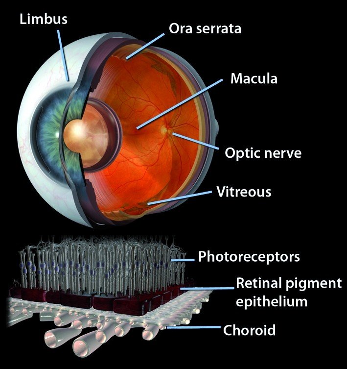
Retinal detachment (RD), defined as the separation of the neurosensory retina from the underlying retinal pigment epithelium (RPE), can cause devastating visual loss. With advances in surgical techniques, the anatomical reattachment rate has greatly increased, especially after repeated surgery [, ]. However, the final visual outcome remains, in many cases, disappointing once the retinal detachment involving the macula. Even with total retinal reattachment, long-term post-operative VA may lie anywhere in the range of 0.2 to 0.4 []. Photoreceptor apoptosis has been postulated to be the main reason for such visual loss [], alongside other structural changes in the retina such as glial scarring []. Neuroprotection has thus become a focus of research to achieve a better visual outcome by preventing or reducing photoreceptor cell death.

Retinal detachment is a disorder of the eye in which the retina separates from the layer underneath.[rx] Symptoms include an increase in the number of floaters, flashes of light, and worsening of the outer part of the visual field.[rx][rx] This may be described as a curtain over part of the field of vision.[rx] In about 7% of cases, both eyes are affected.[rx] Without treatment, permanent loss of vision may occur.[rx]

Retinal detachment

Types of Retinal Detachment

Retinal detachment (RD) is broadly classified into three types based on clinical appearance and underlying etiology.

  • Rhegmatogenous retinal detachment (RRD) – where the RD develops due to a retinal break (‘rhegma’, meaning rent or a fissure) [rx]. Fluid, from the vitreous cavity, passes through the retinal break into the potential space under the retina, leading to separation of the retina from the underlying choroid. This requires surgical treatment.
  • Tractional retinal detachment (TRD) – which occurs due to pre-retinal membrane formation and scarring that pulls the retina from its attachment. This may require surgery depending on the extent of the RD. The commonest causes of TRD are diabetes, Eales’s disease, sickle cell retinopathy, and trauma.
  • Exudative and serous retinal detachments – occur due to abnormalities in water transport across the bed of the retina (retinal pigment epithelium) or in its blood supply.

Tractional and exudative/serous retinal detachments are less common and will not be discussed in this paper.

Causes of Retinal Detachment

Rhegmatogenous

Caused by breaks in the retina, Associated with

  • Age
  • Myopia
  • Cataract surgery
  • Trauma
  • Degenerative retinal lesions
  • Stickler’s syndrome
  • Juvenile X-linked retinoschisis
  • Marfan’s syndrome

Tractional

Caused by chronic traction from scars on the retinal surface and across the vitreous cavity. Associated with

  • Proliferative diabetic retinopathy
  • Proliferative vitreoretinopathy
  • Retinopathy of prematurity
  • Penetrating eye injury
  • Sickle cell retinopathy
  • Retinal vein occlusion

Exudative

It happens when fluid collects under your retina, but there’s no tear. It can affect both eyes. This type of detachment often comes from an eye injury or as a complication of a wide range of diseases. They include various inflammatory and kidney diseases, as well as Lyme disease, eye tumors, and severe high blood pressure.

Caused by leakage of fluid into the subretinal space. Associated conditions

  • Inflammatory (uveitis, scleritis)
  • Hydrostatic (malignant hypertension, toxemia of pregnancy)
  • Neoplastic (choroidal melanoma, haemangioma, metastasis)
  • Vascular (Coat’s disease, retinal microaneurysm)
  • Maculopathy (neovascular macular degeneration, central serous chorioretinopathy)
  • Congenital disorders (nanophthalmos, optic disc pit)
  • Post cataract surgery (aphakia/pseudophakia) especially if the posterior capsule is ruptured during surgery and/or there is a vitreous loss.
  • Yag laser capsulotomy.
  • Lattice degeneration of the retina.
  • Symptomatic (flashes/floaters) retinal tears.
  • Ocular trauma
  • RD in one eye
  • Family history of RD
  • Certain genetic disorders such as Marfan’s syndrome, Stickler’s syndrome.
  • Pre-existing retinal diseases like coloboma choroid, retinoschisis.
  • Following acute retinal infections as in acute retinal necrosis syndrome (ARN) or CMV retinitis.
  • Glaucoma
  • AIDS[rx]
  • Cataract surgery[rx]
  • Diabetic retinopathy[rx][rx]
  • Eclampsia[rx]
  • Family history of retinal detachment[rx]
  • Homocysteinuria[rx]
  • Malignant hypertension[rx]
  • Metastatic cancer, which spreads to the eye (eye cancer)[rx]
  • Retinoblastoma[rx]
  • Severe myopia[rx]
  • Smoking and passive smoking[rx]
  • Stickler syndrome[rx]
  • Von Hippel-Lindau disease[rx]

Symptoms of Retinal Detachment

Retinal detachment

A rhegmatogenous retinal detachment is commonly preceded by a posterior vitreous detachment which gives rise to these symptoms

  • The detachment is visible as a grey, mobile membrane inside the eye.
  • Intraocular pressure may be reduced
  • The red reflex is usually pale, or grey, rather than the normal orange
  • When examining the eye using a slit lamp, you may see pigment cells in the vitreous.
  • Very brief flashes of light (photopsia) in the extreme peripheral (outside of center) part of a vision
  • A sudden dramatic increase in the number of floaters
  • A ring of floaters or hairs just to the temporal (skull) side of the central vision

Although most posterior vitreous detachments do not progress to retinal detachments, those that do produce the following symptoms

  • The sudden appearance of many floaters — tiny specks that seem to drift through your field of vision
  • Flashes of light in one or both eyes (photopsia)
  • Blurred vision
  • Gradually reduced side (peripheral) vision
  • A curtain-like shadow over your visual field
  • The dense shadow that starts in the peripheral vision and slowly progresses towards the central vision
  • The impression that a veil or curtain was drawn over the field of vision
  • Straight lines (scale, the edge of the wall, road, etc.) that suddenly appear curved (positive Amsler grid test)
  • Central visual loss.

In the event of an appearance of sudden flashes of light or floaters, an eye doctor needs to be consulted immediately.[rx] Any loss of vision is also a medical emergency.

Diagnosis of Retinal Detachment

  • A retinal examination – The doctor may use an instrument with a bright light and special lenses to examine the back of your eye, including the retina. This type of device provides a highly detailed view of your whole eye, allowing the doctor to see any retinal holes, tears or detachments.
  • Retinal detachment can be examined – by fundus photography or ophthalmoscopy. Fundus photography generally needs a considerably larger instrument than the ophthalmoscopy but has the advantage of availing the image to be examined by a specialist at another location and/or time, as well as providing photo documentation for future reference.
  • Modern fundus photographs – generally recreate considerably larger areas of the fundus than what can be seen at any one time with handheld ophthalmoscopes.
  • Ultrasound – has diagnostic accuracy similar to that of examination by an ophthalmologist.[rx] The recent meta-analysis shows the diagnostic accuracy of emergency department (ED) ocular ultrasonography is high. The sensitivity and specificity ranged from 97% to 100% and 83% to 100%.[rx]
  • The typical feature of retinal detachment – when viewed on ultrasound, is “flying angel sign”. It shows the detached retina moving with a fixed point under the B mode, linear probe 10 MHz.[rx]

Treatment of Retinal Detachment

There are several methods of treating a detached retina, each of which depends on finding and closing the breaks that have formed in the retina. All three of the procedures follow the same three general principles:

  • Find all retinal breaks
  • Seal all retinal breaks
  • Relieve present (and future) vitreoretinal traction
  • Laser coagulation – the laser light enters the eye via the pupil. The laser energy is absorbed in the retinal pigment epithelium, leading to heat (ca. 60 °C) and coagulation necrosis [, . Cryocoagulation involves freezing of the eyeball all the way from the outside to the retina by application of a cryoprobe (ca. –80 °C). Both procedures are followed after a few days by the formation of a scar, but only if the retina is in contact with the underlying retinal pigment epithelium. Therefore, scar induction by either laser coagulation or cryocoagulation is effective only for prevention of detachment in a still-attached retina; both forms of coagulation are pointless if detachment has already occurred.
  • Funduscopy – A detachment is recognized by the dune-like appearance and mobility of the retina, and the hole responsible for the detachment can often be discerned. The procedures employed for surgical management of retinal detachment are scleral buckling and vitrectomy. Here too laser coagulation or cryocoagulation is used for hole closure, but only after surgery to repair the detachment. Data for both of these surgical options are available from recent prospective randomized clinical trials.
  • Scleral buckling – After precise localization of all retinal breaks and marking of the sclera, the holes are treated with cryopexy for scar induction. The traction exerted on the holes by the vitreous body is then reduced by a foam sponge sutured to the sclera [
  • Scleral buckle surgery – is an established treatment in which the eye surgeon sews one or more silicone bands (or tires) to the sclera (the white outer coat of the eyeball). The bands push the wall of the eye inward against the retinal hole, closing the break or reducing fluid flow through it and reducing the effect of vitreous traction thereby allowing the retina to re-attach. Cryotherapy (freezing) is applied around retinal breaks prior to placing the buckle. Often subretinal fluid is drained as part of the buckling procedure
  • Pneumatic retinopexy – This operation is generally performed in the doctor’s office under local anesthesia. It is another method of repairing a retinal detachment in which a gas bubble (SF6[rx] or C3F8[rx] gas) is injected into the eye after which laser or freezing treatment is applied to the retinal hole. The patient’s head is then positioned so that the bubble rests against the retinal hole. Patients may have to keep their heads tilted for several days to keep the gas bubble in contact with the retinal hole. The surface tension of the gas/water interface seals the hole in the retina, and allows the retinal pigment epithelium to pump the subretinal space dry and “suck the retina back into place”.
  • Vitrectomy – Vitrectomy begins with the removal of the vitreous humor causing the retinal detachment, followed by displacement of the subretinal fluid by means of a heavy tamponade (perfluorodecalin or perfluorocarbon) and scarring of the retina by laser coagulation or cryocoagulation. The vitreous is then replaced by a tamponade[rx], which holds the retina against the underlying retinal pigment epithelium until a firm scar has formed around the retinal hole. A mixture of air and gas or a silicone oil tamponade can be chosen for this purpose. The air/gas mixture is usually chosen in simpler situations (e.g., when the hole is at the top of the eyeball). The advantage of the air/gas tamponade is that it is absorbed and thus does not require removal.
  • Injecting air or gas into your eye – In this procedure, called pneumatic retinopexy, the surgeon injects a bubble of air or gas into the center part of the eye (the vitreous cavity). If positioned properly, the bubble pushes the area of the retina containing the hole or holes against the wall of the eye, stopping the flow of fluid into space behind the retina. Your doctor also uses cryopexy during the procedure to repair the retinal break.
  • Indenting the surface of your eye – This procedure, called scleral (SKLAIR-ul) buckling, involves the surgeon sewing (suturing) a piece of silicone material to the white of your eye (sclera) over the affected area. This procedure indents the wall of the eye and relieves some of the force caused by the vitreous tugging on the retina. If you have several tears or holes or an extensive detachment, your surgeon may create a scleral buckle that encircles your entire eye like a belt. The buckle is placed in a way that doesn’t block your vision, and it usually remains in place permanently.
  • Draining and replacing the fluid in the eye – In this procedure, called vitrectomy, the surgeon removes the vitreous along with any tissue that is tugging on the retina. Air, gas or silicone oil is then injected into the vitreous space to help flatten the retina.

Health Tips  for Home Treatment

Retinal detachment may cause you to lose vision. Depending on your degree of vision loss, your lifestyle might change significantly.

You may find the following ideas useful as you learn to live with impaired vision:

  • Get glasses – Optimize the vision you have with glasses that are specifically tailored for your eyes. Request safety lenses to protect your better-seeing eye.
  • Brighten your home – Have proper light in your home for reading and other activities.
  • Make your home safer – Eliminate throw rugs and place colored tape on the edges of steps. Consider installing motion-activated lights.
  • Enlist the help of others – Tell friends and family members about your vision problems so they can help you.
  • Get help from technology – Digital talking books and computer screen readers can help with reading, and another new technology continues to advance.
  • Check into transportation – Investigate vans and shuttles, volunteer driving networks, or ride shares available in your area for people with impaired vision.
  • Talk to others with impaired vision – Take advantage of online networks, support groups, and resources for people with impaired vision.

References


Loading

If the article is helpful, please Click to Star Icon and Rate This Post!
[Total: 0 Average: 0]

Treatment of Meningitis, Treatment, Prevention

Treatment of Meningitis/Meningitis is inflammation of the meninges covering the brain. It is a pathological definition. The cerebrospinal fluid (CSF) typically exhibits an elevated number of leucocytes (or a pleocytosis). In adults, >5 leucocytes/μL is defined as elevated. Bacterial or viral meningitis is confirmed by the detection of a pathogen in the CSF. Bacterial meningitis may also be suggested by symptoms of meningism and appropriate bacteria in the blood.

Meningitis is an inflammation of the membranes (meninges) surrounding your brain and spinal cord. The inflammation may be caused by infection with viruses, bacteria, or other microorganisms, and less commonly by certain drugs. Meningitis can be life-threatening because of the inflammation’s proximity to the brain and spinal cord; therefore, the condition is classified as a medical emergency.

Types of Meningitis

Bacterial

Meningitis caused by bacteria can be deadly and requires immediate medical attention. Vaccines are available to help protect against some kinds of bacterial meningitis.Streptococcus pneumoniae- A causative bacteria of meningitis.

The types of bacteria that cause bacterial meningitis vary according to the infected individual’s age group.

  • In premature babies and newborns up to three months old, common causes are group B streptococci (subtypes III which normally inhabit the vagina and are mainly a cause during the first week of life) and bacteria that normally inhabit the digestive tract such as Escherichia coli (carrying the K1 antigen). Listeria monocytogenes (serotype IVb) is transmitted by the mother before birth and may cause meningitis in the newborn.
  • Older children are more commonly affected by Neisseria meningitidis (meningococcus) and Streptococcus pneumoniae (serotypes 6, 9, 14, 18 and 23) and those under five by Haemophilus influenzae type B (in countries that do not offer vaccination).
  • In adults, Neisseria meningitidis and Streptococcus pneumoniae together cause 80% of bacterial meningitis cases. Risk of infection with Listeria monocytogenes is increased in persons over 50 years old.The introduction of pneumococcal vaccine has lowered rates of pneumococcal meningitis in both children and adults.
  • Recent skull trauma potentially allows nasal cavity bacteria to enter the meningeal space. Similarly, devices in the brain and meninges, such as cerebral shunts, extraventricular drains or Ommaya reservoirs, carry an increased risk of meningitis. In these cases, the persons are more likely to be infected with Staphylococci, Pseudomonas, and other Gram-negative bacteria.These pathogens are also associated with meningitis in people with an impaired immune system.
  • Tuberculous meningitis, which is meningitis caused by Mycobacterium tuberculosis, is more common in people from countries in which tuberculosis is endemic, but is also encountered in persons with immune problems, such as AIDS.

Viral

  • Meningitis caused by viruses is serious but often is less severe than bacterial meningitis. People with normal immune systems who get viral meningitis usually get better on their own. There are vaccines to prevent some kinds of viral meningitis.
  • Viruses that cause meningitis include enteroviruses, herpes simplex virus (generally type 2, which produces most genital sores; less commonly type 1), varicella zoster virus (known for causing chickenpox and shingles), mumps virus, HIV, and LCMV. Mollaret’s meningitis is a chronic recurrent form of herpes meningitis; it is thought to be caused by herpes simplex virus type 2.

Fungal

  • Meningitis caused by fungi is rare, but people can get it by inhaling fungal spores from the environment. People with certain medical conditions, like diabetes, cancer, or HIV, are at higher risk of fungal meningitis.
  • There are a number of risk factors for fungal meningitis, including the use of immunosuppressants (such as after organ transplantation), HIV/AIDS, and the loss of immunity associated with aging. It is uncommon in those with a normal immune system but has occurred with medication contamination.
  • Symptom onset is typically more gradual, with headaches and fever being present for at least a couple of weeks before diagnosis. The most common fungal meningitis is cryptococcal meningitis due to Cryptococcus neoformans.
  • In Africa, cryptococcal meningitis is now the most common cause of meningitis in multiple studies, and it accounts for 20–25% of AIDS-related deaths in Africa. Other less common fungal pathogens which can cause meningitis include: Coccidioides immitisHistoplasma capsulatumBlastomyces dermatitidis, and Candidaspecies.

Parasitic

  • Various parasites can cause meningitis or can affect the brain or nervous system in other ways. Overall, parasitic meningitis is much less common than viral and bacterial meningitis.
  • A parasitic cause is often assumed when there is a predominance of eosinophils (a type of white blood cell) in the CSF. The most common parasites implicated are Angiostrongylus cantonensisGnathostoma spinigerumSchistosoma, as well as the conditions cysticercosis, toxocariasis, baylisascariasis, paragonimiasis, and a number of rarer infections and noninfective conditions.

Aseptic meningitis

Aseptic meningitis is a term referring to the broad category of meningitis that is not caused by bacteria. Approximately 50% of aseptic meningitis is due to viral infections. Other less common causes include

  • drug reactions or allergies, and
  • inflammatory diseases like lupus.

Non-infectious

  • Meningitis may occur as the result of several non-infectious causes: the spread of cancer to the meninges (malignant or neoplastic meningitis) and certain drugs (mainly non-steroidal anti-inflammatory drugs, antibiotics and intravenous immunoglobulins).
  • It may also be caused by several inflammatory conditions, such as sarcoidosis (which is then called neurosarcoidosis), connective tissue disorders such as systemic lupus erythematosus, and certain forms of vasculitis (inflammatory conditions of the blood vessel wall), such as Behçet’s disease. Epidermoid cysts and dermoid cysts may cause meningitis by releasing irritant matter into the subarachnoid space.

Amebic Meningitis

  • Primary amebic meningoencephalitis (PAM) is a rare and devastating infection of the brain caused by Naegleria fowleriNaegleria fowleri is a free-living microscopic ameba that lives in warm water and soil.

Pathophysiology

Meningitis typically occurs through two routes of inoculation:

Hematogenous Seeding

  • Bacterial droplets colonize the nasopharynx and enter the bloodstream after the mucosal invasion. Upon making their way to the subarachnoid space, the bacteria cross the blood-brain barrier, causing a direct inflammatory and immune-mediated reaction.

Direct Contiguous Spread

  • Organisms can enter the cerebrospinal fluid (CSF) via neighboring anatomic structures (otitis media, sinusitis), foreign objects (medical devices, penetrating trauma) or during operative procedures.

Viruses can penetrate the central nervous system (CNS) via retrograde transmission along neuronal pathways or by hematogenous seeding.

Causes of Meningitis

Causative organisms.

Organism Comment
Streptococcus pneumoniae Commonest organism
Affects healthy children
Additional risk factors: basilar skull or cribriform fracture, asplenism, HIV, and cochlear implants
Neisseria meningitidis Can cause epidemic, endemic, or sporadic infections
Haemophilus influenza type B Reduced incidence after introduction of the vaccination program
Group B streptococcus The less common pathogens
Group B streptococcus, E. Coli and L. monocytogenes more common in neonates
Escherichia coli
Non typeable H. influenzae
Other gram-negative bacilli
Listeria monocytogenes
Group A streptococci
Staphylococcal species Penetrating head trauma and neurosurgery
Streptococci
Aerobic gram-negative bacilli

Several strains of bacteria can cause acute bacterial meningitis, most commonly

  • Streptococcus pneumoniae (pneumococcus) – This bacterium is the most common cause of bacterial meningitis in infants, young children, and adults in the United States. It more commonly causes pneumonia or ear or sinus infections. A vaccine can help prevent this infection.
  • Neisseria meningitidis (meningococcus) – This bacterium is another leading cause of bacterial meningitis. These bacteria commonly cause an upper respiratory infection but can cause meningococcal meningitis when they enter the bloodstream. This is a highly contagious infection that affects mainly teenagers and young adults. It may cause local epidemics in college dormitories, boarding schools, and military bases. A vaccine can help prevent infection.
  • Haemophilus influenzae (Haemophilus) – Haemophilus influenza type b (Hib) bacterium was once the leading cause of bacterial meningitis in children. But new Hib vaccines have greatly reduced the number of cases of this type of meningitis.
  • Listeria monocytogenes (listeria) – These bacteria can be found in unpasteurized cheeses, hot dogs and luncheon meats. Pregnant women, newborns, older adults and people with weakened immune systems are most susceptible.
  • Fungal infection
  • Syphilis
  • Tuberculosis
  • Autoimmune disorders
  • Cancer medications
  • Adults older than 60 years of age
  • Children younger than 5 years of age
  • People with alcoholism
  • People with sickle cell anemia
  • People with cancer, especially those receiving chemotherapy
  • People who have received transplants and are taking drugs that suppress the immune system
  • People with diabetes
  • Those recently exposed to meningitis at home
  • People living in close quarters (military barracks, dormitories)
  • IV drug users
  • People with shunts in place for hydrocephalus

Spreading the bacteria

The meningococcal bacteria that cause meningitis do not live long outside the body, so they are usually only spread through prolonged, close contact. Possible ways to spread the bacteria include:

  • sneezing
  • coughing
  • kissing
  • sharing utensils, such as cutlery
  • sharing personal possessions, such as a toothbrush or cigarette

As most people, particularly adults above 25, have a natural immunity to the meningococcal bacteria, most cases of bacterial meningitis are isolated (single cases).

  • a boarding school
  • a university campus
  • a military base
  • student housing

Symptoms of Meningitis

Possible signs and symptoms in anyone older than the age of 2 include

Signs of Meningitis in newborns 

Newborns and infants may show these signs

The Symptoms By Age

  • Fever;
  • Drowsiness or confusion;
  • Severe headache;
  • A stiff neck;
  • Bright lights hurt the eyes; and
  • Nausea and vomiting.

In babies younger than one year of age, symptoms of meningitis may be more difficult to identify. They may include:

  • Fever;
  • Fretfulness or irritability, especially when handled;
  • Difficulty waking up;
  • Difficulty feeding; and
  • Vomiting.

A stiff neck and bulging of the fontanelle (soft spot on top of the skull) may occur in young babies with meningitis, but usually, these signs are not present early in the illness.

Bacterial Meningitis

Each year there are over 1000 cases of bacterial meningitis in Canada. At least 50 species of bacteria can cause meningitis. Most cases are caused by one of the following bacteria:

  • Meningococcus (scientific name Neisseria meningitis)
  • Pneumococcus (scientific name Streptococcus pneumonia)
  • Group B streptococcus (scientific name Streptococcus agalactiae)
  • E coli (scientific name Escherichia coli)

The following are now uncommon causes of bacterial meningitis in Canada.

  • H flu b or Hib (scientific name Haemophilus influenza type b)
  • Listeria (Scientific name Listeria monocytogenes)
  • Tuberculosis or TB (scientific name Mycobacterium tuberculosis)

Fungal meningitis is quite rare. The following types of fungus can cause meningitis.

  • Candida albicans – is a fungus that normally causes thrush. In rare cases, the fungus can cause a dangerous form of meningitis, primarily in premature babies with very low birth weight or in persons with disorders of the immune system.
  • Cryptococcus neoformans is a fungus that is commonly found in soil. It causes most cases of fungal meningitis. It generally only occurs in people with AIDS, cancer or diabetes. It is life-threatening and requires treatment with antifungal drugs.
  • Histoplasma – is a common fungus commonly found in soil in some parts of Canada. It can cause meningitis and other illnesses in people with disorders of the immune system (AIDS, cancer, etc).

Diagnosis of Meningitis

Lumbar puncture

  • Lumbar puncture is the key investigation. It enables rapid confirmation of meningitis and type of infecting organism. Diagnostic yield of LP can be diminished by collecting small CSF volumes. At least 10 mL can be safely removed.

Cerebrospinal fluid cell count

  • The cerebrospinal fluid remains one of the most rapidly informative tests. Pleocytosis indicates meningeal inflammation, of which infection is the most common cause. Van de Beek and colleagues reported that >90% of adults with bacterial meningitis had a CSF leukocyte count >100 cells/μL.
  • Absence of pleocytosis makes meningitis much less likely, but does not completely rule it out. Approximately 1–2% of patients with bacterial meningitis will have a normal CSF leukocyte count. Positive pathogen detection and an absence of pleocytosis more frequently occurs among children, the immunocompromised, those pretreated with antibiotics or with mycobacteria tuberculosis infection.

Cerebrospinal fluid leukocyte differential

Cerebrospinal fluid leukocyte differential can help predict which type of pathogen is causing infection. Lymphocyte predominance suggests viral, while neutrophil predominance suggests bacterial infection. There are several exceptions to this general guide, including CSF neutrophil predominance observed in association with tuberculous meningitis

Laboratory Investigations

  • Initial blood tests – should be performed for full blood count, coagulation studies, and electrolytes to assess for complications of sepsis and to guide fluid management. Serum glucose should be routinely measured as it may be low in the child with meningitis, contributing to seizures. Its measurement is also needed to accurately interpret the CSF glucose.
  • Blood cultures – should be performed in all patients with suspected bacterial meningitis. They may be of particular value if a lumbar puncture is contraindicated. The likelihood of a positive blood culture result varies with the infecting organism; 40% of children with meningococcal meningitis will have a positive blood culture, whereas 50–90% of H. influenzae and 75% of S. pneumonia meningitis patients will have a positive culture result [].
  • Both CRP and procalcitonin – have been evaluated to distinguish between viral and bacterial meningitis. Several studies have shown procalcitonin to have better diagnostic accuracy than CRP in differentiating between aseptic and bacterial meningitis [, ]. Procalcitonin levels in combination with other clinical scoring systems have also been studied to evaluate the risk of bacterial meningitis [, ]. Although potentially increasing the sensitivity of scoring systems, the use of procalcitonin in association with clinical scores to exclude the diagnosis of bacterial meningitis is not currently recommended

Cerebrospinal fluid biochemistry

  • Cerebrospinal fluid glucose is normally approximately two-thirds of the blood (plasma) concentration. It is often lower in bacterial and tuberculous meningitis. As CSF glucose is influenced by the plasma glucose, it is essential to measure blood glucose at LP, to obtain an accurate CSF – blood glucose ratio. A CSF – blood glucose ratio <0.36 is an accurate (93%) marker for distinguishing bacterial from viral meningitis.
  • Cerebrospinal fluid protein is normally <0.4 g/L. Elevated protein suggests inflammation. A CSF protein < 0.6 g/L largely rules out bacterial infection.

Cerebrospinal fluid microscopy with Gram stain

  • Cerebrospinal fluid microscopy with Gram stain (or an acid-fast stain for M tuberculosis) can rapidly detect bacteria. It has a sensitivity between 50% and 99%. Detection, particularly for M tuberculosis, is enhanced by a collection of >10 mL of CSF and subsequent cytospin.

Cerebrospinal fluid culture

  • Cerebrospinal fluid culture is historically regarded as the ‘gold standard’ for the diagnosis of bacterial meningitis. It is diagnostic in 70–85% of cases prior to antibiotic exposure. Sensitivity decreases by 20% following antibiotic pretreatment. Cerebrospinal fluid sterilization can occur within 2–4 hours of antibiotic administration for meningococci and pneumococci respectively. Lumbar puncture should be performed as soon as possible to maximize pathogen detection.

Cerebrospinal fluid polymerase chain reaction

  • Cerebrospinal fluid polymerase chain reaction (PCR), using pathogen-specific nucleic acid sequences, can detect both bacteria and viruses with high sensitivity. The polymerase chain reaction is the ‘gold standard’ for the diagnosis of viral meningitis. The polymerase chain reaction is increasingly relied upon in bacterial meningitis. It has far greater sensitivity than culture in invasive meningococcal disease.17 Cerebrospinal fluid PCR is particularly valuable in patients who receive antibiotics before LP. Polymerase chain reaction for 16S ribosomal RNA (present in almost all bacteria) enables a broad screen for bacteria, but has lower sensitivity than pathogen-specific PCR.

Blood tests

  • Blood cultures should always be taken on admission and are helpful when antibiotics are started before LP. Blood cultures are positive in 50–80% of bacterial meningitis cases.
  • Blood PCR is increasingly important, especially as PCR detects bacteria several days after antibiotic initiation. Blood PCR substantially increases the confirmation in meningococcal disease.
  • Despite these tests, many patients will not have a cause identified for their meningitis.
  • Blood biomarkers, such as procalcitonin and C-reactive protein, can help distinguish bacterial from viral meningitis in adults and can be used to help guide treatment if no aetiology is found. Host biomarkers for detecting bacterial meningitis are being actively investigated by our Liverpool group and others. To date, there is insufficient evidence to recommend their routine use in the NHS.

Swabs

  • Throat, nasopharyngeal, and stool swabs are useful for detecting enteroviruses if the CSF PCR is negative.

Brain imaging

  • Brain imaging is neither obligatory in the management of meningitis nor a prerequisite to LP. Performing neuroimaging before LP is associated with delays in commencing antibiotics, which in turn can lead to an increase in mortality. An urgent CT scan should be performed if there are clinical signs of brain shift. Clinical features indicative of a brain shift include focal neurological signs and reduced Glasgow Coma Score (GCS) [rx].
  • The 2016 UK meningitis guidelines recommend an LP be performed without prior neuroimaging if the GCS is >12. Patients with a GCS ≤12 should be considered for critical care, intubation assessment, and neuroimaging. Imaging, particularly when contrast is used, may exhibit meningeal enhancement in meningitis. When brain shift is identified liaison with critical care and neurosurgical teams are essential.

Common Tests Include The Following

Lumbar puncture findings [, ].

CSF finding Normal2 Viral Bacterial Partially treated bacterial
White cell count (cells/mm3) <5 <1000 >1000 >1000
PMNs 0 20–40% >85–90% >80%
Protein (mg/dL) <40 Normal or <100 >100–200 60–100+
Glucose (mmol/L) ≥2.5 Normal Undetectable–<2.2 <2.2
Blood to glucose ratio ≥0.6 Normal <0.4 <0.4
Positive gram stain 75–90% (depending on organism) 55–70%
Positive culture >70–85% <85%

Other investigations may also be performed on CSF to exclude nonbacterial causes of meningitis depending on the clinical scenario; including India Ink staining or antigen testing for Cryptococcus neoformans, Herpes simplex virus (HSV), cytomegalovirus (CMV) and enterovirus PCR.

 Values for pediatric patients >1 month of age; some values vary for neonates [].

Neonates: white cell count may be higher (<20 in the form of lymphocytes); normally zero PMNs, however some studies have found up to 5% PMNs in neonates without meningitis.

Neonates: normal protein <100 mg/dL.

Investigations for suspected bacterial meningitis.

Investigation Comment
Blood:
 Full blood count Neutrophilia suggestive of bacterial infection
 Serum glucose Often low; allows interpretation of CSF glucose
 Electrolytes, urea, and creatinine To assess for complications and fluid management
 Coagulation studies To assess for complications
 Blood cultures Positive in 40–90% depending on organism
 Inflammatory markers Elevation suggestive of bacterial infection; procalcitonin of more value; neither can establish nor exclude diagnosis
  CRP, procalcitonin
CSF:
 Protein and glucose
 Microscopy, culture, and sensitivities Gram stain:
 S. pneumoniae—gram +ve cocci
 N. menigitidis—gram −ve cocci
 H. influenzae—gram −ve rod
 Latex agglutination Rapid; not 100% specific or diagnostic
 PCR Rapid; good sensitivity, techniques improving
 Lactate Routine use not currently recommended
Imaging:
Computed tomography of the head
Indicated for focal neurology, signs of increased intracranial pressure (ICP), deteriorating neurological function, previous neurosurgical procedures, or immunocompromised
May show evidence of hydrocephalus, abscess, subdural empyema, or infarction
Normal scan does not entirely exclude risk of raised ICP
Other:
PCR on blood or urine
Useful if CSF not obtainable

1Latex agglutination depends on laboratory availability; including N. meningitidis, S. pneumoniae, H. influenzae type B, Escherichia coli and group B streptococci.

2PCR depends on laboratory availability; including N. meningitidisS. pneumoniaeH. influenzae type b, L. monocytogenes, HSV, CMV, Enterovirus and Mycobacterium tuberculosis.

Treatment

Immediate treatment

Blood tests may be used to monitor the patient’s levels of blood sugar, sodium, and other vital chemicals.

Treatment / Management

Antibiotics and supportive care are critical in all infectious resuscitations.

Managing the airway, maintaining oxygenation, giving sufficient intra-venous fluids while providing fever control are parts of the foundation of sepsis management.

The type of antibiotic is based on the presumed organism causing the infection. The clinician must take into account patient demographics and past medical history in order to provide the best antimicrobial coverage.

Current Empiric Therapy

Neonates – Up to 1 month old

  • Ampicillin 100 mg/kg intravenously (IV) and
  • Cefotaxime 75 mg/kg IV or Gentamicin 2.5 mg/kg IV and
  • Acyclovir IV 40 mg/kg

More than 1 month old

  • Ampicillin 50 mg/kg IV and
  • Ceftriaxone 2 g IV  and
  • Acyclovir IV 40 mg/kg

Adults (18 to 49 years old)

  • Ceftriaxone 2 g IV and
  • Vancomycin 20 mg/kg IV

Adults older than 50 years old and the immunocompromised

  • Ceftriaxone 2 g IV and
  • Vancomycin 20 mg/kg IV and
  • Ampicillin 2 g IV

Meningitis associated with a foreign body (post-procedure, penetrating trauma)

  • Cefepime 2 g IV or Ceftazidime 2 g IV or Meropenem 2 g IV and
  • Vancomycin 20 mg/kg IV

Meningitis with severe penicillin allergy

  • Chloramphenicol 1 g IV and
  • Vancomycin 20 mg/kg IV

Fungal (Cryptococcal) meningitis

  • Amphotericin B 1 mg/kg IV and
  • Flucytosine 25 mg/kg by mouth

Antibiotics

Ceftriaxone

  • Third-generation cephalosporin
  • Gram-negative coverage
  • Very effective against S. pneumoniae and N. meningitides
  • Better CNS penetration than Piperacillin-Tazobactam (typically used in gram-negative sepsis coverage)

Vancomycin

  • Gram-positive coverage (MRSA)
  • Also used for resistant pneumococcus

Ampicillin

  • Listeria coverage (gram-positive bacilli)
  • Is an aminopenicillin

Cefepime

  • Fourth generation cephalosporin
  • Increased activity against pseudomonas

Cefotaxime

  • Third generation Cephalosporin
  • Safe for neonates

Steroid Therapy

Administration of dexamethasone 10 mg IV before or with the first dose of antibiotics has been shown to reduce the risk of morbidity and mortality, especially in the setting of S. pneumoniae infection.

It is important to note; the Infectious Disease Society of America recommends against dexamethasone if the patient has already received antibiotics.

Increased Intracranial Pressure

If the patient develops clinical signs of increased intracranial pressure, interventions to maintain cerebral perfusion include:

  • Elevating the head of the bed to 30 degrees
  • Inducing mild hyperventilation in the intubated patient
  • Osmotic diuretics such as 25% mannitol or 3% saline

Chemoprophylaxis

The transmission rate of N. meningitidis is 5% for close contacts, but chemoprophylaxis within 24 hours decreases that by 89%. Thus, chemoprophylaxis is indicated for close contacts of a patient suspected of having bacterial meningitis.

Close contacts include housemates, significant others, those who have shared utensils and health care providers in proximity to secretions (providing mouth-to-mouth resuscitation, intubating without a facemask).

Antibiotic chemoprophylaxis options include:

  • Rifampin 10 mg/kg (max 200 mg/dose) every 12 hours for 4 doses or
  • Ciprofloxacin 500 g orally once, or
  • Ceftriaxone 250 mg intramuscularly once

Prevention of Meningitis

As several types of bacteria can cause bacterial meningitis, so a range of vaccines is necessary to prevent infection.

  • Haemophilus influenzae type b – can be prevented with Hib immunisation, which is available in combination vaccines free on the National Immunisation Program Schedule. It is routinely offered tor babies and needs to be purchased on prescription for some groups at high risk of bacterial disease.
  • Meningococcal group A, B, C, W135 and Y – can be prevented with a range of vaccines. Some immunisation is available free on the National Immunisation Program Schedule routinely for 12 month old babies or childhood catch-up and some vaccine needs to be purchased with prescription for some groups at high risk of bacterial disease or some travellers
  • Pneumococcal – can be prevented with two types of pneumococcal vaccine.. They are available free on the National Immunisation Schedule to all babies and adults 50 years of age, if the person is an Aboriginal or Torres Strait Islander, or at 65 years of age and over. They need to be purchased on prescription for some groups at high risk of bacterial disease.
  • A survey of 17 million people in the U.S. found that the incidence of all types of meningitis fell by 31 percent from 1998 to 2007, after the introduction of routine vaccinations against meningitis-causing bacteria.
  • The meningococcal vaccine is the primary vaccine in the U.S. All children should have this at the age of 11 to 12 years and again at 16 years, when the risk of infection is higher.
  • The Hib vaccine protects children against H. Influenzae. Before its introduction in the U.S. in 1985, H. Influenzae infected over 20,000 children under 5 years annually, with a 3 to 6 percent mortality rate. Widespread vaccination has reduced the incidence of bacterial meningitis by over 99 percent.
  • The Hib vaccine is given in four doses at the ages of 2, 4, 6, and 12 to 15 months.

Complication

For patients treated promptly, the prognosis is good. However, patients who present with an altered state of consciousness have a high morbidity and mortality. Some patients may develop seizures during the illness, which are very difficult to control or are prolonged. Any patient with a residual neurological deficit after meningitis treatment is also left with a disability. Patients art the greatest risk for death usually have the following features:

  • Advanced aged
  • Low GCS
  • CSF WBC count which is low
  • Tachycardia
  • Gram-positive cocci in the CSF

Serious complications in survivors include:

  • Ataxia
  • Hearing loss
  • Cranial nerve palsies
  • Cognitive dysfunction
  • Cortical blindness
  • Hydrocephalus
  • Seizures
  • Focal paralysis

References

Treatment of Meningitis

Loading

If the article is helpful, please Click to Star Icon and Rate This Post!
[Total: 0 Average: 0]

Parasitic Meningitis, Symptoms, Treatment

Parasitic Meningitis/Meningitis is inflammation of the meninges covering the brain. It is a pathological definition. The cerebrospinal fluid (CSF) typically exhibits an elevated number of leucocytes (or a pleocytosis). In adults, >5 leucocytes/μL is defined as elevated. Bacterial or viral meningitis is confirmed by the detection of a pathogen in the CSF. Bacterial meningitis may also be suggested by symptoms of meningism and appropriate bacteria in the blood.

Meningitis is an inflammation of the membranes (meninges) surrounding your brain and spinal cord. The inflammation may be caused by infection with viruses, bacteria, or other microorganisms, and less commonly by certain drugs. Meningitis can be life-threatening because of the inflammation’s proximity to the brain and spinal cord; therefore, the condition is classified as a medical emergency.

Types of Meningitis

Bacterial

Meningitis caused by bacteria can be deadly and requires immediate medical attention. Vaccines are available to help protect against some kinds of bacterial meningitis. Streptococcus pneumoniae- A causative bacteria of meningitis.

The types of bacteria that cause bacterial meningitis vary according to the infected individual’s age group.

  • In premature babies and newborns up to three months old – common causes are group B streptococci (subtypes III which normally inhabit the vagina and are mainly a cause during the first week of life) and bacteria that normally inhabit the digestive tract such as Escherichia coli (carrying the K1 antigen). Listeria monocytogenes (serotype IVb) is transmitted by the mother before birth and may cause meningitis in the newborn.
  • Older children are more commonly affected byNeisseria meningitidis (meningococcus) and Streptococcus pneumoniae (serotypes 6, 9, 14, 18 and 23) and those under five by Haemophilus influenzae type B (in countries that do not offer vaccination).
  • In adults –  Neisseria meningitidis and Streptococcus pneumoniae together cause 80% of bacterial meningitis cases. Risk of infection with Listeria monocytogenes is increased in persons over 50 years old. The introduction of the pneumococcal vaccine has lowered rates of pneumococcal meningitis in both children and adults.
  • Recent skull trauma potentially – allows nasal cavity bacteria to enter the meningeal space. Similarly, devices in the brain and meninges, such as cerebral shunts, extraventricular drains or Ommaya reservoirs, carry an increased risk of meningitis. In these cases, the persons are more likely to be infected with Staphylococci, Pseudomonas, and other Gram-negative bacteria. These pathogens are also associated with meningitis in people with an impaired immune system.
  • Tuberculous meningitis – which is meningitis caused by Mycobacterium tuberculosis, is more common in people from countries in which tuberculosis is endemic, but is also encountered in persons with immune problems, such as AIDS.

Viral

  • Meningitis caused by viruses is serious but often is less severe than bacterial meningitis. People with normal immune systems who get viral meningitis usually get better on their own. There are vaccines to prevent some kinds of viral meningitis.
  • Viruses that cause meningitis include enteroviruses, herpes simplex virus (generally type 2, which produces most genital sores; less commonly type 1), varicella zoster virus (known for causing chickenpox and shingles), mumps virus, HIV, and LCMV. Mollaret’s meningitis is a chronic recurrent form of herpes meningitis; it is thought to be caused by herpes simplex virus type 2.

Fungal

  • Meningitis caused by fungi is rare, but people can get it by inhaling fungal spores from the environment. People with certain medical conditions, like diabetes, cancer, or HIV, are at higher risk of fungal meningitis.
  • There are a number of risk factors for fungal meningitis, including the use of immunosuppressants (such as after organ transplantation), HIV/AIDS, and the loss of immunity associated with aging. It is uncommon in those with a normal immune system but has occurred with medication contamination.
  • Symptom onset is typically more gradual, with headaches and fever being present for at least a couple of weeks before diagnosis. The most common fungal meningitis is cryptococcal meningitis due to Cryptococcus neoformans.
  • In Africa, cryptococcal meningitis is now the most common cause of meningitis in multiple studies, and it accounts for 20–25% of AIDS-related deaths in Africa. Other less common fungal pathogens which can cause meningitis include: Coccidioides immitisHistoplasma capsulatumBlastomyces dermatitidis, and Candidaspecies.

Parasitic

  • Various parasites can cause meningitis or can affect the brain or nervous system in other ways. Overall, parasitic meningitis is much less common than viral and bacterial meningitis.
  • A parasitic cause is often assumed when there is a predominance of eosinophils (a type of white blood cell) in the CSF. The most common parasites implicated are Angiostrongylus cantonensisGnathostoma spinigerumSchistosoma, as well as the conditions cysticercosis, toxocariasis, baylisascariasis, paragonimiasis, and a number of rarer infections and noninfective conditions.

Aseptic meningitis

Aseptic meningitis is a term referring to the broad category of meningitis that is not caused by bacteria. Approximately 50% of aseptic meningitis is due to viral infections. Other less common causes include

  • drug reactions or allergies, and
  • inflammatory diseases like lupus.

Non-infectious

  • Meningitis may occur as the result of several non-infectious causes: the spread of cancer to the meninges (malignant or neoplastic meningitis) and certain drugs (mainly non-steroidal anti-inflammatory drugs, antibiotics and intravenous immunoglobulins).
  • It may also be caused by several inflammatory conditions, such as sarcoidosis (which is then called neurosarcoidosis), connective tissue disorders such as systemic lupus erythematosus, and certain forms of vasculitis (inflammatory conditions of the blood vessel wall), such as Behçet’s disease. Epidermoid cysts and dermoid cysts may cause meningitis by releasing irritant matter into the subarachnoid space.

Amebic Meningitis

  • Primary amebic meningoencephalitis (PAM) is a rare and devastating infection of the brain caused by Naegleria fowleriNaegleria fowleri is a free-living microscopic ameba that lives in warm water and soil.

Pathophysiology

Meningitis typically occurs through two routes of inoculation:

Hematogenous Seeding

  • Bacterial droplets colonize the nasopharynx and enter the bloodstream after the mucosal invasion. Upon making their way to the subarachnoid space, the bacteria cross the blood-brain barrier, causing a direct inflammatory and immune-mediated reaction.

Direct Contiguous Spread

  • Organisms can enter the cerebrospinal fluid (CSF) via neighboring anatomic structures (otitis media, sinusitis), foreign objects (medical devices, penetrating trauma) or during operative procedures.

Viruses can penetrate the central nervous system (CNS) via retrograde transmission along neuronal pathways or by hematogenous seeding.

Causes of Meningitis

Causative organisms.

Organism Comment
Streptococcus pneumoniae Commonest organism
Affects healthy children
Additional risk factors: basilar skull or cribriform fracture, asplenism, HIV, and cochlear implants
Neisseria meningitidis Can cause epidemic, endemic, or sporadic infections
Haemophilus influenza type B Reduced incidence after introduction of the vaccination program
Group B streptococcus The less common pathogens
Group B streptococcus, E. Coli and L. monocytogenes more common in neonates
Escherichia coli
Non typeable H. influenzae
Other gram-negative bacilli
Listeria monocytogenes
Group A streptococci
Staphylococcal species Penetrating head trauma and neurosurgery
Streptococci
Aerobic gram-negative bacilli

Several strains of bacteria can cause acute bacterial meningitis, most commonly

  • Streptococcus pneumoniae (pneumococcus) – This bacterium is the most common cause of bacterial meningitis in infants, young children, and adults in the United States. It more commonly causes pneumonia or ear or sinus infections. A vaccine can help prevent this infection.
  • Neisseria meningitidis (meningococcus) – This bacterium is another leading cause of bacterial meningitis. These bacteria commonly cause an upper respiratory infection but can cause meningococcal meningitis when they enter the bloodstream. This is a highly contagious infection that affects mainly teenagers and young adults. It may cause local epidemics in college dormitories, boarding schools, and military bases. A vaccine can help prevent infection.
  • Haemophilus influenzae (Haemophilus) – Haemophilus influenza type b (Hib) bacterium was once the leading cause of bacterial meningitis in children. But new Hib vaccines have greatly reduced the number of cases of this type of meningitis.
  • Listeria monocytogenes (listeria) – These bacteria can be found in unpasteurized cheeses, hot dogs and luncheon meats. Pregnant women, newborns, older adults and people with weakened immune systems are most susceptible.
  • Fungal infection
  • Syphilis
  • Tuberculosis
  • Autoimmune disorders
  • Cancer medications
  • Adults older than 60 years of age
  • Children younger than 5 years of age
  • People with alcoholism
  • People with sickle cell anemia
  • People with cancer, especially those receiving chemotherapy
  • People who have received transplants and are taking drugs that suppress the immune system
  • People with diabetes
  • Those recently exposed to meningitis at home
  • People living in close quarters (military barracks, dormitories)
  • IV drug users
  • People with shunts in place for hydrocephalus

Spreading the bacteria

The meningococcal bacteria that cause meningitis do not live long outside the body, so they are usually only spread through prolonged, close contact. Possible ways to spread the bacteria include:

  • sneezing
  • coughing
  • kissing
  • sharing utensils, such as cutlery
  • sharing personal possessions, such as a toothbrush or cigarette

As most people, particularly adults above 25, have a natural immunity to the meningococcal bacteria, most cases of bacterial meningitis are isolated (single cases).

  • a boarding school
  • a university campus
  • a military base
  • student housing

Symptoms of Meningitis

Possible signs and symptoms in anyone older than the age of 2 include

Signs of Meningitis in newborns 

Newborns and infants may show these signs

The Symptoms By Age

  • Fever;
  • Drowsiness or confusion;
  • Severe headache;
  • A stiff neck;
  • Bright lights hurt the eyes; and
  • Nausea and vomiting.

In babies younger than one year of age, symptoms of meningitis may be more difficult to identify. They may include:

  • Fever;
  • Fretfulness or irritability, especially when handled;
  • Difficulty waking up;
  • Difficulty feeding; and
  • Vomiting.

A stiff neck and bulging of the fontanelle (soft spot on top of the skull) may occur in young babies with meningitis, but usually, these signs are not present early in the illness.

Bacterial Meningitis

Each year there are over 1000 cases of bacterial meningitis in Canada. At least 50 species of bacteria can cause meningitis. Most cases are caused by one of the following bacteria:

  • Meningococcus (scientific name Neisseria meningitis)
  • Pneumococcus (scientific name Streptococcus pneumonia)
  • Group B streptococcus (scientific name Streptococcus agalactiae)
  • E coli (scientific name Escherichia coli)

The following are now uncommon causes of bacterial meningitis in Canada.

  • H flu b or Hib (scientific name Haemophilus influenza type b)
  • Listeria (Scientific name Listeria monocytogenes)
  • Tuberculosis or TB (scientific name Mycobacterium tuberculosis)

Fungal meningitis is quite rare. The following types of fungus can cause meningitis.

  • Candida albicans – is a fungus that normally causes thrush. In rare cases, the fungus can cause a dangerous form of meningitis, primarily in premature babies with very low birth weight or in persons with disorders of the immune system.
  • Cryptococcus neoformans is a fungus that is commonly found in soil. It causes most cases of fungal meningitis. It generally only occurs in people with AIDS, cancer or diabetes. It is life-threatening and requires treatment with antifungal drugs.
  • Histoplasma – is a common fungus commonly found in soil in some parts of Canada. It can cause meningitis and other illnesses in people with disorders of the immune system (AIDS, cancer, etc).

Diagnosis of Meningitis

Lumbar puncture

  • Lumbar puncture is the key investigation. It enables rapid confirmation of meningitis and type of infecting organism. Diagnostic yield of LP can be diminished by collecting small CSF volumes. At least 10 mL can be safely removed.

Cerebrospinal fluid cell count

  • The cerebrospinal fluid remains one of the most rapidly informative tests. Pleocytosis indicates meningeal inflammation, of which infection is the most common cause. Van de Beek and colleagues reported that >90% of adults with bacterial meningitis had a CSF leukocyte count >100 cells/μL.
  • Absence of pleocytosis makes meningitis much less likely, but does not completely rule it out. Approximately 1–2% of patients with bacterial meningitis will have a normal CSF leukocyte count. Positive pathogen detection and an absence of pleocytosis more frequently occurs among children, the immunocompromised, those pretreated with antibiotics or with mycobacteria tuberculosis infection.

Cerebrospinal fluid leukocyte differential

Cerebrospinal fluid leukocyte differential can help predict which type of pathogen is causing infection. Lymphocyte predominance suggests viral, while neutrophil predominance suggests bacterial infection. There are several exceptions to this general guide, including CSF neutrophil predominance observed in association with tuberculous meningitis

Laboratory Investigations

  • Initial blood tests – should be performed for full blood count, coagulation studies, and electrolytes to assess for complications of sepsis and to guide fluid management. Serum glucose should be routinely measured as it may be low in the child with meningitis, contributing to seizures. Its measurement is also needed to accurately interpret the CSF glucose.
  • Blood cultures – should be performed in all patients with suspected bacterial meningitis. They may be of particular value if a lumbar puncture is contraindicated. The likelihood of a positive blood culture result varies with the infecting organism; 40% of children with meningococcal meningitis will have a positive blood culture, whereas 50–90% of H. influenzae and 75% of S. pneumonia meningitis patients will have a positive culture result [].
  • Both CRP and procalcitonin – have been evaluated to distinguish between viral and bacterial meningitis. Several studies have shown procalcitonin to have better diagnostic accuracy than CRP in differentiating between aseptic and bacterial meningitis [, ]. Procalcitonin levels in combination with other clinical scoring systems have also been studied to evaluate the risk of bacterial meningitis [, ]. Although potentially increasing the sensitivity of scoring systems, the use of procalcitonin in association with clinical scores to exclude the diagnosis of bacterial meningitis is not currently recommended

Cerebrospinal fluid biochemistry

  • Cerebrospinal fluid glucose is normally approximately two-thirds of the blood (plasma) concentration. It is often lower in bacterial and tuberculous meningitis. As CSF glucose is influenced by the plasma glucose, it is essential to measure blood glucose at LP, to obtain an accurate CSF – blood glucose ratio. A CSF – blood glucose ratio <0.36 is an accurate (93%) marker for distinguishing bacterial from viral meningitis.
  • Cerebrospinal fluid protein is normally <0.4 g/L. Elevated protein suggests inflammation. A CSF protein < 0.6 g/L largely rules out bacterial infection.

Cerebrospinal fluid microscopy with Gram stain

  • Cerebrospinal fluid microscopy with Gram stain (or an acid-fast stain for M tuberculosis) can rapidly detect bacteria. It has a sensitivity between 50% and 99%. Detection, particularly for M tuberculosis, is enhanced by a collection of >10 mL of CSF and subsequent cytospin.

Cerebrospinal fluid culture

  • Cerebrospinal fluid culture is historically regarded as the ‘gold standard’ for the diagnosis of bacterial meningitis. It is diagnostic in 70–85% of cases prior to antibiotic exposure. Sensitivity decreases by 20% following antibiotic pretreatment. Cerebrospinal fluid sterilization can occur within 2–4 hours of antibiotic administration for meningococci and pneumococci respectively. Lumbar puncture should be performed as soon as possible to maximize pathogen detection.

Cerebrospinal fluid polymerase chain reaction

  • Cerebrospinal fluid polymerase chain reaction (PCR), using pathogen-specific nucleic acid sequences, can detect both bacteria and viruses with high sensitivity. The polymerase chain reaction is the ‘gold standard’ for the diagnosis of viral meningitis. The polymerase chain reaction is increasingly relied upon in bacterial meningitis. It has far greater sensitivity than culture in invasive meningococcal disease.17 Cerebrospinal fluid PCR is particularly valuable in patients who receive antibiotics before LP. Polymerase chain reaction for 16S ribosomal RNA (present in almost all bacteria) enables a broad screen for bacteria, but has lower sensitivity than pathogen-specific PCR.

Blood tests

  • Blood cultures should always be taken on admission and are helpful when antibiotics are started before LP. Blood cultures are positive in 50–80% of bacterial meningitis cases.
  • Blood PCR is increasingly important, especially as PCR detects bacteria several days after antibiotic initiation. Blood PCR substantially increases the confirmation in meningococcal disease.
  • Despite these tests, many patients will not have a cause identified for their meningitis.
  • Blood biomarkers, such as procalcitonin and C-reactive protein, can help distinguish bacterial from viral meningitis in adults and can be used to help guide treatment if no aetiology is found. Host biomarkers for detecting bacterial meningitis are being actively investigated by our Liverpool group and others. To date, there is insufficient evidence to recommend their routine use in the NHS.

Swabs

  • Throat, nasopharyngeal, and stool swabs are useful for detecting enteroviruses if the CSF PCR is negative.

Brain imaging

  • Brain imaging is neither obligatory in the management of meningitis nor a prerequisite to LP. Performing neuroimaging before LP is associated with delays in commencing antibiotics, which in turn can lead to an increase in mortality. An urgent CT scan should be performed if there are clinical signs of brain shift. Clinical features indicative of a brain shift include focal neurological signs and reduced Glasgow Coma Score (GCS) [rx].
  • The 2016 UK meningitis guidelines recommend an LP be performed without prior neuroimaging if the GCS is >12. Patients with a GCS ≤12 should be considered for critical care, intubation assessment, and neuroimaging. Imaging, particularly when contrast is used, may exhibit meningeal enhancement in meningitis. When brain shift is identified liaison with critical care and neurosurgical teams are essential.

Common Tests Include The Following

Lumbar puncture findings [, ].

CSF finding Normal2 Viral Bacterial Partially treated bacterial
White cell count (cells/mm3) <5 <1000 >1000 >1000
PMNs 0 20–40% >85–90% >80%
Protein (mg/dL) <40 Normal or <100 >100–200 60–100+
Glucose (mmol/L) ≥2.5 Normal Undetectable–<2.2 <2.2
Blood to glucose ratio ≥0.6 Normal <0.4 <0.4
Positive gram stain 75–90% (depending on organism) 55–70%
Positive culture >70–85% <85%

Other investigations may also be performed on CSF to exclude nonbacterial causes of meningitis depending on the clinical scenario; including India Ink staining or antigen testing for Cryptococcus neoformans, Herpes simplex virus (HSV), cytomegalovirus (CMV) and enterovirus PCR.

 Values for pediatric patients >1 month of age; some values vary for neonates [].

Neonates: white cell count may be higher (<20 in the form of lymphocytes); normally zero PMNs, however some studies have found up to 5% PMNs in neonates without meningitis.

Neonates: normal protein <100 mg/dL.

Investigations for suspected bacterial meningitis.

Investigation Comment
Blood:
 Full blood count Neutrophilia suggestive of bacterial infection
 Serum glucose Often low; allows interpretation of CSF glucose
 Electrolytes, urea, and creatinine To assess for complications and fluid management
 Coagulation studies To assess for complications
 Blood cultures Positive in 40–90% depending on organism
 Inflammatory markers Elevation suggestive of bacterial infection; procalcitonin of more value; neither can establish nor exclude diagnosis
  CRP, procalcitonin
CSF:
 Protein and glucose
 Microscopy, culture, and sensitivities Gram stain:
 S. pneumoniae—gram +ve cocci
 N. menigitidis—gram −ve cocci
 H. influenzae—gram −ve rod
 Latex agglutination Rapid; not 100% specific or diagnostic
 PCR Rapid; good sensitivity, techniques improving
 Lactate Routine use not currently recommended
Imaging:
Computed tomography of the head
Indicated for focal neurology, signs of increased intracranial pressure (ICP), deteriorating neurological function, previous neurosurgical procedures, or immunocompromised
May show evidence of hydrocephalus, abscess, subdural empyema, or infarction
Normal scan does not entirely exclude risk of raised ICP
Other:
PCR on blood or urine
Useful if CSF not obtainable

1Latex agglutination depends on laboratory availability; including N. meningitidis, S. pneumoniae, H. influenzae type B, Escherichia coli and group B streptococci.

2PCR depends on laboratory availability; including N. meningitidisS. pneumoniaeH. influenzae type b, L. monocytogenes, HSV, CMV, Enterovirus and Mycobacterium tuberculosis.

Treatment

Immediate treatment

Blood tests may be used to monitor the patient’s levels of blood sugar, sodium, and other vital chemicals.

Treatment / Management

Antibiotics and supportive care are critical in all infectious resuscitations.

Managing the airway, maintaining oxygenation, giving sufficient intra-venous fluids while providing fever control are parts of the foundation of sepsis management.

The type of antibiotic is based on the presumed organism causing the infection. The clinician must take into account patient demographics and past medical history in order to provide the best antimicrobial coverage.

Current Empiric Therapy

Neonates – Up to 1 month old

  • Ampicillin 100 mg/kg intravenously (IV) and
  • Cefotaxime 75 mg/kg IV or Gentamicin 2.5 mg/kg IV and
  • Acyclovir IV 40 mg/kg

More than 1 month old

  • Ampicillin 50 mg/kg IV and
  • Ceftriaxone 2 g IV  and
  • Acyclovir IV 40 mg/kg

Adults (18 to 49 years old)

  • Ceftriaxone 2 g IV and
  • Vancomycin 20 mg/kg IV

Adults older than 50 years old and the immunocompromised

  • Ceftriaxone 2 g IV and
  • Vancomycin 20 mg/kg IV and
  • Ampicillin 2 g IV

Meningitis associated with a foreign body (post-procedure, penetrating trauma)

  • Cefepime 2 g IV or Ceftazidime 2 g IV or Meropenem 2 g IV and
  • Vancomycin 20 mg/kg IV

Meningitis with severe penicillin allergy

  • Chloramphenicol 1 g IV and
  • Vancomycin 20 mg/kg IV

Fungal (Cryptococcal) meningitis

  • Amphotericin B 1 mg/kg IV and
  • Flucytosine 25 mg/kg by mouth

Antibiotics

Ceftriaxone

  • Third-generation cephalosporin
  • Gram-negative coverage
  • Very effective against S. pneumoniae and N. meningitides
  • Better CNS penetration than Piperacillin-Tazobactam (typically used in gram-negative sepsis coverage)

Vancomycin

  • Gram-positive coverage (MRSA)
  • Also used for resistant pneumococcus

Ampicillin

  • Listeria coverage (gram-positive bacilli)
  • Is an aminopenicillin

Cefepime

  • Fourth generation cephalosporin
  • Increased activity against pseudomonas

Cefotaxime

  • Third generation Cephalosporin
  • Safe for neonates

Steroid Therapy

Administration of dexamethasone 10 mg IV before or with the first dose of antibiotics has been shown to reduce the risk of morbidity and mortality, especially in the setting of S. pneumoniae infection.

It is important to note; the Infectious Disease Society of America recommends against dexamethasone if the patient has already received antibiotics.

Increased Intracranial Pressure

If the patient develops clinical signs of increased intracranial pressure, interventions to maintain cerebral perfusion include:

  • Elevating the head of the bed to 30 degrees
  • Inducing mild hyperventilation in the intubated patient
  • Osmotic diuretics such as 25% mannitol or 3% saline

Chemoprophylaxis

The transmission rate of N. meningitidis is 5% for close contacts, but chemoprophylaxis within 24 hours decreases that by 89%. Thus, chemoprophylaxis is indicated for close contacts of a patient suspected of having bacterial meningitis.

Close contacts include housemates, significant others, those who have shared utensils and health care providers in proximity to secretions (providing mouth-to-mouth resuscitation, intubating without a facemask).

Antibiotic chemoprophylaxis options include:

  • Rifampin 10 mg/kg (max 200 mg/dose) every 12 hours for 4 doses or
  • Ciprofloxacin 500 g orally once, or
  • Ceftriaxone 250 mg intramuscularly once

Prevention of Meningitis

As several types of bacteria can cause bacterial meningitis, so a range of vaccines is necessary to prevent infection.

  • Haemophilus influenzae type b – can be prevented with Hib immunisation, which is available in combination vaccines free on the National Immunisation Program Schedule. It is routinely offered tor babies and needs to be purchased on prescription for some groups at high risk of bacterial disease.
  • Meningococcal group A, B, C, W135 and Y – can be prevented with a range of vaccines. Some immunisation is available free on the National Immunisation Program Schedule routinely for 12 month old babies or childhood catch-up and some vaccine needs to be purchased with prescription for some groups at high risk of bacterial disease or some travellers
  • Pneumococcal – can be prevented with two types of pneumococcal vaccine.. They are available free on the National Immunisation Schedule to all babies and adults 50 years of age, if the person is an Aboriginal or Torres Strait Islander, or at 65 years of age and over. They need to be purchased on prescription for some groups at high risk of bacterial disease.
  • A survey of 17 million people in the U.S. found that the incidence of all types of meningitis fell by 31 percent from 1998 to 2007, after the introduction of routine vaccinations against meningitis-causing bacteria.
  • The meningococcal vaccine is the primary vaccine in the U.S. All children should have this at the age of 11 to 12 years and again at 16 years, when the risk of infection is higher.
  • The Hib vaccine protects children against H. Influenzae. Before its introduction in the U.S. in 1985, H. Influenzae infected over 20,000 children under 5 years annually, with a 3 to 6 percent mortality rate. Widespread vaccination has reduced the incidence of bacterial meningitis by over 99 percent.
  • The Hib vaccine is given in four doses at the ages of 2, 4, 6, and 12 to 15 months.

Complication

For patients treated promptly, the prognosis is good. However, patients who present with an altered state of consciousness have a high morbidity and mortality. Some patients may develop seizures during the illness, which are very difficult to control or are prolonged. Any patient with a residual neurological deficit after meningitis treatment is also left with a disability. Patients art the greatest risk for death usually have the following features:

  • Advanced aged
  • Low GCS
  • CSF WBC count which is low
  • Tachycardia
  • Gram-positive cocci in the CSF

Serious complications in survivors include:

  • Ataxia
  • Hearing loss
  • Cranial nerve palsies
  • Cognitive dysfunction
  • Cortical blindness
  • Hydrocephalus
  • Seizures
  • Focal paralysis

References

Parasitic Meningitis[wpedon id=”117664″ align=”center”][wpedon id=”117664″ align=”center”]

Loading

If the article is helpful, please Click to Star Icon and Rate This Post!
[Total: 0 Average: 0]

Fungal Meningitis, Causes, Diagnosis, Treatment

Fungal Meningitis/Meningitis is inflammation of the meninges covering the brain. It is a pathological definition. The cerebrospinal fluid (CSF) typically exhibits an elevated number of leucocytes (or a pleocytosis). In adults, >5 leucocytes/μL is defined as elevated. Bacterial or viral meningitis is confirmed by the detection of a pathogen in the CSF. Bacterial meningitis may also be suggested by symptoms of meningism and appropriate bacteria in the blood.

Meningitis is an inflammation of the membranes (meninges) surrounding your brain and spinal cord. The inflammation may be caused by infection with viruses, bacteria, or other microorganisms, and less commonly by certain drugs. Meningitis can be life-threatening because of the inflammation’s proximity to the brain and spinal cord; therefore, the condition is classified as a medical emergency.

Types of Meningitis

Bacterial

Meningitis caused by bacteria can be deadly and requires immediate medical attention. Vaccines are available to help protect against some kinds of bacterial meningitis. Streptococcus pneumoniae- A causative bacteria of meningitis.

The types of bacteria that cause bacterial meningitis vary according to the infected individual’s age group.

  • In premature babies and newborns up to three months old – common causes are group B streptococci (subtypes III which normally inhabit the vagina and are mainly a cause during the first week of life) and bacteria that normally inhabit the digestive tract such as Escherichia coli (carrying the K1 antigen). Listeria monocytogenes (serotype IVb) is transmitted by the mother before birth and may cause meningitis in the newborn.
  • Older children are more commonly affected byNeisseria meningitidis (meningococcus) and Streptococcus pneumoniae (serotypes 6, 9, 14, 18 and 23) and those under five by Haemophilus influenzae type B (in countries that do not offer vaccination).
  • In adults –  Neisseria meningitidis and Streptococcus pneumoniae together cause 80% of bacterial meningitis cases. Risk of infection with Listeria monocytogenes is increased in persons over 50 years old. The introduction of the pneumococcal vaccine has lowered rates of pneumococcal meningitis in both children and adults.
  • Recent skull trauma potentially – allows nasal cavity bacteria to enter the meningeal space. Similarly, devices in the brain and meninges, such as cerebral shunts, extraventricular drains or Ommaya reservoirs, carry an increased risk of meningitis. In these cases, the persons are more likely to be infected with Staphylococci, Pseudomonas, and other Gram-negative bacteria. These pathogens are also associated with meningitis in people with an impaired immune system.
  • Tuberculous meningitis – which is meningitis caused by Mycobacterium tuberculosis, is more common in people from countries in which tuberculosis is endemic, but is also encountered in persons with immune problems, such as AIDS.

Viral

  • Meningitis caused by viruses is serious but often is less severe than bacterial meningitis. People with normal immune systems who get viral meningitis usually get better on their own. There are vaccines to prevent some kinds of viral meningitis.
  • Viruses that cause meningitis include enteroviruses, herpes simplex virus (generally type 2, which produces most genital sores; less commonly type 1), varicella zoster virus (known for causing chickenpox and shingles), mumps virus, HIV, and LCMV. Mollaret’s meningitis is a chronic recurrent form of herpes meningitis; it is thought to be caused by herpes simplex virus type 2.

Fungal

  • Meningitis caused by fungi is rare, but people can get it by inhaling fungal spores from the environment. People with certain medical conditions, like diabetes, cancer, or HIV, are at higher risk of fungal meningitis.
  • There are a number of risk factors for fungal meningitis, including the use of immunosuppressants (such as after organ transplantation), HIV/AIDS, and the loss of immunity associated with aging. It is uncommon in those with a normal immune system but has occurred with medication contamination.
  • Symptom onset is typically more gradual, with headaches and fever being present for at least a couple of weeks before diagnosis. The most common fungal meningitis is cryptococcal meningitis due to Cryptococcus neoformans.
  • In Africa, cryptococcal meningitis is now the most common cause of meningitis in multiple studies, and it accounts for 20–25% of AIDS-related deaths in Africa. Other less common fungal pathogens which can cause meningitis include: Coccidioides immitisHistoplasma capsulatumBlastomyces dermatitidis, and Candidaspecies.

Parasitic

  • Various parasites can cause meningitis or can affect the brain or nervous system in other ways. Overall, parasitic meningitis is much less common than viral and bacterial meningitis.
  • A parasitic cause is often assumed when there is a predominance of eosinophils (a type of white blood cell) in the CSF. The most common parasites implicated are Angiostrongylus cantonensisGnathostoma spinigerumSchistosoma, as well as the conditions cysticercosis, toxocariasis, baylisascariasis, paragonimiasis, and a number of rarer infections and noninfective conditions.

Aseptic meningitis

Aseptic meningitis is a term referring to the broad category of meningitis that is not caused by bacteria. Approximately 50% of aseptic meningitis is due to viral infections. Other less common causes include

  • drug reactions or allergies, and
  • inflammatory diseases like lupus.

Non-infectious

  • Meningitis may occur as the result of several non-infectious causes: the spread of cancer to the meninges (malignant or neoplastic meningitis) and certain drugs (mainly non-steroidal anti-inflammatory drugs, antibiotics and intravenous immunoglobulins).
  • It may also be caused by several inflammatory conditions, such as sarcoidosis (which is then called neurosarcoidosis), connective tissue disorders such as systemic lupus erythematosus, and certain forms of vasculitis (inflammatory conditions of the blood vessel wall), such as Behçet’s disease. Epidermoid cysts and dermoid cysts may cause meningitis by releasing irritant matter into the subarachnoid space.

Amebic Meningitis

  • Primary amebic meningoencephalitis (PAM) is a rare and devastating infection of the brain caused by Naegleria fowleriNaegleria fowleri is a free-living microscopic ameba that lives in warm water and soil.

Pathophysiology

Meningitis typically occurs through two routes of inoculation:

Hematogenous Seeding

  • Bacterial droplets colonize the nasopharynx and enter the bloodstream after the mucosal invasion. Upon making their way to the subarachnoid space, the bacteria cross the blood-brain barrier, causing a direct inflammatory and immune-mediated reaction.

Direct Contiguous Spread

  • Organisms can enter the cerebrospinal fluid (CSF) via neighboring anatomic structures (otitis media, sinusitis), foreign objects (medical devices, penetrating trauma) or during operative procedures.

Viruses can penetrate the central nervous system (CNS) via retrograde transmission along neuronal pathways or by hematogenous seeding.

Causes of Meningitis

Causative organisms.

Organism Comment
Streptococcus pneumoniae Commonest organism
Affects healthy children
Additional risk factors: basilar skull or cribriform fracture, asplenism, HIV, and cochlear implants
Neisseria meningitidis Can cause epidemic, endemic, or sporadic infections
Haemophilus influenza type B Reduced incidence after introduction of the vaccination program
Group B streptococcus The less common pathogens
Group B streptococcus, E. Coli and L. monocytogenes more common in neonates
Escherichia coli
Non typeable H. influenzae
Other gram-negative bacilli
Listeria monocytogenes
Group A streptococci
Staphylococcal species Penetrating head trauma and neurosurgery
Streptococci
Aerobic gram-negative bacilli

Several strains of bacteria can cause acute bacterial meningitis, most commonly

  • Streptococcus pneumoniae (pneumococcus) – This bacterium is the most common cause of bacterial meningitis in infants, young children, and adults in the United States. It more commonly causes pneumonia or ear or sinus infections. A vaccine can help prevent this infection.
  • Neisseria meningitidis (meningococcus) – This bacterium is another leading cause of bacterial meningitis. These bacteria commonly cause an upper respiratory infection but can cause meningococcal meningitis when they enter the bloodstream. This is a highly contagious infection that affects mainly teenagers and young adults. It may cause local epidemics in college dormitories, boarding schools, and military bases. A vaccine can help prevent infection.
  • Haemophilus influenzae (Haemophilus) – Haemophilus influenza type b (Hib) bacterium was once the leading cause of bacterial meningitis in children. But new Hib vaccines have greatly reduced the number of cases of this type of meningitis.
  • Listeria monocytogenes (listeria) – These bacteria can be found in unpasteurized cheeses, hot dogs and luncheon meats. Pregnant women, newborns, older adults and people with weakened immune systems are most susceptible.
  • Fungal infection
  • Syphilis
  • Tuberculosis
  • Autoimmune disorders
  • Cancer medications
  • Adults older than 60 years of age
  • Children younger than 5 years of age
  • People with alcoholism
  • People with sickle cell anemia
  • People with cancer, especially those receiving chemotherapy
  • People who have received transplants and are taking drugs that suppress the immune system
  • People with diabetes
  • Those recently exposed to meningitis at home
  • People living in close quarters (military barracks, dormitories)
  • IV drug users
  • People with shunts in place for hydrocephalus

Spreading the bacteria

The meningococcal bacteria that cause meningitis do not live long outside the body, so they are usually only spread through prolonged, close contact. Possible ways to spread the bacteria include:

  • sneezing
  • coughing
  • kissing
  • sharing utensils, such as cutlery
  • sharing personal possessions, such as a toothbrush or cigarette

As most people, particularly adults above 25, have a natural immunity to the meningococcal bacteria, most cases of bacterial meningitis are isolated (single cases).

  • a boarding school
  • a university campus
  • a military base
  • student housing

Symptoms of Meningitis

Possible signs and symptoms in anyone older than the age of 2 include

Signs of Meningitis in newborns 

Newborns and infants may show these signs

The Symptoms By Age

  • Fever;
  • Drowsiness or confusion;
  • Severe headache;
  • A stiff neck;
  • Bright lights hurt the eyes; and
  • Nausea and vomiting.

In babies younger than one year of age, symptoms of meningitis may be more difficult to identify. They may include:

  • Fever;
  • Fretfulness or irritability, especially when handled;
  • Difficulty waking up;
  • Difficulty feeding; and
  • Vomiting.

A stiff neck and bulging of the fontanelle (soft spot on top of the skull) may occur in young babies with meningitis, but usually, these signs are not present early in the illness.

Bacterial Meningitis

Each year there are over 1000 cases of bacterial meningitis in Canada. At least 50 species of bacteria can cause meningitis. Most cases are caused by one of the following bacteria:

  • Meningococcus (scientific name Neisseria meningitis)
  • Pneumococcus (scientific name Streptococcus pneumonia)
  • Group B streptococcus (scientific name Streptococcus agalactiae)
  • E coli (scientific name Escherichia coli)

The following are now uncommon causes of bacterial meningitis in Canada.

  • H flu b or Hib (scientific name Haemophilus influenza type b)
  • Listeria (Scientific name Listeria monocytogenes)
  • Tuberculosis or TB (scientific name Mycobacterium tuberculosis)

Fungal meningitis is quite rare. The following types of fungus can cause meningitis.

  • Candida albicans – is a fungus that normally causes thrush. In rare cases, the fungus can cause a dangerous form of meningitis, primarily in premature babies with very low birth weight or in persons with disorders of the immune system.
  • Cryptococcus neoformans is a fungus that is commonly found in soil. It causes most cases of fungal meningitis. It generally only occurs in people with AIDS, cancer or diabetes. It is life-threatening and requires treatment with antifungal drugs.
  • Histoplasma – is a common fungus commonly found in soil in some parts of Canada. It can cause meningitis and other illnesses in people with disorders of the immune system (AIDS, cancer, etc).

Diagnosis of Meningitis

Lumbar puncture

  • Lumbar puncture is the key investigation. It enables rapid confirmation of meningitis and type of infecting organism. Diagnostic yield of LP can be diminished by collecting small CSF volumes. At least 10 mL can be safely removed.

Cerebrospinal fluid cell count

  • The cerebrospinal fluid remains one of the most rapidly informative tests. Pleocytosis indicates meningeal inflammation, of which infection is the most common cause. Van de Beek and colleagues reported that >90% of adults with bacterial meningitis had a CSF leukocyte count >100 cells/μL.
  • Absence of pleocytosis makes meningitis much less likely, but does not completely rule it out. Approximately 1–2% of patients with bacterial meningitis will have a normal CSF leukocyte count. Positive pathogen detection and an absence of pleocytosis more frequently occurs among children, the immunocompromised, those pretreated with antibiotics or with mycobacteria tuberculosis infection.

Cerebrospinal fluid leukocyte differential

Cerebrospinal fluid leukocyte differential can help predict which type of pathogen is causing infection. Lymphocyte predominance suggests viral, while neutrophil predominance suggests bacterial infection. There are several exceptions to this general guide, including CSF neutrophil predominance observed in association with tuberculous meningitis

Laboratory Investigations

  • Initial blood tests – should be performed for full blood count, coagulation studies, and electrolytes to assess for complications of sepsis and to guide fluid management. Serum glucose should be routinely measured as it may be low in the child with meningitis, contributing to seizures. Its measurement is also needed to accurately interpret the CSF glucose.
  • Blood cultures – should be performed in all patients with suspected bacterial meningitis. They may be of particular value if a lumbar puncture is contraindicated. The likelihood of a positive blood culture result varies with the infecting organism; 40% of children with meningococcal meningitis will have a positive blood culture, whereas 50–90% of H. influenzae and 75% of S. pneumonia meningitis patients will have a positive culture result [].
  • Both CRP and procalcitonin – have been evaluated to distinguish between viral and bacterial meningitis. Several studies have shown procalcitonin to have better diagnostic accuracy than CRP in differentiating between aseptic and bacterial meningitis [, ]. Procalcitonin levels in combination with other clinical scoring systems have also been studied to evaluate the risk of bacterial meningitis [, ]. Although potentially increasing the sensitivity of scoring systems, the use of procalcitonin in association with clinical scores to exclude the diagnosis of bacterial meningitis is not currently recommended

Cerebrospinal fluid biochemistry

  • Cerebrospinal fluid glucose is normally approximately two-thirds of the blood (plasma) concentration. It is often lower in bacterial and tuberculous meningitis. As CSF glucose is influenced by the plasma glucose, it is essential to measure blood glucose at LP, to obtain an accurate CSF – blood glucose ratio. A CSF – blood glucose ratio <0.36 is an accurate (93%) marker for distinguishing bacterial from viral meningitis.
  • Cerebrospinal fluid protein is normally <0.4 g/L. Elevated protein suggests inflammation. A CSF protein < 0.6 g/L largely rules out bacterial infection.

Cerebrospinal fluid microscopy with Gram stain

  • Cerebrospinal fluid microscopy with Gram stain (or an acid-fast stain for M tuberculosis) can rapidly detect bacteria. It has a sensitivity between 50% and 99%. Detection, particularly for M tuberculosis, is enhanced by a collection of >10 mL of CSF and subsequent cytospin.

Cerebrospinal fluid culture

  • Cerebrospinal fluid culture is historically regarded as the ‘gold standard’ for the diagnosis of bacterial meningitis. It is diagnostic in 70–85% of cases prior to antibiotic exposure. Sensitivity decreases by 20% following antibiotic pretreatment. Cerebrospinal fluid sterilization can occur within 2–4 hours of antibiotic administration for meningococci and pneumococci respectively. Lumbar puncture should be performed as soon as possible to maximize pathogen detection.

Cerebrospinal fluid polymerase chain reaction

  • Cerebrospinal fluid polymerase chain reaction (PCR), using pathogen-specific nucleic acid sequences, can detect both bacteria and viruses with high sensitivity. The polymerase chain reaction is the ‘gold standard’ for the diagnosis of viral meningitis. The polymerase chain reaction is increasingly relied upon in bacterial meningitis. It has far greater sensitivity than culture in invasive meningococcal disease.17 Cerebrospinal fluid PCR is particularly valuable in patients who receive antibiotics before LP. Polymerase chain reaction for 16S ribosomal RNA (present in almost all bacteria) enables a broad screen for bacteria, but has lower sensitivity than pathogen-specific PCR.

Blood tests

  • Blood cultures should always be taken on admission and are helpful when antibiotics are started before LP. Blood cultures are positive in 50–80% of bacterial meningitis cases.
  • Blood PCR is increasingly important, especially as PCR detects bacteria several days after antibiotic initiation. Blood PCR substantially increases the confirmation in meningococcal disease.
  • Despite these tests, many patients will not have a cause identified for their meningitis.
  • Blood biomarkers, such as procalcitonin and C-reactive protein, can help distinguish bacterial from viral meningitis in adults and can be used to help guide treatment if no aetiology is found. Host biomarkers for detecting bacterial meningitis are being actively investigated by our Liverpool group and others. To date, there is insufficient evidence to recommend their routine use in the NHS.

Swabs

  • Throat, nasopharyngeal, and stool swabs are useful for detecting enteroviruses if the CSF PCR is negative.

Brain imaging

  • Brain imaging is neither obligatory in the management of meningitis nor a prerequisite to LP. Performing neuroimaging before LP is associated with delays in commencing antibiotics, which in turn can lead to an increase in mortality. An urgent CT scan should be performed if there are clinical signs of brain shift. Clinical features indicative of a brain shift include focal neurological signs and reduced Glasgow Coma Score (GCS) [rx].
  • The 2016 UK meningitis guidelines recommend an LP be performed without prior neuroimaging if the GCS is >12. Patients with a GCS ≤12 should be considered for critical care, intubation assessment, and neuroimaging. Imaging, particularly when contrast is used, may exhibit meningeal enhancement in meningitis. When brain shift is identified liaison with critical care and neurosurgical teams are essential.

Common Tests Include The Following

Lumbar puncture findings [, ].

CSF finding Normal2 Viral Bacterial Partially treated bacterial
White cell count (cells/mm3) <5 <1000 >1000 >1000
PMNs 0 20–40% >85–90% >80%
Protein (mg/dL) <40 Normal or <100 >100–200 60–100+
Glucose (mmol/L) ≥2.5 Normal Undetectable–<2.2 <2.2
Blood to glucose ratio ≥0.6 Normal <0.4 <0.4
Positive gram stain 75–90% (depending on organism) 55–70%
Positive culture >70–85% <85%

Other investigations may also be performed on CSF to exclude nonbacterial causes of meningitis depending on the clinical scenario; including India Ink staining or antigen testing for Cryptococcus neoformans, Herpes simplex virus (HSV), cytomegalovirus (CMV) and enterovirus PCR.

 Values for pediatric patients >1 month of age; some values vary for neonates [].

Neonates: white cell count may be higher (<20 in the form of lymphocytes); normally zero PMNs, however some studies have found up to 5% PMNs in neonates without meningitis.

Neonates: normal protein <100 mg/dL.

Investigations for suspected bacterial meningitis.

Investigation Comment
Blood:
 Full blood count Neutrophilia suggestive of bacterial infection
 Serum glucose Often low; allows interpretation of CSF glucose
 Electrolytes, urea, and creatinine To assess for complications and fluid management
 Coagulation studies To assess for complications
 Blood cultures Positive in 40–90% depending on organism
 Inflammatory markers Elevation suggestive of bacterial infection; procalcitonin of more value; neither can establish nor exclude diagnosis
  CRP, procalcitonin
CSF:
 Protein and glucose
 Microscopy, culture, and sensitivities Gram stain:
 S. pneumoniae—gram +ve cocci
 N. menigitidis—gram −ve cocci
 H. influenzae—gram −ve rod
 Latex agglutination Rapid; not 100% specific or diagnostic
 PCR Rapid; good sensitivity, techniques improving
 Lactate Routine use not currently recommended
Imaging:
Computed tomography of the head
Indicated for focal neurology, signs of increased intracranial pressure (ICP), deteriorating neurological function, previous neurosurgical procedures, or immunocompromised
May show evidence of hydrocephalus, abscess, subdural empyema, or infarction
Normal scan does not entirely exclude risk of raised ICP
Other:
PCR on blood or urine
Useful if CSF not obtainable

1Latex agglutination depends on laboratory availability; including N. meningitidis, S. pneumoniae, H. influenzae type B, Escherichia coli and group B streptococci.

2PCR depends on laboratory availability; including N. meningitidisS. pneumoniaeH. influenzae type b, L. monocytogenes, HSV, CMV, Enterovirus and Mycobacterium tuberculosis.

Treatment

Immediate treatment

Blood tests may be used to monitor the patient’s levels of blood sugar, sodium, and other vital chemicals.

Treatment / Management

Antibiotics and supportive care are critical in all infectious resuscitations.

Managing the airway, maintaining oxygenation, giving sufficient intra-venous fluids while providing fever control are parts of the foundation of sepsis management.

The type of antibiotic is based on the presumed organism causing the infection. The clinician must take into account patient demographics and past medical history in order to provide the best antimicrobial coverage.

Current Empiric Therapy

Neonates – Up to 1 month old

  • Ampicillin 100 mg/kg intravenously (IV) and
  • Cefotaxime 75 mg/kg IV or Gentamicin 2.5 mg/kg IV and
  • Acyclovir IV 40 mg/kg

More than 1 month old

  • Ampicillin 50 mg/kg IV and
  • Ceftriaxone 2 g IV  and
  • Acyclovir IV 40 mg/kg

Adults (18 to 49 years old)

  • Ceftriaxone 2 g IV and
  • Vancomycin 20 mg/kg IV

Adults older than 50 years old and the immunocompromised

  • Ceftriaxone 2 g IV and
  • Vancomycin 20 mg/kg IV and
  • Ampicillin 2 g IV

Meningitis associated with a foreign body (post-procedure, penetrating trauma)

  • Cefepime 2 g IV or Ceftazidime 2 g IV or Meropenem 2 g IV and
  • Vancomycin 20 mg/kg IV

Meningitis with severe penicillin allergy

  • Chloramphenicol 1 g IV and
  • Vancomycin 20 mg/kg IV

Fungal (Cryptococcal) meningitis

  • Amphotericin B 1 mg/kg IV and
  • Flucytosine 25 mg/kg by mouth

Antibiotics

Ceftriaxone

  • Third-generation cephalosporin
  • Gram-negative coverage
  • Very effective against S. pneumoniae and N. meningitides
  • Better CNS penetration than Piperacillin-Tazobactam (typically used in gram-negative sepsis coverage)

Vancomycin

  • Gram-positive coverage (MRSA)
  • Also used for resistant pneumococcus

Ampicillin

  • Listeria coverage (gram-positive bacilli)
  • Is an aminopenicillin

Cefepime

  • Fourth generation cephalosporin
  • Increased activity against pseudomonas

Cefotaxime

  • Third generation Cephalosporin
  • Safe for neonates

Steroid Therapy

Administration of dexamethasone 10 mg IV before or with the first dose of antibiotics has been shown to reduce the risk of morbidity and mortality, especially in the setting of S. pneumoniae infection.

It is important to note; the Infectious Disease Society of America recommends against dexamethasone if the patient has already received antibiotics.

Increased Intracranial Pressure

If the patient develops clinical signs of increased intracranial pressure, interventions to maintain cerebral perfusion include:

  • Elevating the head of the bed to 30 degrees
  • Inducing mild hyperventilation in the intubated patient
  • Osmotic diuretics such as 25% mannitol or 3% saline

Chemoprophylaxis

The transmission rate of N. meningitidis is 5% for close contacts, but chemoprophylaxis within 24 hours decreases that by 89%. Thus, chemoprophylaxis is indicated for close contacts of a patient suspected of having bacterial meningitis.

Close contacts include housemates, significant others, those who have shared utensils and health care providers in proximity to secretions (providing mouth-to-mouth resuscitation, intubating without a facemask).

Antibiotic chemoprophylaxis options include:

  • Rifampin 10 mg/kg (max 200 mg/dose) every 12 hours for 4 doses or
  • Ciprofloxacin 500 g orally once, or
  • Ceftriaxone 250 mg intramuscularly once

Prevention of Meningitis

As several types of bacteria can cause bacterial meningitis, so a range of vaccines is necessary to prevent infection.

  • Haemophilus influenzae type b – can be prevented with Hib immunisation, which is available in combination vaccines free on the National Immunisation Program Schedule. It is routinely offered tor babies and needs to be purchased on prescription for some groups at high risk of bacterial disease.
  • Meningococcal group A, B, C, W135 and Y – can be prevented with a range of vaccines. Some immunisation is available free on the National Immunisation Program Schedule routinely for 12 month old babies or childhood catch-up and some vaccine needs to be purchased with prescription for some groups at high risk of bacterial disease or some travellers
  • Pneumococcal – can be prevented with two types of pneumococcal vaccine.. They are available free on the National Immunisation Schedule to all babies and adults 50 years of age, if the person is an Aboriginal or Torres Strait Islander, or at 65 years of age and over. They need to be purchased on prescription for some groups at high risk of bacterial disease.
  • A survey of 17 million people in the U.S. found that the incidence of all types of meningitis fell by 31 percent from 1998 to 2007, after the introduction of routine vaccinations against meningitis-causing bacteria.
  • The meningococcal vaccine is the primary vaccine in the U.S. All children should have this at the age of 11 to 12 years and again at 16 years, when the risk of infection is higher.
  • The Hib vaccine protects children against H. Influenzae. Before its introduction in the U.S. in 1985, H. Influenzae infected over 20,000 children under 5 years annually, with a 3 to 6 percent mortality rate. Widespread vaccination has reduced the incidence of bacterial meningitis by over 99 percent.
  • The Hib vaccine is given in four doses at the ages of 2, 4, 6, and 12 to 15 months.

Complication

For patients treated promptly, the prognosis is good. However, patients who present with an altered state of consciousness have a high morbidity and mortality. Some patients may develop seizures during the illness, which are very difficult to control or are prolonged. Any patient with a residual neurological deficit after meningitis treatment is also left with a disability. Patients art the greatest risk for death usually have the following features:

  • Advanced aged
  • Low GCS
  • CSF WBC count which is low
  • Tachycardia
  • Gram-positive cocci in the CSF

Serious complications in survivors include:

  • Ataxia
  • Hearing loss
  • Cranial nerve palsies
  • Cognitive dysfunction
  • Cortical blindness
  • Hydrocephalus
  • Seizures
  • Focal paralysis

References

Fungal Meningitis[wpedon id=”117664″ align=”center”][wpedon id=”117664″ align=”center”]

Loading

If the article is helpful, please Click to Star Icon and Rate This Post!
[Total: 0 Average: 0]

Viral Meningitis, Causes, Symptoms, Treatment

Viral Meningitis, Causes, Symptoms, Treatment/Meningitis is inflammation of the meninges covering the brain. It is a pathological definition. The cerebrospinal fluid (CSF) typically exhibits an elevated number of leucocytes (or a pleocytosis). In adults, >5 leucocytes/μL is defined as elevated. Bacterial or viral meningitis is confirmed by the detection of a pathogen in the CSF. Bacterial meningitis may also be suggested by symptoms of meningism and appropriate bacteria in the blood.

Meningitis is an inflammation of the membranes (meninges) surrounding your brain and spinal cord. The inflammation may be caused by infection with viruses, bacteria, or other microorganisms, and less commonly by certain drugs. Meningitis can be life-threatening because of the inflammation’s proximity to the brain and spinal cord; therefore, the condition is classified as a medical emergency.

Types of Meningitis

Bacterial

Meningitis caused by bacteria can be deadly and requires immediate medical attention. Vaccines are available to help protect against some kinds of bacterial meningitis. Streptococcus pneumoniae- A causative bacteria of meningitis.

The types of bacteria that cause bacterial meningitis vary according to the infected individual’s age group.

  • In premature babies and newborns up to three months old – common causes are group B streptococci (subtypes III which normally inhabit the vagina and are mainly a cause during the first week of life) and bacteria that normally inhabit the digestive tract such as Escherichia coli (carrying the K1 antigen). Listeria monocytogenes (serotype IVb) is transmitted by the mother before birth and may cause meningitis in the newborn.
  • Older children are more commonly affected byNeisseria meningitidis (meningococcus) and Streptococcus pneumoniae (serotypes 6, 9, 14, 18 and 23) and those under five by Haemophilus influenzae type B (in countries that do not offer vaccination).
  • In adults –  Neisseria meningitidis and Streptococcus pneumoniae together cause 80% of bacterial meningitis cases. Risk of infection with Listeria monocytogenes is increased in persons over 50 years old. The introduction of the pneumococcal vaccine has lowered rates of pneumococcal meningitis in both children and adults.
  • Recent skull trauma potentially – allows nasal cavity bacteria to enter the meningeal space. Similarly, devices in the brain and meninges, such as cerebral shunts, extraventricular drains or Ommaya reservoirs, carry an increased risk of meningitis. In these cases, the persons are more likely to be infected with Staphylococci, Pseudomonas, and other Gram-negative bacteria. These pathogens are also associated with meningitis in people with an impaired immune system.
  • Tuberculous meningitis – which is meningitis caused by Mycobacterium tuberculosis, is more common in people from countries in which tuberculosis is endemic, but is also encountered in persons with immune problems, such as AIDS.

Viral

  • Meningitis caused by viruses is serious but often is less severe than bacterial meningitis. People with normal immune systems who get viral meningitis usually get better on their own. There are vaccines to prevent some kinds of viral meningitis.
  • Viruses that cause meningitis include enteroviruses, herpes simplex virus (generally type 2, which produces most genital sores; less commonly type 1), varicella zoster virus (known for causing chickenpox and shingles), mumps virus, HIV, and LCMV. Mollaret’s meningitis is a chronic recurrent form of herpes meningitis; it is thought to be caused by herpes simplex virus type 2.

Fungal

  • Meningitis caused by fungi is rare, but people can get it by inhaling fungal spores from the environment. People with certain medical conditions, like diabetes, cancer, or HIV, are at higher risk of fungal meningitis.
  • There are a number of risk factors for fungal meningitis, including the use of immunosuppressants (such as after organ transplantation), HIV/AIDS, and the loss of immunity associated with aging. It is uncommon in those with a normal immune system but has occurred with medication contamination.
  • Symptom onset is typically more gradual, with headaches and fever being present for at least a couple of weeks before diagnosis. The most common fungal meningitis is cryptococcal meningitis due to Cryptococcus neoformans.
  • In Africa, cryptococcal meningitis is now the most common cause of meningitis in multiple studies, and it accounts for 20–25% of AIDS-related deaths in Africa. Other less common fungal pathogens which can cause meningitis include: Coccidioides immitisHistoplasma capsulatumBlastomyces dermatitidis, and Candidaspecies.

Parasitic

  • Various parasites can cause meningitis or can affect the brain or nervous system in other ways. Overall, parasitic meningitis is much less common than viral and bacterial meningitis.
  • A parasitic cause is often assumed when there is a predominance of eosinophils (a type of white blood cell) in the CSF. The most common parasites implicated are Angiostrongylus cantonensisGnathostoma spinigerumSchistosoma, as well as the conditions cysticercosis, toxocariasis, baylisascariasis, paragonimiasis, and a number of rarer infections and noninfective conditions.

Aseptic meningitis

Aseptic meningitis is a term referring to the broad category of meningitis that is not caused by bacteria. Approximately 50% of aseptic meningitis is due to viral infections. Other less common causes include

  • drug reactions or allergies, and
  • inflammatory diseases like lupus.

Non-infectious

  • Meningitis may occur as the result of several non-infectious causes: the spread of cancer to the meninges (malignant or neoplastic meningitis) and certain drugs (mainly non-steroidal anti-inflammatory drugs, antibiotics and intravenous immunoglobulins).
  • It may also be caused by several inflammatory conditions, such as sarcoidosis (which is then called neurosarcoidosis), connective tissue disorders such as systemic lupus erythematosus, and certain forms of vasculitis (inflammatory conditions of the blood vessel wall), such as Behçet’s disease. Epidermoid cysts and dermoid cysts may cause meningitis by releasing irritant matter into the subarachnoid space.

Amebic Meningitis

  • Primary amebic meningoencephalitis (PAM) is a rare and devastating infection of the brain caused by Naegleria fowleriNaegleria fowleri is a free-living microscopic ameba that lives in warm water and soil.

Pathophysiology

Meningitis typically occurs through two routes of inoculation:

Hematogenous Seeding

  • Bacterial droplets colonize the nasopharynx and enter the bloodstream after the mucosal invasion. Upon making their way to the subarachnoid space, the bacteria cross the blood-brain barrier, causing a direct inflammatory and immune-mediated reaction.

Direct Contiguous Spread

  • Organisms can enter the cerebrospinal fluid (CSF) via neighboring anatomic structures (otitis media, sinusitis), foreign objects (medical devices, penetrating trauma) or during operative procedures.

Viruses can penetrate the central nervous system (CNS) via retrograde transmission along neuronal pathways or by hematogenous seeding.

Causes of Meningitis

Causative organisms.

Organism Comment
Streptococcus pneumoniae Commonest organism
Affects healthy children
Additional risk factors: basilar skull or cribriform fracture, asplenism, HIV, and cochlear implants
Neisseria meningitidis Can cause epidemic, endemic, or sporadic infections
Haemophilus influenza type B Reduced incidence after introduction of the vaccination program
Group B streptococcus The less common pathogens
Group B streptococcus, E. Coli and L. monocytogenes more common in neonates
Escherichia coli
Non typeable H. influenzae
Other gram-negative bacilli
Listeria monocytogenes
Group A streptococci
Staphylococcal species Penetrating head trauma and neurosurgery
Streptococci
Aerobic gram-negative bacilli

Several strains of bacteria can cause acute bacterial meningitis, most commonly

  • Streptococcus pneumoniae (pneumococcus) – This bacterium is the most common cause of bacterial meningitis in infants, young children, and adults in the United States. It more commonly causes pneumonia or ear or sinus infections. A vaccine can help prevent this infection.
  • Neisseria meningitidis (meningococcus) – This bacterium is another leading cause of bacterial meningitis. These bacteria commonly cause an upper respiratory infection but can cause meningococcal meningitis when they enter the bloodstream. This is a highly contagious infection that affects mainly teenagers and young adults. It may cause local epidemics in college dormitories, boarding schools, and military bases. A vaccine can help prevent infection.
  • Haemophilus influenzae (Haemophilus) – Haemophilus influenza type b (Hib) bacterium was once the leading cause of bacterial meningitis in children. But new Hib vaccines have greatly reduced the number of cases of this type of meningitis.
  • Listeria monocytogenes (listeria) – These bacteria can be found in unpasteurized cheeses, hot dogs and luncheon meats. Pregnant women, newborns, older adults and people with weakened immune systems are most susceptible.
  • Fungal infection
  • Syphilis
  • Tuberculosis
  • Autoimmune disorders
  • Cancer medications
  • Adults older than 60 years of age
  • Children younger than 5 years of age
  • People with alcoholism
  • People with sickle cell anemia
  • People with cancer, especially those receiving chemotherapy
  • People who have received transplants and are taking drugs that suppress the immune system
  • People with diabetes
  • Those recently exposed to meningitis at home
  • People living in close quarters (military barracks, dormitories)
  • IV drug users
  • People with shunts in place for hydrocephalus

Spreading the bacteria

The meningococcal bacteria that cause meningitis do not live long outside the body, so they are usually only spread through prolonged, close contact. Possible ways to spread the bacteria include:

  • sneezing
  • coughing
  • kissing
  • sharing utensils, such as cutlery
  • sharing personal possessions, such as a toothbrush or cigarette

As most people, particularly adults above 25, have a natural immunity to the meningococcal bacteria, most cases of bacterial meningitis are isolated (single cases).

  • a boarding school
  • a university campus
  • a military base
  • student housing

Symptoms of Meningitis

Possible signs and symptoms in anyone older than the age of 2 include

Signs of Meningitis in newborns 

Newborns and infants may show these signs

The Symptoms By Age

  • Fever;
  • Drowsiness or confusion;
  • Severe headache;
  • A stiff neck;
  • Bright lights hurt the eyes; and
  • Nausea and vomiting.

In babies younger than one year of age, symptoms of meningitis may be more difficult to identify. They may include:

  • Fever;
  • Fretfulness or irritability, especially when handled;
  • Difficulty waking up;
  • Difficulty feeding; and
  • Vomiting.

A stiff neck and bulging of the fontanelle (soft spot on top of the skull) may occur in young babies with meningitis, but usually, these signs are not present early in the illness.

Bacterial Meningitis

Each year there are over 1000 cases of bacterial meningitis in Canada. At least 50 species of bacteria can cause meningitis. Most cases are caused by one of the following bacteria:

  • Meningococcus (scientific name Neisseria meningitis)
  • Pneumococcus (scientific name Streptococcus pneumonia)
  • Group B streptococcus (scientific name Streptococcus agalactiae)
  • E coli (scientific name Escherichia coli)

The following are now uncommon causes of bacterial meningitis in Canada.

  • H flu b or Hib (scientific name Haemophilus influenza type b)
  • Listeria (Scientific name Listeria monocytogenes)
  • Tuberculosis or TB (scientific name Mycobacterium tuberculosis)

Fungal meningitis is quite rare. The following types of fungus can cause meningitis.

  • Candida albicans – is a fungus that normally causes thrush. In rare cases, the fungus can cause a dangerous form of meningitis, primarily in premature babies with very low birth weight or in persons with disorders of the immune system.
  • Cryptococcus neoformans is a fungus that is commonly found in soil. It causes most cases of fungal meningitis. It generally only occurs in people with AIDS, cancer or diabetes. It is life-threatening and requires treatment with antifungal drugs.
  • Histoplasma – is a common fungus commonly found in soil in some parts of Canada. It can cause meningitis and other illnesses in people with disorders of the immune system (AIDS, cancer, etc).

Diagnosis of Meningitis

Lumbar puncture

  • Lumbar puncture is the key investigation. It enables rapid confirmation of meningitis and type of infecting organism. Diagnostic yield of LP can be diminished by collecting small CSF volumes. At least 10 mL can be safely removed.

Cerebrospinal fluid cell count

  • The cerebrospinal fluid remains one of the most rapidly informative tests. Pleocytosis indicates meningeal inflammation, of which infection is the most common cause. Van de Beek and colleagues reported that >90% of adults with bacterial meningitis had a CSF leukocyte count >100 cells/μL.
  • Absence of pleocytosis makes meningitis much less likely, but does not completely rule it out. Approximately 1–2% of patients with bacterial meningitis will have a normal CSF leukocyte count. Positive pathogen detection and an absence of pleocytosis more frequently occurs among children, the immunocompromised, those pretreated with antibiotics or with mycobacteria tuberculosis infection.

Cerebrospinal fluid leukocyte differential

Cerebrospinal fluid leukocyte differential can help predict which type of pathogen is causing infection. Lymphocyte predominance suggests viral, while neutrophil predominance suggests bacterial infection. There are several exceptions to this general guide, including CSF neutrophil predominance observed in association with tuberculous meningitis

Laboratory Investigations

  • Initial blood tests – should be performed for full blood count, coagulation studies, and electrolytes to assess for complications of sepsis and to guide fluid management. Serum glucose should be routinely measured as it may be low in the child with meningitis, contributing to seizures. Its measurement is also needed to accurately interpret the CSF glucose.
  • Blood cultures – should be performed in all patients with suspected bacterial meningitis. They may be of particular value if a lumbar puncture is contraindicated. The likelihood of a positive blood culture result varies with the infecting organism; 40% of children with meningococcal meningitis will have a positive blood culture, whereas 50–90% of H. influenzae and 75% of S. pneumonia meningitis patients will have a positive culture result [].
  • Both CRP and procalcitonin – have been evaluated to distinguish between viral and bacterial meningitis. Several studies have shown procalcitonin to have better diagnostic accuracy than CRP in differentiating between aseptic and bacterial meningitis [, ]. Procalcitonin levels in combination with other clinical scoring systems have also been studied to evaluate the risk of bacterial meningitis [, ]. Although potentially increasing the sensitivity of scoring systems, the use of procalcitonin in association with clinical scores to exclude the diagnosis of bacterial meningitis is not currently recommended

Cerebrospinal fluid biochemistry

  • Cerebrospinal fluid glucose is normally approximately two-thirds of the blood (plasma) concentration. It is often lower in bacterial and tuberculous meningitis. As CSF glucose is influenced by the plasma glucose, it is essential to measure blood glucose at LP, to obtain an accurate CSF – blood glucose ratio. A CSF – blood glucose ratio <0.36 is an accurate (93%) marker for distinguishing bacterial from viral meningitis.
  • Cerebrospinal fluid protein is normally <0.4 g/L. Elevated protein suggests inflammation. A CSF protein < 0.6 g/L largely rules out bacterial infection.

Cerebrospinal fluid microscopy with Gram stain

  • Cerebrospinal fluid microscopy with Gram stain (or an acid-fast stain for M tuberculosis) can rapidly detect bacteria. It has a sensitivity between 50% and 99%. Detection, particularly for M tuberculosis, is enhanced by a collection of >10 mL of CSF and subsequent cytospin.

Cerebrospinal fluid culture

  • Cerebrospinal fluid culture is historically regarded as the ‘gold standard’ for the diagnosis of bacterial meningitis. It is diagnostic in 70–85% of cases prior to antibiotic exposure. Sensitivity decreases by 20% following antibiotic pretreatment. Cerebrospinal fluid sterilization can occur within 2–4 hours of antibiotic administration for meningococci and pneumococci respectively. Lumbar puncture should be performed as soon as possible to maximize pathogen detection.

Cerebrospinal fluid polymerase chain reaction

  • Cerebrospinal fluid polymerase chain reaction (PCR), using pathogen-specific nucleic acid sequences, can detect both bacteria and viruses with high sensitivity. The polymerase chain reaction is the ‘gold standard’ for the diagnosis of viral meningitis. The polymerase chain reaction is increasingly relied upon in bacterial meningitis. It has far greater sensitivity than culture in invasive meningococcal disease.17 Cerebrospinal fluid PCR is particularly valuable in patients who receive antibiotics before LP. Polymerase chain reaction for 16S ribosomal RNA (present in almost all bacteria) enables a broad screen for bacteria, but has lower sensitivity than pathogen-specific PCR.

Blood tests

  • Blood cultures should always be taken on admission and are helpful when antibiotics are started before LP. Blood cultures are positive in 50–80% of bacterial meningitis cases.
  • Blood PCR is increasingly important, especially as PCR detects bacteria several days after antibiotic initiation. Blood PCR substantially increases the confirmation in meningococcal disease.
  • Despite these tests, many patients will not have a cause identified for their meningitis.
  • Blood biomarkers, such as procalcitonin and C-reactive protein, can help distinguish bacterial from viral meningitis in adults and can be used to help guide treatment if no aetiology is found. Host biomarkers for detecting bacterial meningitis are being actively investigated by our Liverpool group and others. To date, there is insufficient evidence to recommend their routine use in the NHS.

Swabs

  • Throat, nasopharyngeal, and stool swabs are useful for detecting enteroviruses if the CSF PCR is negative.

Brain imaging

  • Brain imaging is neither obligatory in the management of meningitis nor a prerequisite to LP. Performing neuroimaging before LP is associated with delays in commencing antibiotics, which in turn can lead to an increase in mortality. An urgent CT scan should be performed if there are clinical signs of brain shift. Clinical features indicative of a brain shift include focal neurological signs and reduced Glasgow Coma Score (GCS) [rx].
  • The 2016 UK meningitis guidelines recommend an LP be performed without prior neuroimaging if the GCS is >12. Patients with a GCS ≤12 should be considered for critical care, intubation assessment, and neuroimaging. Imaging, particularly when contrast is used, may exhibit meningeal enhancement in meningitis. When brain shift is identified liaison with critical care and neurosurgical teams are essential.

Common Tests Include The Following

Lumbar puncture findings [, ].

CSF finding Normal2 Viral Bacterial Partially treated bacterial
White cell count (cells/mm3) <5 <1000 >1000 >1000
PMNs 0 20–40% >85–90% >80%
Protein (mg/dL) <40 Normal or <100 >100–200 60–100+
Glucose (mmol/L) ≥2.5 Normal Undetectable–<2.2 <2.2
Blood to glucose ratio ≥0.6 Normal <0.4 <0.4
Positive gram stain 75–90% (depending on organism) 55–70%
Positive culture >70–85% <85%

Other investigations may also be performed on CSF to exclude nonbacterial causes of meningitis depending on the clinical scenario; including India Ink staining or antigen testing for Cryptococcus neoformans, Herpes simplex virus (HSV), cytomegalovirus (CMV) and enterovirus PCR.

 Values for pediatric patients >1 month of age; some values vary for neonates [].

Neonates: white cell count may be higher (<20 in the form of lymphocytes); normally zero PMNs, however some studies have found up to 5% PMNs in neonates without meningitis.

Neonates: normal protein <100 mg/dL.

Investigations for suspected bacterial meningitis.

Investigation Comment
Blood:
 Full blood count Neutrophilia suggestive of bacterial infection
 Serum glucose Often low; allows interpretation of CSF glucose
 Electrolytes, urea, and creatinine To assess for complications and fluid management
 Coagulation studies To assess for complications
 Blood cultures Positive in 40–90% depending on organism
 Inflammatory markers Elevation suggestive of bacterial infection; procalcitonin of more value; neither can establish nor exclude diagnosis
  CRP, procalcitonin
CSF:
 Protein and glucose
 Microscopy, culture, and sensitivities Gram stain:
 S. pneumoniae—gram +ve cocci
 N. menigitidis—gram −ve cocci
 H. influenzae—gram −ve rod
 Latex agglutination Rapid; not 100% specific or diagnostic
 PCR Rapid; good sensitivity, techniques improving
 Lactate Routine use not currently recommended
Imaging:
Computed tomography of the head
Indicated for focal neurology, signs of increased intracranial pressure (ICP), deteriorating neurological function, previous neurosurgical procedures, or immunocompromised
May show evidence of hydrocephalus, abscess, subdural empyema, or infarction
Normal scan does not entirely exclude risk of raised ICP
Other:
PCR on blood or urine
Useful if CSF not obtainable

1Latex agglutination depends on laboratory availability; including N. meningitidis, S. pneumoniae, H. influenzae type B, Escherichia coli and group B streptococci.

2PCR depends on laboratory availability; including N. meningitidisS. pneumoniaeH. influenzae type b, L. monocytogenes, HSV, CMV, Enterovirus and Mycobacterium tuberculosis.

Treatment

Immediate treatment

Blood tests may be used to monitor the patient’s levels of blood sugar, sodium, and other vital chemicals.

Treatment / Management

Antibiotics and supportive care are critical in all infectious resuscitations.

Managing the airway, maintaining oxygenation, giving sufficient intra-venous fluids while providing fever control are parts of the foundation of sepsis management.

The type of antibiotic is based on the presumed organism causing the infection. The clinician must take into account patient demographics and past medical history in order to provide the best antimicrobial coverage.

Current Empiric Therapy

Neonates – Up to 1 month old

  • Ampicillin 100 mg/kg intravenously (IV) and
  • Cefotaxime 75 mg/kg IV or Gentamicin 2.5 mg/kg IV and
  • Acyclovir IV 40 mg/kg

More than 1 month old

  • Ampicillin 50 mg/kg IV and
  • Ceftriaxone 2 g IV  and
  • Acyclovir IV 40 mg/kg

Adults (18 to 49 years old)

  • Ceftriaxone 2 g IV and
  • Vancomycin 20 mg/kg IV

Adults older than 50 years old and the immunocompromised

  • Ceftriaxone 2 g IV and
  • Vancomycin 20 mg/kg IV and
  • Ampicillin 2 g IV

Meningitis associated with a foreign body (post-procedure, penetrating trauma)

  • Cefepime 2 g IV or Ceftazidime 2 g IV or Meropenem 2 g IV and
  • Vancomycin 20 mg/kg IV

Meningitis with severe penicillin allergy

  • Chloramphenicol 1 g IV and
  • Vancomycin 20 mg/kg IV

Fungal (Cryptococcal) meningitis

  • Amphotericin B 1 mg/kg IV and
  • Flucytosine 25 mg/kg by mouth

Antibiotics

Ceftriaxone

  • Third-generation cephalosporin
  • Gram-negative coverage
  • Very effective against S. pneumoniae and N. meningitides
  • Better CNS penetration than Piperacillin-Tazobactam (typically used in gram-negative sepsis coverage)

Vancomycin

  • Gram-positive coverage (MRSA)
  • Also used for resistant pneumococcus

Ampicillin

  • Listeria coverage (gram-positive bacilli)
  • Is an aminopenicillin

Cefepime

  • Fourth generation cephalosporin
  • Increased activity against pseudomonas

Cefotaxime

  • Third generation Cephalosporin
  • Safe for neonates

Steroid Therapy

Administration of dexamethasone 10 mg IV before or with the first dose of antibiotics has been shown to reduce the risk of morbidity and mortality, especially in the setting of S. pneumoniae infection.

It is important to note; the Infectious Disease Society of America recommends against dexamethasone if the patient has already received antibiotics.

Increased Intracranial Pressure

If the patient develops clinical signs of increased intracranial pressure, interventions to maintain cerebral perfusion include:

  • Elevating the head of the bed to 30 degrees
  • Inducing mild hyperventilation in the intubated patient
  • Osmotic diuretics such as 25% mannitol or 3% saline

Chemoprophylaxis

The transmission rate of N. meningitidis is 5% for close contacts, but chemoprophylaxis within 24 hours decreases that by 89%. Thus, chemoprophylaxis is indicated for close contacts of a patient suspected of having bacterial meningitis.

Close contacts include housemates, significant others, those who have shared utensils and health care providers in proximity to secretions (providing mouth-to-mouth resuscitation, intubating without a facemask).

Antibiotic chemoprophylaxis options include:

  • Rifampin 10 mg/kg (max 200 mg/dose) every 12 hours for 4 doses or
  • Ciprofloxacin 500 g orally once, or
  • Ceftriaxone 250 mg intramuscularly once

Prevention of Meningitis

As several types of bacteria can cause bacterial meningitis, so a range of vaccines is necessary to prevent infection.

  • Haemophilus influenzae type b – can be prevented with Hib immunisation, which is available in combination vaccines free on the National Immunisation Program Schedule. It is routinely offered tor babies and needs to be purchased on prescription for some groups at high risk of bacterial disease.
  • Meningococcal group A, B, C, W135 and Y – can be prevented with a range of vaccines. Some immunisation is available free on the National Immunisation Program Schedule routinely for 12 month old babies or childhood catch-up and some vaccine needs to be purchased with prescription for some groups at high risk of bacterial disease or some travellers
  • Pneumococcal – can be prevented with two types of pneumococcal vaccine.. They are available free on the National Immunisation Schedule to all babies and adults 50 years of age, if the person is an Aboriginal or Torres Strait Islander, or at 65 years of age and over. They need to be purchased on prescription for some groups at high risk of bacterial disease.
  • A survey of 17 million people in the U.S. found that the incidence of all types of meningitis fell by 31 percent from 1998 to 2007, after the introduction of routine vaccinations against meningitis-causing bacteria.
  • The meningococcal vaccine is the primary vaccine in the U.S. All children should have this at the age of 11 to 12 years and again at 16 years, when the risk of infection is higher.
  • The Hib vaccine protects children against H. Influenzae. Before its introduction in the U.S. in 1985, H. Influenzae infected over 20,000 children under 5 years annually, with a 3 to 6 percent mortality rate. Widespread vaccination has reduced the incidence of bacterial meningitis by over 99 percent.
  • The Hib vaccine is given in four doses at the ages of 2, 4, 6, and 12 to 15 months.

Complication

For patients treated promptly, the prognosis is good. However, patients who present with an altered state of consciousness have a high morbidity and mortality. Some patients may develop seizures during the illness, which are very difficult to control or are prolonged. Any patient with a residual neurological deficit after meningitis treatment is also left with a disability. Patients art the greatest risk for death usually have the following features:

  • Advanced aged
  • Low GCS
  • CSF WBC count which is low
  • Tachycardia
  • Gram-positive cocci in the CSF

Serious complications in survivors include:

  • Ataxia
  • Hearing loss
  • Cranial nerve palsies
  • Cognitive dysfunction
  • Cortical blindness
  • Hydrocephalus
  • Seizures
  • Focal paralysis

References

Viral Meningitis[wpedon id=”117664″ align=”center”][wpedon id=”117664″ align=”center”]

Loading

If the article is helpful, please Click to Star Icon and Rate This Post!
[Total: 0 Average: 0]
Translate »