The nervous system is a complex collection of nerves and specialized cells known as neurons that transmit signals between different parts of the body. It is essentially the body’s electrical wiring. Structurally, the nervous system has two components: the central nervous system and the peripheral nervous system.
The nervous system is a complex network that enables an organism to interact with its surroundings. Sensory components that detect environmental stimuli, and motor components that provide skeletal, cardiac, and smooth muscle control, as well as control of glandular secretions, are coordinated in a system to compel appropriate motor responses to the stimuli or sensory inputs that have been received, stored, and processed.
The nervous system is made up of vast neural networks; signaling within these circuits enables thinking, language, feeling, learning, memory, and all function and sensation. It is well-established that through the plasticity of existing cells our nervous systems can adapt to situations not previously encountered, but it also has been shown that neural stem cells (NSCs) are plastic and involved in creating new connections in adaptation and response to injury. NSCs play a fundamental role in development, and in the ability to respond to stimuli in the environment and injury.[1][2]
The nervous system is the part of an animal’s body that coordinates its behavior and transmits signals between different body areas. Invertebrates it consists of two main parts, called the central nervous system (CNS) and the peripheral nervous system (PNS). The CNS contains the brain and spinal cord.
Organization of the Nervous System
The nervous system is a network of cells called neurons that coordinate actions and transmit signals between different parts of the body.
Key Points
Neurons (specialized cells of the nervous system ) send signals along thin fibers called axons and communicate with other cells by releasing chemicals called neurotransmitters at cell-cell junctions called synapses.
Glial cells are non-neuronal cells that provide support and nutrition in the nervous system.
In humans, the nervous system consists of both the central and peripheral nervous systems.
The human central nervous system contains the brain, spinal cord, and retina.
The peripheral nervous system consists of sensory neurons, clusters of neurons called ganglia, and nerves connecting them to each other and to the central nervous system.
Key Terms
sensory receptor: A nerve ending that recognizes stimulus in the internal or external environment of an organism.
peripheral nervous system: This system consists of the nerves and ganglia outside of the brain and spinal cord.
glia: Non-neuronal cells that maintain homeostasis, form myelin, and provide support and protection for neurons in the brain and other parts of the nervous system.
EXAMPLES
A nervous system allows us to react to the changing environment around us.
The nervous system is an organ system that coordinates voluntary and involuntary actions and responses by transmitting signals between different parts of our bodies.
Neurons
Central to the functioning of the nervous system is an extensive network of specialized cells called neurons. Neurons feature many thin projecting fibers called axons, which penetrate deep into tissues. They are able to communicate with other cells by chemical or electrical means at synapses. Neuronal function is supported by neuroglia, specialized cells which provide nutrition, mechanical support, and protection.

Major elements in neuron-to-neuron communication: Electrical impulses travel along the axon of a neuron. When this signal reaches a synapse, it provokes release of neurotransmitter molecules, which bind to receptor molecules located in the the target cell.
Divisions of the Nervous System
In most animals, including humans, the nervous system consists of two parts: central and peripheral. The central nervous system (CNS) is composed of the brain, spinal cord, and cerebellum. The peripheral nervous system (PNS) consists of sensory neurons, motor neurons, and neurons that communicate either between subdivisions of the PNS or connect the PNS to the CNS

The Human Nervous System: The major organs and nerves of the human nervous system. The CNS is comprised of the brain, cerebellum and spinal cord. Remaining neurons, and associated cells, distributed throughout the body form the PNS.
The nervous system has three broad functions: sensory input, information processing, and motor output. In the PNS, sensory receptor neurons respond to physical stimuli in our environment, like touch or temperature, and send signals that inform the CNS of the state of the body and the external environment. This sensory information is then processed by the CNS, predominantly by the brain.
After information is processed, motor neurons return signals to the muscles and glands of the PNS, which responds with motor output. Central neurons, which in humans greatly outnumber the sensory and motor neurons, make all of their input and output connections with other neurons. The connections of these neurons form neural circuits that are responsible for our perceptions of the world and determine our behavior. Along with neurons, the nervous system relies on the function of other specialized cells called glial cells, or glia, that provide structural and metabolic support to the nervous system.
Functions of the Nervous System
The primary function of the nervous system is to coordinate and control the various body functions.
Key Points
The nervous system is a highly integrated system. The nervous system has three overlapping functions based on sensory input, integration, and motor output.
At a more integrative level, the primary function of the nervous system is to control and communicate information throughout the body.
Key Terms
hormone: A molecule released by a cell or a gland in one part of the body that sends out messages affecting cells in other parts of the organism.
nervous system: The organ system that coordinates the activities of muscles, monitors organs, constructs and processes data received from the senses, and initiates actions.
The nervous system has three overlapping functions based on the sensory input, integration, and motor output. The nervous system is a highly integrated system.
Sensory Input
Sensory input comes from the many sensory receptors that monitor changes occurring both inside and outside the body. The total sum of the information gathered by these receptors is called sensory input. The nervous system processes and interprets sensory input and decides what actions should be taken. The nervous system activates effector organs such as muscles and glands to cause a response called motor output.
Integration
At a more integrative level, the primary function of the nervous system is to control and communicate information throughout the body. It does this by extracting information from the environment using sensory receptors. This sensory input is sent to the central nervous system, which determines an appropriate response.
Motor Response
Once the response is activated, the nervous system sends signals via motor output to muscles or glands to initiate the response.
In humans, the sophistication of the nervous system allows for language, abstract representation of concepts, transmission of culture, and many other features of society that would not otherwise exist.

Major elements in neuron-to-neuron communication: Electrical impulses travel along the axon of a neuron. When this signal reaches a synapse, it provokes release of neurotransmitter molecules, which bind to receptor molecules located in the the target cell.
Subdivisions of the Nervous System
The CNS includes the brain and spinal cord, while the PNS is a network of nerves linking the body to the brain and spinal cord.
Key Points
The nervous system is often divided into components called gray matter and white matter. Gray matter contains a relatively high proportion of neuron cell bodies and white matter is composed mainly of axons.
The peripheral nervous system is subdivided into nerves, the autonomic system, and the somatic system. The autonomic nervous system is further subdivided into the parasympathetic and sympathetic nervous systems.
The enteric nervous system is an independent subsystem of the peripheral nervous system.
The central nervous system includes the brain and spinal cord and has various centers that integrate of all the information in the body. These can be subdivided into lower centers that carry out essential body functions and higher centers that control more sophisticated information processing.
Key Terms
gray matter: A major component of the central nervous system, consisting of neuronal cell bodies, neuropil (dendrites and unmyelinated axons), glial cells (astroglia and oligodendrocytes), and capillaries.
central nervous system: In vertebrates, the part of the nervous system comprising the brain, brainstem, and spinal cord.
white matter: A region of the central nervous system containing myelinated nerve fibers and no dendrites.
peripheral nervous system: This system consists of the nerves and ganglia outside of the brain and spinal cord.
The nervous system is comprised of two major subdivisions, the central nervous system (CNS) and the peripheral nervous system (PNS).
Central Nervous System

The Central Nervous System: The central nervous system (2) is a combination of the brain (1) and the spinal cord (3).
The CNS includes the brain and spinal cord along with various centers that integrate all the sensory and motor information in the body. These centers can be broadly subdivided into lower centers, including the spinal cord and brain stem, that carry out essential body and organ-control functions and higher centers within the brain that control more sophisticated information processing, including our thoughts and perceptions. Further subdivisions of the brain will be discussed in a later section.
Gray Matter and White Matter
The nervous system is often divided into components called gray matter and white matter. Gray matter, which is gray in preserved tissue but pink or light brown in living tissue, contains a relatively high proportion of neuron cell bodies. Conversely, white matter is composed mainly of axons and is named because of the color of the fatty insulation called myelin that coats many axons. White matter includes all of the nerves of the PNS and much of the interior of the brain and spinal cord. Gray matter is found in clusters of neurons in the brain and spinal cord and in cortical layers that line their surfaces.
By convention, a cluster of neuron cell bodies in the gray matter of the brain or spinal cord is called a nucleus, whereas a cluster of neuron cell bodies in the periphery is called a ganglion. However, there are a few notable exceptions to this rule, including a part of the brain called the basal ganglia, which will be discussed later.
Peripheral Nervous System
The PNS is a vast network of nerves consisting of bundles of axons that link the body to the brain and the spinal cord. Sensory nerves of the PNS contain sensory receptors that detect changes in the internal and external environment. This information is sent to the CNS via afferent sensory nerves. Following information processing in the CNS, signals are relayed back to the PNS by way of efferent peripheral nerves.
Autonomic and Somatic Nervous Systems
The PNS is further subdivided into the autonomic nervous system (ANS) and the somatic nervous system. The autonomic system has involuntary control of internal organs, blood vessels, and smooth and cardiac muscles. The somatic system has voluntary control of our movements via skeletal muscle.
As mentioned, the autonomic nervous system acts as a control system and most functions occur without conscious thought. The ANS affects heart rate, digestion, respiratory rate, salivation, perspiration, pupil diameter, urination, and sexual arousal. While most of its actions are involuntary, some, such as breathing, work in tandem with the conscious mind. The ANS is classically divided into two subsystems: the parasympathetic nervous system (PSNS) and sympathetic nervous system (SNS).
Parasympathetic and Sympathetic Nervous Systems
Broadly, the parasympathetic system is responsible for stimulation of “rest-and-digest” activities that occur when the body is at rest, including sexual arousal, salivation, lacrimation (tears), urination, digestion, and defecation. The sympathetic nervous syste is responsible for stimulating activities associated with the “fight-or-flight” response: mobilizing the systems of the body for escape or attacking sources of danger. In truth, the functions of both the parasympathetic and sympathetic nervous systems are not so straightforward, but this division is a useful rule of thumb.
The enteric nervous system (ENS) controls the gastrointestinal system and is sometimes considered part of the autonomic nervous system. However, it is sometimes considered an independent system because it can operate independently of the brain and the spinal cord.

The Nervous System of a Vertebrate: The brain and the spinal cord are the central nervous system (CNS) (shown in yellow). The left-right pair of cranial nerves, spinal nerves, and ganglia make up the peripheral nervous system (shown in dark gold).
Enteric nervous system
Special part of the nervous system is the enteric nervous system (ENS; lat. enter = inside)—the ‘brain of the gut’. The ENS consists of approximately 100 million neurons in the enteric plexus, which is an intramural nervous system in the gastrointestinal tract made up of sympathetic and parasympathetic fiber networks and parasympathetic cells and small ganglia.
The enteric plexus stretches along the entire gastrointestinal tract. To a certain extent, the neurons of the enteric plexus act independently from the ANS and CNS, although they communicate with the CNS through sympathetic and parasympathetic neurons.
The sensory neurons of the ENS are responsible for chemical changes in the gastrointestinal tract.
The motor neurons of the ENS regulate the contractions of the smooth muscles of the gastrointestinal tract, in order to move the food through the intestines. Controlling the secretions of organs of the gastrointestinal tract, such as gastric acid in the stomach, is also a function of enteric motor neurons.
Structure of a nerve in the nervous system
At a cellular level, the nervous tissue is made up of nerve cells (neurons) and their processes, as well as neuroglia, i.e., glial cells, which are the cells that form the support structure of the nervous system.
There are about 86 billion nerve cells in the human brain. Their specific functions include receiving stimuli from changes in the environment, transmitting these impulses over very long distances, processing the transmitted information, and passing impulses on to other nerve cells or effector organs, like the muscles or glandular cells.
The nerve cell, also called the neuron, is the smallest functional unit of the nervous system. It consists of 3 parts:
- Cell body (also called soma or perikaryon)
- Dendrites
- Axon
Cell body
The cell body contains the nucleus, which has a large vesicular form and is located in the center of the cell body. The perikaryon is the trophic center of the nerve cell.
The cytoplasm surrounding the nucleus houses many different cell organelles, typical ones being the lysosome, mitochondria, and the Golgi apparatus. Free ribosomes, which serve as the site of protein synthesis, and distinctive clusters of the rough endoplasmic reticulum (Nissl bodies) can also be found in the cell body.
The Nissl bodies use the newly synthesized proteins to replace cellular components that serve as material for neuron growth and to repair damaged axons in the PNS.
Most neurons possess 2 types of processes (appendages):
- Several dendrites
- One axon
Dendrites
Dendrites are ‘small trees’ whose purpose is receiving information from other nerve cells. In many neurons, they form a tree-like structure of processes with numerous branches that extend from the cell body.
Their cytoplasm contains Nissl bodies, mitochondria, and other organelles. Highly responsive peripheral nerves have especially long dendrites that run from the spinal cord all the way to peripheral organs like the skin.
Axon
Axon (Lat. axis) has a long, thin, and cylindrical cable-like form. It is connected to the cell body by a small elevation, the axon hillock. The axonal function is to conduct nerve impulses to another neuron, muscle fiber, or a glandular cell.
Axons are enclosed in a multilayered shell (myelin) composed of lipids and proteins. The myelin sheath increases the velocity of nerve impulses and serves as insulation for the axon.
The initial segment is the part of the axon that is adjacent to the axon hillock. In most neurons, the junction between the axon hillock and the initial segment is the site where nerve impulses are initiated. It is also called a trigger zone; from here, the nerve impulses start their way along the axon towards their final destination.
An axon contains mitochondria, which is the site of cellular respiration and production of ATP. The axon also contains microtubules, which are responsible for transporting material between the cell body and the axon, and neurofilaments, which provide structural support to the cell.
The axoplasm in the cytoplasm of an axon and is covered by a plasma membrane, the axolemma (lat. lemma = shell). The axon ends in numerous thin processes: the axon terminals and the telodendria.
At the end of some axon terminals, synaptic boutons form. This is the area we call a synapse, where the communication between 2 neurons, or a neuron and an effector cell, takes place. Many neurons contain 2 or even more types of neurotransmitters, each having different effects on the postsynaptic cell. The molecules of the neurotransmitter inhibit or excite other neurons, muscle fibers, or glandular cells.

Neuroglia of the nervous system
The neuroglia, or glial cells (gr. glia = glue), are the second most important components of the PNS and CNS. These cells are crucial to the functioning of the nervous system. They are involved in all transport processes, contribute to the alimentation of the nerve cells, and serve as protection and insulation. Unlike the neurons, the glial cells do not initiate or transmit action potentials.
In a mature nervous system, glial cells reproduce and divide. Particularly in the case of injuries or diseases, they reproduce in order to fill up the space that was formerly occupied by neurons.
For every nerve cell, there exist about 10 glial cells. Still, the glial cells only make up half of the total volume of the nervous system since glial cells are much smaller than nerve cells. The glial cells in the CNS and PNS differ from each other structurally and functionally.
The action potential
The resting membrane potential is an electric potential across the plasma membrane (-70 mV). The inside of the cell is more negative than the outside. The gradient is maintained by the Na+ / K+ ATPase pumps (3 Na+ out for 2 K+ in).
A depolarization wave of the plasma membrane travels down the axon. Mediated by voltage-gated Na+ channels. The electric potential across the plasma membrane is quickly restored to a resting state by voltage-gated K+ channels. The ATP travels by a process termed saltatory conduction.
Saltatory conduction
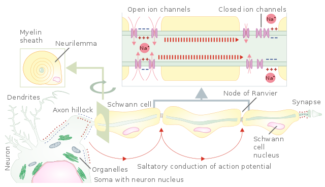
Image: Propagation of action potential along the myelinated nerve fiber. Schematic representation of action potential propagation through a myelinated nerve fiber of the peripheral nervous system. From the axon hillock of the neuron body (soma), an action potential propagates from one part of the unmyelinated fiber to the next one. The unmyelinated parts of the nerve fiber are the nodes of Ranvier. This type of action potential propagation is called saltatory conduction (red arrows in the diagram). Ion channels open, allowing potassium ions to enter the cell, leading to membrane depolarization and the generation of the action potential. Myelination of nerve fibers in the peripheral nervous system is achieved by Schwann cells wrapping around an axon part (cross-section). The nucleus and most of the Schwan cell cytoplasm are contained in the outermost layer called the neurilemma. By: Helixitta. License: CC BY-SA 4.0
Functions of the glial cells in the central nervous system
Astrocytes (macroglia):
- Exchange of substances
- Storage of glycogen and providing adjacent neurons with glucose
- Building the blood-brain-barrier
- Phagocytosis of dead synapses
- Forming glial scar tissue in the CNS, e.g., following a stroke or multiple sclerosis
Oligodendrocytes:
- Building the myelin sheath in the CNS
Microglia (Hortega cells):
- Phagocytosis of foreign bodies and of endogenous dead tissue
Ependymal cells:
- The lining of the ventricles of the brain and of the central canal of the spinal cord
- Part of the cerebrospinal fluid
Functions of the glial cells of the peripheral nervous system
The Schwann cells are responsible for myelination in the PNS. The satellite cells serve as helpers for the neurons and substitute cerebral astrocytes in the peripheral ganglia.
The Myelin Sheath of a Neuron in the Nervous System
As mentioned above, the myelin sheath protects the axon of a neuron against electrical shocks and speeds up the transmission of nerve impulses. From birth to maturity, the amount of myelin grows constantly, increasing transmission speed significantly.
Two types of neuroglia form the myelin sheath:
- Schwann cells in the PNS
- Oligodendrocytes in the CNS
Schwann cells only exist in the PNS. In contrast to the CNS where all nerve cells possess a myelin sheath, some nerves in the PNS do not have a myelin sheath. The formation of myelin sheaths around the axons begins as early as during fetal development.
Every Schwann cell forms multiple layers around a segment of an axon that is about 1-mm long and delimited on each side by a node. The interior side, consisting of up to 100 membrane layers, is the myelin sheath (Schwann sheath).
The outer cytoplasmic layer of the Schwann cell that contains the nucleus and covers the myelin sheath is the neurolemma. At the junction between 2 adjacent Schwann cells, there is a gap or node in the myelin sheath. These gaps in the myelin sheath are called the nodes of Ranvier.
The oligodendrocytes are responsible for building the myelin sheaths in the CNS. They have longer processes than the Schwann cells and therefore, they not only cover one but up to 50 adjacent axons. Consequently, the oligodendrocytes have fewer nodes of Ranvier. Since the cell body and the nucleus of the oligodendrocyte do not cover the axon, there is no neurolemma.
Demyelination
Demyelination is the loss or damage of the myelin sheath around the axon and can be the consequence of diseases like multiple sclerosis or Tay-Sachs disease. Furthermore, a vitamin B12 deficiency can cause demyelination, particularly in the spinal cord, leading to sensory dysfunctions and even motor paralysis.
References
2 Comments
Comments are closed.
Visitor Rating: 4 Stars
Visitor Rating: 5 Stars