Movement and Function of Hip Joint/The hip joint is a ball and socket joint that is the point of articulation between the head of the femur and the acetabulum of the pelvis. The joint is a diarthrodial joint with its inherent stability dictated primarily by its osseous components/articulations. The primary function of the hip joint is to provide dynamic support the weight of the body/trunk while facilitating force and load transmission from the axial skeleton to the lower extremities, allowing mobility.[rx][rx][rx]
The hip joint, scientifically referred to as the acetabulum femoral joint, is the joint between the femur and acetabulum of the pelvis, and its primary function is to support the weight of the body in both static (e.g. standing) and dynamic (e.g. walking or running) postures. The hip joints have very important roles in retaining balance, and for maintaining the pelvic inclination angle.
Muscle Attachment of Hip Joint
Biceps Femoris Long Head
-
Origin: Common (conjoint) tendon from the superior medial quadrant of the posterior ischial tuberosity (with semitendinosus)
-
Insertion: Majority onto the fibular head; also the lateral collateral ligament of the knee and lateral tibial condyle
-
Action: Flexion of the knee, and lateral rotation of the tibia; extension of the hip joint
-
Innervation: Tibial nerve (a portion of the sciatic nerve)
-
Arterial Supply: Perforating (muscular) branches of profunda femoris artery, inferior gluteal artery, and the superior muscular branches of the popliteal artery
Biceps Femoris Short Head
-
Origin: Lateral lip of linea aspera, the lateral intermuscular septum of the thigh, and lateral supracondylar ridge of femur
-
Insertion: Majority on the fibular head; and lateral collateral ligament of the knee, and lateral tibial condyle
-
Action: Flexion of the knee, and lateral rotation of the tibia
-
Innervation: Common peroneal nerve (a portion of the sciatic nerve)
-
Arterial Supply: Perforating (muscular) branches of profunda femoris artery, inferior gluteal artery, and the superior muscular branches of the popliteal artery
Semimembranosus
-
Origin: Superior lateral aspect of the ischial tuberosity
-
Insertion: The posterior surface of the medial tibial condyle
-
Action: Extension of the hip, flexion of the knee, and medial rotation of the tibia (specifically with knee flexion)
-
Innervation: Tibial nerve (a portion of the sciatic nerve)
-
Arterial Supply: Perforating (muscular) branches of profunda femoris artery, inferior gluteal artery, and the superior muscular branches of the popliteal artery
Semitendinosus
-
Origin: The common (conjoint) tendon from the superior medial quadrant of the posterior ischial tuberosity (with biceps femoris long head)
-
Insertion: Superior aspect of the medial tibial shaft (into the distal portion of the pes anserinus along with the gracious and sartorius muscles)
-
Action: Extension of the hip and flexion of the knee, medial rotation of the tibia (specifically with knee flexion)
-
Innervation: Tibial nerve (a portion of the sciatic nerve)
-
Arterial Supply: Perforating (muscular) branches of profunda femoris artery, inferior gluteal artery, and the superior muscular branches of the popliteal artery
The rectus femoris is responsible for thigh flexion at the hip and knee extension.[rx]
-
Vastus Lateralis – The largest of the four muscles. Origin is from the greater trochanter and lateral lip of linea Aspera. It inserts at the lateral base and border of the patella, forming the lateral patellar retinaculum and the lateral side of the quadriceps femoris tendon.[rx]
-
Vastus Medialis – Originates at the inferior portion of the intertrochanteric line and medial lip of the linea Aspera. Inserts at the medial base and border of the patella, forming the medial patellar retinaculum and the medial side of the quadriceps femoris tendon.[rx]
-
Vastus Intermedius – Originates at the anterior and lateral surfaces of the femoral shaft. It inserts at the lateral border of the patella, forming the deep portion of the quadriceps tendon.[rx]
-
Rectus Femoris – Comprised of two proximal heads: The straight head consists of the anterior inferior iliac spine (ASIS) of the ilium. The reflected head consists of the ilium superior to the acetabulum. Inserts at the quadriceps femoris tendon.[rx]
Hip Flexors
Psoas major
-
Origin: T12-L5 vertebrae
-
Insertion: Lesser trochanter
-
Innervation: Femoral nerve
Psoas minor (present in 50% of the population)
-
Origin: T12-L1 vertebrae
-
Insertion: Iliopubic eminence
-
Innervation: L1 ventral ramus
Pectineus (flexes and adducts thigh)
-
Origin: Pectineal line of the pubis
-
Insertion: Pectineal line of femur
-
Innervation: Femoral nerve
Iliacus
-
Origin: Iliac fossa/ Sacral ala
-
Insertion: Lesser trochanter
-
Innervation: Femoral nerve
-
Hip Extensors and External Rotators-
Gluteus maximus
-
Origin: Ilium, dorsal sacrum
-
Insertion: ITB, gluteal tuberosity
-
Innervation: Inferior gluteal nerve
Obturator externus
-
Origin: Ischiopubic rami, obturator membrane
-
Insertion: Trochanteric fossa
-
Innervation: Obturator nerve
Short External Rotators
Piriformis
-
Origin: Anterior sacrum
-
Insertion: Superior greater trochanter
-
Innervation: Nerve to Piriformis (S2, posterior division of lumbosacral plexus)
Superior gemellus
-
Origin: Ischial spine
-
Insertion: Medial greater trochanter
-
Innervation: Nerve to obturator internus (L5-S2, anterior division of lumbosacral plexus)
Obturator internus
-
Origin: Ischiopubic rami, obturator membrane
-
Insertion: Medial greater trochanter
-
Innervation: Nerve to obturator internus (L5-S2, anterior division of lumbosacral plexus)
Inferior gemellus
-
Origin: Ischial tuberosity
-
Insertion: Medial greater trochanter
-
Innervation: Nerve to quadratus femoris (L4-S1, anterior division of lumbosacral plexus)
Quadratus femoris
-
Origin: Ischial tuberosity
-
Insertion: Intertrochanteric crest
-
Innervation: Nerve to quadratus femoris (L4-S1, anterior division of lumbosacral plexus)
Hip Abductors
Tensor fascia latae
-
Origin: Iliac crest, ASIS
-
Insertion: Iliotibial band/proximal tibia
-
Innervation: Superior gluteal nerve
Gluteus medius
-
Origin: Ilium between anterior and posterior gluteal lines
-
Insertion: Greater trochanter
-
Innervation: Superior gluteal nerve
Gluteus minimus
-
Origin: Ilium between anterior and posterior gluteal lines
-
Insertion: Greater trochanter
-
Innervation: Superior gluteal nerve
Hip Adductors
Adductor Magnus
-
Origin: Pubic ramus, ischial tuberosity
-
Insertion: Linea Aspera, adductor tubercle
-
Innervation: Obturator nerve, the sciatic nerve
Adductor longus
-
Origin: Body of pubis
-
Insertion: Linea Aspera
-
Innervation: Obturator nerve
Adductor Brevis
-
Origin: Body and inferior pubic ramus
-
Insertion: Pectineal line, Linea Aspera
-
Innervation: Obturator nerve
Gracilis
-
Origin: Body and inferior pubic ramus
-
Insertion: Proximal medial tibia (pes anserinus)
-
Innervation: Obturator nerve
There are many muscles involved in the movement of the hip joint, these include (in alphabetical order) the
-
Adductor longus, brevis, and magnus
-
Gluteus maximus, medius, and minimus
-
Gracilis
-
Hamstring muscles: semimembranosus, semitendinosus, and the biceps femoris
-
Iliacus
-
Obturator
-
Pectineus
-
Piriformis
-
Psoas major
-
Quadriceps muscles: rectus femoris, vastus intermedius, vastus lateralis, and vastus medialis
-
Quadratus femoris
-
Sartorius
-
Tensor fascia latae
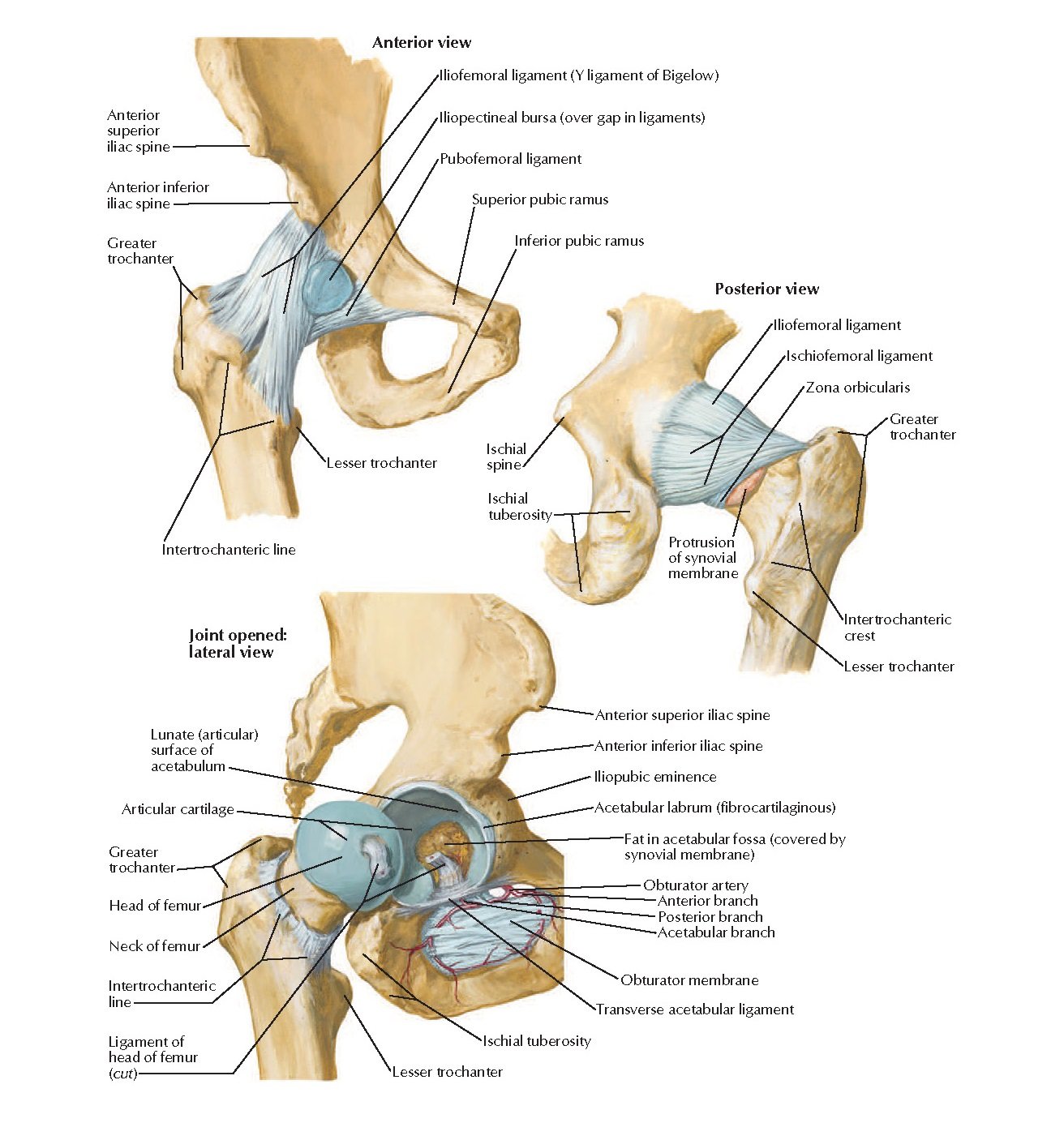
Ligament of Hip Joint
The Hip Joint Ligament
- Ischiofemoral ligament – It attaches to the posterior surface of the acetabular rim and labrum and courses circumferentially around the joint to its insertion on the anterior aspect of the femur. The ischial femoral ligament limits internal rotation and hip adduction with flexion.
- Iliofemoral ligament (Y Ligament of Bigelow) – It is a triangle-shaped ligament that attaches along the intertrochanteric line of the femur and converges into its attachment on the anterior inferior iliac spine (AIIS). This is the strongest ligament in the body. The iliofemoral ligament limits extension and external rotation of the hip and assists in the maintenance of a static erect posture with minimal muscular activity. [rx],[rx]
- Pubofemoral ligament – Located on the anterior aspect of the hip joint, this ligament extends from the anterior portion of the pubic ramus to the anterior surface of the intertrochanteric fossa often blending with the inferior fibers of the iliofemoral ligament. The pubofemoral ligament limits hip abduction and extension.
- Zona orbicularis (annular ligament) – Not visible externally, it encircles the femoral neck like a buttonhole and acts as a biomechanical locking ring wrapped around the femoral neck. The zona orbicular forms a locking ring around the femur which resists distraction forces on the hip.
- Ligamentum teres – Located deep in the hip, it has a pyramidal shape with a broad origin from nearly the entire transverse acetabular ligament attaching to the ischial and pubic bases by two bundles, with the posterior bundle being stronger than the anterior bundle. [rx]
- Acetabular labrum – This is a fibrocartilaginous rim, composed of circumferential collagen fibers, that spans the entirety of the acetabulum and is continuous with the transverse acetabular ligament. The labrum contributes approximately 22% of the articulating surface of the hip and increases the volume of the acetabulum by 33%. [rx]

Blood Supply and Lymphatics of Hip Joint
From age 0 to 4 years, the femoral head receives significant blood supply from the
- Medial femoral circumflex artery (MFCA),
- Lateral femoral circumflex artery (LFCA), and
- The artery of ligamentum teres.
From age 4 to 8 years, the MFCA provides the majority of the blood supply with supplementary contributions from the LFCA and artery of ligamentum teres. After 8 years of age, the MFCA predominates with a negligible contribution from the LFCA and artery of ligamentum teres.
The following list includes the branches of the anterior trunk of the internal iliac artery
-
Obturator artery
-
Umbilical artery, which branches to form the superior vesical artery
-
Inferior vesical artery
-
Vaginal artery (female)
-
Uterine artery (female)
-
Middle rectal artery
-
Internal pudendal artery
-
Inferior gluteal artery
The following list includes the branches of the posterior trunk of the internal iliac artery
-
Superior gluteal artery
-
Lateral sacral arteries
-
Iliolumbar artery
Most of the arteries of the hip region originate from the external iliac artery and include
-
Femoral artery
-
Superficial circumflex iliac artery
-
External pudendal artery
-
Superficial femoral artery
-
Profunda femoral artery (the deep artery of the thigh)
-
The lateral femoral circumflex artery
-
The medial femoral circumflex artery
Nerves Supply of Hip Joint
- Obturator nerve – Originates from nerve roots L2-L4 and exits through the obturator canal before splitting into an anterior division that runs anterior to obturator externus and a posterior division which runs posterior to obturator externus. The obturator nerve supplies sensory innervation to the inferomedial thigh via the cutaneous branch of the obturator nerve and motor innervation to gracilis (anterior division), adductor longus (anterior division), adductor brevis (anterior/posterior divisions), and adductor Magnus (posterior division).
- Genitofemoral nerve – Originates from nerve roots L1-L2. It pierces the psoas muscle and continues down the anteromedial surface of psoas before dividing it into femoral and genital branches. The femoral branch provides sensory innervation to the proximal anterior thigh over the femoral triangle.
- Lateral femoral cutaneous nerve – Originates from nerve roots L2-L3. Crosses inferior to the anterior superior iliac spine (ASIS) and provides sensory innervation to the lateral thigh. It has no motor function.
- The femoral nerve – originates from nerve roots (L2-L4). It lies between the psoas major and iliacus and branches in the femoral triangle. The femoral nerve provides sensory innervation to the anteromedial thigh via anterior cutaneous branches and motor innervation to the psoas, pectineus, Sartorius, quadriceps (rectus femoris, vastus lateralis, vastus intermedius, vastus medialis).
- Sciatic nerve – originates from the sacral plexus and projects through the greater sciatic foramen descending down the posterior thigh deep to the hamstrings and superficial to adductor Magnus. The sciatic nerve has two distinct divisions: tibial division and common peroneal division.
- Posterior femoral cutaneous nerve – Originates from nerve roots S1-S3 and passes through the greater sciatic foramen medial to the sciatic nerve. The posterior femoral cutaneous nerve provides sensory innervation to the posterior thigh and has no motor function.

Movement and Function of Hip Joint
Muscles of the hip joint can be grouped based upon their functions relative to the movements of the hip.[rx]
-
Flexion – Primarily accomplished via the psoas major and the iliacus, with some assistance from the pectineus, rectus femoris, and the sartorius.
-
Extension – Primarily accomplished via the gluteus maximus as well as the hamstring muscles.
-
Medial rotation – Primarily accomplished by the tensor fascia lata and fibers of the gluteus medius and minimus.
-
Lateral rotation – Primarily accomplished by the obturator muscles, the quadratus femoris, and the Gemelli with assistance from the gluteus maximus, sartorius, and piriformis.
-
Adduction – Primarily accomplished by the adductor longus, brevis, and Magnus with assistance from the gracilis and pectineus
-
Abduction – Primarily accomplished by the gluteus medius and minimus with assistance from the tensor fascia lata and sartorius.

