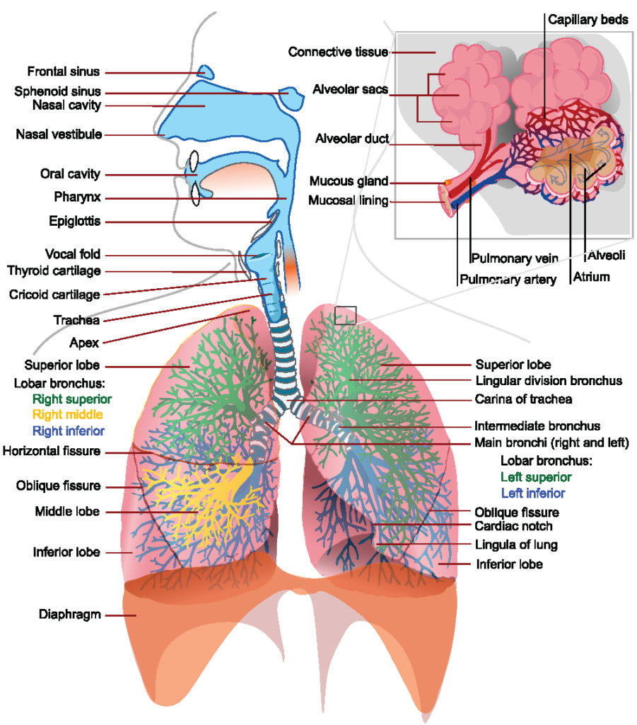
Breathing (or ventilation) is the process of moving air out and in the lungs to facilitate gas exchange with the internal environment, mostly to flush out carbon dioxide and bring in oxygen. All aerobic creatures need oxygen for cellular respiration, which uses the oxygen to break down foods for energy and produces carbon dioxide as a waste product. Breathing, or “external respiration”, brings air into the lungs where gas exchange takes place in the alveoli through diffusion. The body’s circulatory system transports these gases to and from the cells, where “cellular respiration” takes place.[rx][rx]
Inhaled air is by volume 78% nitrogen, 20.95% oxygen and small amounts of other gases including argon, carbon dioxide, neon, helium, and hydrogen.[rx]
The gas exhaled is 4% to 5% by volume of carbon dioxide, about a 100 fold increase over the inhaled amount. The volume of oxygen is reduced by a small amount, 4% to 5%, compared to the oxygen inhaled. The typical composition is:[17]
- 5.0–6.3% water vapor
- 79% nitrogen [rx]
- 13.6–16.0% oxygen
- 4.0–5.3% carbon dioxide
- 1% argon
- parts per million (ppm) of hydrogen, from the metabolic activity of microorganisms in the large intestine.[19]
- ppm of carbon monoxide from the degradation of heme proteins.
- 1 ppm of ammonia.
- Trace many hundreds of volatile organic compounds especially isoprene and acetone. The presence of certain organic compounds indicates disease.[rx][rx]
In addition to air, underwater divers practicing technical diving may breathe oxygen-rich, oxygen-depleted or helium-rich breathing gas mixtures. Oxygen and analgesic gases are sometimes given to patients under medical care. The atmosphere in space suits is pure oxygen. However, this is kept at around 20% of Earthbound atmospheric pressure to regulate the rate of inspiration.
Pressure Changes During Pulmonary Ventilation
Ventilation is the rate at which gas enters or leaves the lung.
Key Points
Ventilation is the rate at which gas enters or leaves the lung.
The three types of ventilation are minute ventilation, alveolar ventilation, and dead space ventilation.
The alveolar ventilation rate changes according to the frequency of breath, tidal volume, and amount of dead space.
PA refers to the alveolar partial pressure of a gas, while Pa refers to the partial pressure of that gas in arterial blood.
Gas exchange occurs from passive diffusion because PAO2 is greater than PaO2 in deoxygenated blood.
Key Terms
- ventilation: The bodily process of breathing, the inhalation of air to provide oxygen, and the exhalation of spent air to remove carbon dioxide.
- partial pressure: The pressure exerted by a gas, either in air or dissolved, that indicates the concentration of that gas.
The Types of Ventilation Rates
In respiratory physiology, the ventilation rate is the rate at which gas enters or leaves the lung. Ventilation is generally expressed as volume of air times a respiratory rate.
The volume of air can refer to tidal volume (the amount inhaled in an average breath) or something more specific, such as the volume of dead space in the airways. The three main types of ventilation rates used in respiratory physiology are:
- Minute ventilation (VE): The amount of air entering the lungs per minute. It can be defined as VE=Tidal Volume×Breaths Per Minute
- Alveolar ventilation (VA): The amount of gas per unit of time that reaches the alveoli and becomes involved in gas exchange. It is defined as VA=(Tidal Volume−Dead Space Volume)×Respiratory Rate
- Dead space ventilation (VD): The amount of air per unit of time that is not involved in gas exchange, such as the air that remains in the conducting zones. It is defined as VD=Dead Space Volume×Respiratory Rate.
Additionally, minute ventilation can be described as the sum of alveolar and dead space ventilation, provided that the respiratory rate used to derive them is in terms of breaths per minute.
The three types of ventilation are mathematically linked to one another, so changes in one ventilation rate can cause the change of the other. This is most apparent in changes of the dead space volume. Breathing through a snorkeling tube and having a pulmonary embolism both increase the amount of dead space volume (through anatomical versus alveolar dead space respectively), which will reduce alveolar ventilation.
Alveolar ventilation is the most important type of ventilation for measuring how much oxygen actually gets into the body, which can initiate negative feedback mechanisms to try and increase alveolar ventilation despite the increase in dead space. In particular, the body will generally attempt to combat increased dead space by raising the frequency of breaths to try and maintain sufficient levels of alveolar ventilation.
Partial Pressure of Gasses
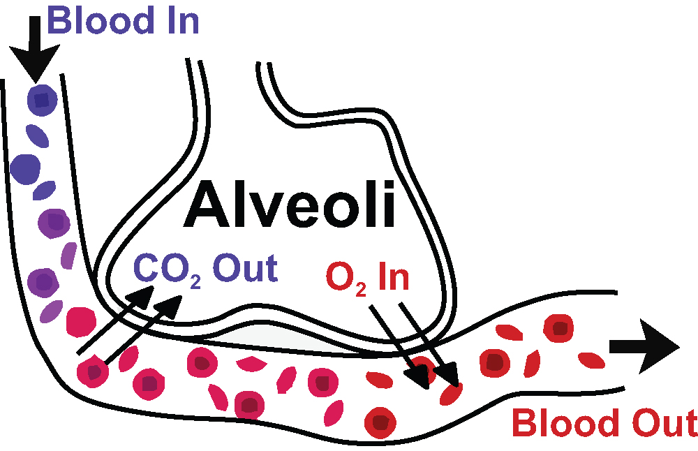
Gaseous Exchange in the Lungs: Diagram of gas exchange in the lungs.
When gasses dissolve in the bloodstream during ventilation, they are generally described by the partial pressure of the gasses. Partial pressure more specifically refers to the relative concentration of those gasses by the pressure they exert in a dissolved state.
In respiratory physiology, PAO2 and PACO2, refer to the partial pressures of oxygen and carbon dioxide in the alveoli.
PaO2 and PaCO2 refer to the partial pressures of oxygen and carbon dioxide within arterial blood. Differences in partial pressures of gasses between the alveolar air and the bloodstream are the reason that gas exchange occurs by passive diffusion.
Under normal conditions, PAO2 is about 100 mmHg, while PaO2 is 80–100 mmHg in systemic arteries, but 40–50 mmHg in the deoxygenated blood of the pulmonary artery going to the lungs.
Recall that gasses travel from areas of high pressure to areas of low pressure, so the greater pressure of oxygen in the alveoli compared to that of the deoxygenated blood explains why oxygen can passively diffuse into the bloodstream during gas exchange.
Conversely, PACO2 is 35 mmHg, while PaCO2 is about 40–45 mmHG in systemic arteries and 50 mmHg in the pulmonary artery. The partial pressure, and thus the concentration of carbon dioxide, is greater in the capillaries of the alveoli compared to the alveolar air, so carbon dioxide will passively diffuse from the bloodstream into the alveoli during gas exchange.
Additionally, because PaCO2 is an indicator of the concentration of carbon dioxide in arterial blood, it can be used to measure blood pH and identify cases of respiratory acidosis and alkalosis.
Inspiration
Inhalation is the flow of air into an organism that is due to a pressure difference between the atmosphere and alveolus.
Key Points
In humans, inspiration is the flow of air into an organism from the external environment, through the airways, and into the alveoli.
Inhalation begins with the onset of a contraction of the diaphragm, which results in expansion of the thoracic and pleural cavities and a decrease in pressure (also called an increase in negative pressure).
There are many accessory muscles involved in inhalation—such as external intercostal muscles, scalene muscles, the sternocleidomastoid muscle, and the trapezius muscle.
Breathing only with the accessory muscles instead of the diaphragm is considered inefficient, and provides much less air during inhalation.
The negative pressure in the pleural cavity is enough to hold the lungs open in spite of the inherent elasticity of the tissue. The thoracic cavity increases in volume causing a drop in the pressure (a partial vacuum) within the lung itself.
As long as the pressure within the alveoli is lower than atmospheric pressure, air will continue to move inwardly, but as soon as the pressure is stabilized air movement stops.
Key Terms
- inspiration: The drawing of air into the lungs, accomplished in mammals by elevation of the chest walls and flattening of the diaphragm.
- accessory muscles: Muscles that help expand small parts of the thoracic cavity, either working in addition to the diaphragm or substituting for it if the diaphragm becomes injured.
- intrapleural pressure: The pressure inside the pleural cavity, which is negative compared to outside air and becomes even more negative during inspiration.
Inspiration refers to inhalation—it is the flow of the respiratory current into an organism. In humans, it is the movement of ambient air through the airways and into the alveoli of the lungs.
The Process of Inspiration
Inspiration begins with the contraction of the diaphragm, which results in the expansion of the thoracic cavity and the pleural cavity. The pleural cavity normally has a lower pressure compared to ambient air (–3 mmHg normally and typically –6 mmHg during inspiration), so when it expands, the pressure inside the lungs drops.
Pressure and volume are inversely related to each other, so the drop in pressure inside the lung increases the volume of air inside the lung by drawing outside air into the lung. As the volume of air inside the lung increases, the lung pushes back against the expanded pleural cavity as a result of the drop in intrapleural pressure (pressure inside the pleural cavity).
The force of the intrapleural pressure is even enough to hold the lungs open during inspiration despite the natural elastic recoil of the lung. The alveolar sacs also expand as a result of being filled with air during inspiration, which contributes to the expansion inside the lung.
Eventually, the pressure inside the lung becomes less negative as the volume inside the lung increases and, when pressure and volume stabilize, air movement stops, inspiration ends, and expiration (exhalation) will begin. Deeper breaths have higher tidal volumes and require a greater drop in intrapleural pressure compared to shallower breaths.
Respiratory System: Resistance in any part of the respiratory tract can cause problems.
Accessory Muscles of Inspiration
The diaphragm is the primary muscle involved in breathing, however, several other muscles play a role in certain circumstances. These muscles are referred to as accessory muscles of inhalation.
- External intercostal muscles: Muscles located between the ribs that help the thoracic cavity and pleural cavity expand during quiet and forced inspiration.
- Scalene muscles: Muscles in the neck that lift the upper ribs (and the thoracic cavity around the upper ribs) to help with breathing. They provide a mechanism for inspiration when the diaphragm is injured and can’t contract normally.
- Sternocleidomastoid muscle: Muscles that connect the sternum to the neck and allow for rotation and turning of the head. They can lift the upper ribs as the scalene muscles can.
- Trapezius muscle: Muscles in the shoulders that retract the scapula and expand the upper part of the thoracic cavity.
The accessory muscles assist breathing by expanding the thoracic cavity in a similar way to the diaphragm. However, they expand a much smaller part of the thoracic cavity compared to the diaphragm. Therefore they should not be used as the primary mechanism of inhalation, because they take in much less air compared to the diaphragm resulting in a much lower tidal volume.
For example, singers need a lot of air to support the powerful voice production needed for singing. A common problem in novice singers is breathing with the accessory muscles of the neck, shoulder, and ribs instead of the diaphragm, which gives them a much smaller air supply than what is needed to sing properly.
Expiration
Exhalation (or expiration) is the flow of the respiratory current out of the organism.
Key Points
In humans, exhalation is the movement of air out of the bronchial tubes, through the airways, to the external environment during breathing.
Exhalation is a passive process because of the elastic properties of the lungs.
During forced exhalation, internal intercostal muscles lower the rib cage and decrease the thoracic volume while the abdominal muscles push up on the diaphragm which causes the thoracic cavity to contract.
Relaxation of the thoracic diaphragm causes contraction of the pleural cavity which puts pressure on the lungs to expel the air.
Brain control of exhalation can be broken down into voluntary control and involuntary control.
Key Terms
- Intercostal muscles: Intercostal muscles are several groups of muscles that run between the ribs, and help form and move the chest wall.
- exhalation: The act or process of exhaling, or sending forth in the form of steam or vapor; evaporation.
Expiration, also called exhalation, is the flow of the respiratory current out of the organism. The purpose of exhalation is to remove metabolic waste, primarily carbon dioxide from the body from gas exchange. The pathway for exhalation is the movement of air out of the conducting zone, to the external environment during breathing.
Respiratory System: As the diaphragm relaxes, the pleural cavity contracts, which exerts pressure on the lungs, which reduces the volume of the lungs as air is passively pushed out of the lungs.
The Process of Expiration
Expiration is typically a passive process that happens from the relaxation of the diaphragm muscle (that contracted during inspiration). The primary reason that expiration is passive is due to the elastic recoil of the lungs. The elasticity of the lungs is due to molecules called elastins in the extracellular matrix of lung tissues and is maintained by surfactant, a chemical that prevents the elasticity of the lungs from becoming too great by reducing surface tension from water. Without surfactant the lungs would collapse at the end of expiration, making it much more difficult to inhale again. Because the lung is elastic, it will automatically return to its smaller size as air leaves the lung.
Exhalation begins when inhalation ends. Just as the pleural cavity’s increased negative pressure leads to air uptake during inhalation, the pleural cavity will contract during the exhalation (due to relaxation of the diaphragm), which exerts pressure on the lungs and causes the pressure inside the cavity to be less negative. An increase in pressure leads to a decrease in volume inside the lung, and the air is pushed out into the airways as the lung returns to its smaller size. The pleural cavity is so important to breathing because its pressure changes the volume of the lungs, and it provides a frictionless space for the lung to expand and contract during breathing.
While expiration is generally a passive process, it can also be an active and forced process. There are two groups of muscles that are involved in forced exhalation.
- Internal Intercostal Muscles: Muscles of the ribcage that help lower the ribcage, which pushes down on the thoracic cavity, causing forced exhalation. Note that these are not the same as the external intercostal muscles involved in inspiration.
- Abdominal Muscles: Any number of muscles in the abdomen that exert pressure on the diaphragm from below to expand it, which in turn contracts the thoracic cavity, causing forced exhalation.
This happens due to elastic properties of the lungs, as well as the internal intercostal muscles that lower the rib cage and decrease thoracic volume. As the thoracic diaphragm relaxes during exhalation it causes the tissue it has depressed to rise superiorly and put pressure on the lungs to expel the air.
Control of Expiration
Expiration can be either voluntary or involuntary in order to serve different purposes for the body. These two types of expiration are controlled by different centers within the body.
Voluntary expiration is actively controlled. It is generally defined by holding air in the lungs and releasing it at a fixed rate, which enables control over when and how much air to exhale. It is required for voice production during speech or singing, which requires very specific control over air, or even simpler activities, like blowing out a candle on one’s birthday. The nervous system component that controls voluntary expiration is the motor cortex (the ascending respiratory pathway), because it controls muscle movements, but this pathway isn’t fully understood, and there are many other possible sites in the brain that may also be involved.
Involuntary expiration is not under conscious control and is an important component of metabolic function. Examples include breathing during sleep or meditation. Changes in breathing patterns may also occur for metabolic reasons, such as through increased breathing rate in people with acidosis from negative feedback. The principal neural control center for involuntary expiration consists of the medulla oblongata and the pons, which are located in the brainstem directly beneath the brain. While these two structures are involved in neural respiratory control, they also have other metabolic regulatory functions for other body systems, such as the cardiovascular system.
Breathing Patterns
Breathing is an autonomic process that moves air in and out of the lungs.
Key Points
Breathing patterns consist of tidal volume and respiratory rate in an individual.
An average breathing pattern is 12 breaths per minute and 500 mL per breath.
Eupnea is normal breathing at rest.
There are types of altered breathing patterns that are symptoms of many diseases.
Altered breathing patterns refer to changes in respiratory rate or amount of air exchanged during breathing, and do not always indicate changes in alveolar ventilation.
The mechanism of generation of the ventilatory pattern involves the integration of neural signals by respiratory control centers in the medulla and pons.
Key Terms
- altered breathing patterns: Abnormal breathing patterns that indicate typically indicate either too fast or too slow respiratory rate or too much or too little tidal volume.
- tidal volume: The amount of air displaced or exchanged in a single breath.
Breathing patterns refer to the respiratory rate, which is defined as the frequency of breaths over a period of time, as well as the amount of air cycled during breathing (tidal volume). Breathing patterns are important diagnostic criteria for many diseases, including some which involve more than the respiratory system itself.
Characteristics of the Breathing Patterns
The respiratory rate is the frequency of breaths over time. The time period is variable but usually expressed in breaths per minute because it that time period allows for estimation of minute ventilation. During normal breathing, the volume of air cycled through inhalation and exhalation is called tidal volume (VT), and is the amount of air exchanged in a single breath. Tidal volume multiplied by the respiratory rate is minute ventilation, which is one of the most important indicators of lung function. In an average human adult, the average respiratory rate is 12 breaths per minute, with a tidal volume of .5 liters and minute ventilation of 6 liters per minute, though these numbers vary from person to person. Infants and children have considerably higher respiratory rates than adults.
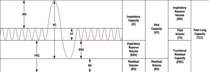
Spirometry curve: The normal respiratory rate refers to the cyclical inhalation and exhalation of tidal volume (VT).
The respiratory rate is controlled by involuntary processes of the autonomic nervous system. In particular, the respiratory centers of the medulla and the pons control the overall respiratory rate based on a variety of chemical stimuli from within the body. The hypothalamus can also influence the respiratory rate during emotional and stress responses.
Normal and Altered Breathing Patterns
Eupnea is the term for the normal respiratory rate of an individual at rest. Several other terms describe abnormal breathing patterns that are indicative of symptoms of many diseases, many of which aren’t mainly respiratory diseases. Some of the more common terms for altered breathing patterns include:
- Dyspnea: commonly called shortness of breath. It describes dramatically decreased tidal volume and sometimes increased respiratory rate, leading to a sensation of breathlessness. It is a common symptom of anxiety attacks, pulmonary embolisms, heart attacks, and emphysema, among other things.
- Hypernea: refers to increased volume of air cycled to meet the body’s metabolic needs, which may or may not involve a change in frequency of breathing. It is a symptom of exercise and adjustment to high altitude, which are generally not problematic but can also be seen in those with anemia or septic shock, which is problematic.
- Tachypnea: describes increased respiratory rate. Often a symptom of carbon monoxide poisoning or pneumonia.
- Bradypnea: describes decreased respiratory rate. Often a symptom of hypertension, heart arrhythmias, or slow metabolic rate from hypothyroidism.
- Apnea: Transient stopped breathing that begins again soon afterward. It is the main symptom of sleep apnea, in which breathing temporarily stops during sleep.
These terms all describe an altered breathing pattern through increased or decreased (or stopped) tidal volume or respiratory rate. It is important to distinguish these terms from hyperventilation and hypoventilation, which refer to abnormalities in alveolar gas exchange (and thus blood pH) instead of an altered breathing pattern, but they may be associated with an altered breathing pattern. For example, dyspnea or tachypnea often occurs together with hyperventilation during anxiety attacks, though not always.
How do you do breathing exercises?
There are lots of breathing exercises you can do to help relax. The first exercise below—belly breathing—is simple to learn and easy to do. It’s best to start there if you have never done breathing exercises before. The other exercises are more advanced. All of these exercises can help you relax and relieve stress.
Belly breathing
Belly breathing is easy to do and very relaxing. Try this basic exercise anytime you need to relax or relieve stress.
- Sit or lie flat in a comfortable position.
- Put one hand on your belly just below your ribs and the other hand on your chest.
- Take a deep breath in through your nose, and let your belly push your hand out. Your chest should not move.
- Breathe out through pursed lips as if you were whistling. Feel the hand on your belly go in, and use it to push all the air out.
- Do this breathing 3 to 10 times. Take your time with each breath.
- Notice how you feel at the end of the exercise.
Next steps
After you have mastered belly breathing, you may want to try one of these more advanced breathing exercises. Try all three, and see which one works best for you:
- 4-7-8 breathing
- Roll breathing
- Morning breathing
4-7-8 breathing
This exercise also uses belly breathing to help you relax. You can do this exercise either sitting or lying down.
- To start, put one hand on your belly and the other on your chest as in the belly breathing exercise.
- Take a deep, slow breath from your belly, and silently count to 4 as you breathe in.
- Hold your breath, and silently count from 1 to 7.
- Breathe out completely as you silently count from 1 to 8. Try to get all the air out of your lungs by the time you count to 8.
- Repeat 3 to 7 times or until you feel calm.
- Notice how you feel at the end of the exercise.
Roll breathing
Roll breathing helps you to develop full use of your lungs and to focus on the rhythm of your breathing. You can do it in any position. But while you are learning, it is best to lie on your back with your knees bent.
- Put your left hand on your belly and your right hand on your chest. Notice how your hands move as you breathe in and out.
- Practice filling your lower lungs by breathing so that your “belly” (left) hand goes up when you inhale and your “chest” (right) hand remains still. Always breathe in through your nose and breathe out through your mouth. Do this 8 to 10 times.
- When you have filled and emptied your lower lungs 8 to 10 times, add the second step to your breathing: inhale first into your lower lungs as before, and then continue inhaling into your upper chest. Breathe slowly and regularly. As you do so, your right hand will rise and your left hand will fall a little as your belly falls.
- As you exhale slowly through your mouth, make a quiet, whooshing sound as first your left hand and then your right hand fall. As you exhale, feel the tension leaving your body as you become more and more relaxed.
- Practice breathing in and out in this way for 3 to 5 minutes. Notice that the movement of your belly and chest rises and falls like the motion of rolling waves.
- Notice how you feel at the end of the exercise.
Practice roll breathing daily for several weeks until you can do it almost anywhere. You can use it as an instant relaxation tool anytime you need one.
Caution: Some people get dizzy the first few times they try roll breathing. If you begin to breathe too fast or feel lightheaded, slow your breathing. Get up slowly.
Morning breathing
Try this exercise when you first get up in the morning to relieve muscle stiffness and clear clogged breathing passages. Then use it throughout the day to relieve back tension.
- From a standing position, bend forward from the waist with your knees slightly bent, letting your arms dangle close to the floor.
- As you inhale slowly and deeply, return to a standing position by rolling up slowly, lifting your head last.
- Hold your breath for just a few seconds in this standing position.
- Exhale slowly as you return to the original position, bending forward from the waist.
- Notice how you feel at the end of the exercise.
Relaxation Techniques and Breathing Exercise
Box Breathing
While there are many different forms of deep breathing exercises, box breathing can be particularly helpful with relaxation. Box breathing is a breathing exercise to assist patients with stress management and can be implemented before, during, and/or after stressful experiences. Box breathing uses four simple steps. Its title is intended to help the patient visualize a box with four equal sides as they perform the exercise. This exercise can be implemented in a variety of circumstances and does not require a calm environment to be effective.
-
Step One: Breathe in through the nose for a count of 4.
-
Step Two: Hold breath for a count of 4.
-
Step Three: Breath out for a count of 4.
-
Step Four: Hold breath for a count of 4.
-
Repeat
Note: The length of the steps can be adjusted to accommodate the individual (e.g., 2 seconds instead of 4 seconds for each step).
Guided Imagery
Guided imagery is a relaxation exercise intended to assist patients with visualizing a calming environment. Visualization of tranquil settings assists patients with managing stress via distraction from intrusive thoughts. Cognitive-behavioral theory suggests that emotions are derived from thoughts, therefore, if intrusive thoughts can be managed, the emotional consequence is more manageable. Imagery employs all five senses to create a deeper sense of relaxation. Guided imagery can be practiced individually or with the support of a narrator.
-
Step One: Sit or lie down comfortably. Ideally, space will have minimal distractions.
-
Step Two: Visualize a relaxing environment by either recalling one from memory or created one through imagination (e.g., a day at the beach). Elicit elements of the environment using each of the five senses using the following prompts:
-
What do you see? (e.g., deep, blue color of the water)
-
What do you hear? (e.g., waves crashing along the shore)
-
What do you smell? (e.g., fruity aromas from sunscreen)
-
What do you taste? (e.g., salty sea air)
-
What do you feel? (e.g., warmth of the sun)
-
Step Three: Sustain the visualization as long as needed or able, focusing on taking slow, deep breaths throughout the exercise. Focus on the feelings of calm associated with being in a relaxing environment.
Progressive Muscle Relaxation
Progressive Muscle Relaxation (PMR) is a relaxation technique targeting the symptom of tension associated with anxiety. The exercise involves tensing and releasing muscles, progressing throughout the body, with the focus on the release of the muscle as the relaxation phase. Progressive muscle relaxation can be practiced individually or with the support of a narrator.
-
Step One: Sit or lie down comfortably. Ideally, the space will have minimal distractions.
-
Step Two: Starting at the feet, curl the toes under and tense the muscles in the foot. Hold for 5 seconds, then slowly release for 10 seconds. During the release, focus attention on the alleviation of tension and the experience of relaxation.
-
Step Three: Tense the muscles in the lower legs. Hold for 5 seconds, then slowly release for 10 seconds. During the release, focus attention on the alleviation of tension and the experience of relaxation.
-
Step Four: Tense the muscles in the hips and buttocks. Hold for 5 seconds, then slowly release for 10 seconds. During the release, focus attention on the alleviation of tension and the experience of relaxation.
-
Step Five: Tense the muscles in the stomach and chest. Hold for 5 seconds, then slowly release for 10 seconds. During the release, focus attention on the alleviation of tension and the experience of relaxation.
-
Step Six: Tense the muscles in the shoulders. Hold for 5 seconds, then slowly release for 10 seconds. During the release, focus attention on the alleviation of tension and the experience of relaxation.
-
Step Seven: Tense the muscles in the face (e.g., squeezing eyes shut). Hold for 5 seconds, then slowly release for 10 seconds. During the release, focus attention on the alleviation of tension and the experience of relaxation.
-
Step Eight: Tense the muscles in the hand, creating a fist. Hold for 5 seconds, then slowly release for 10 seconds. During the release, focus attention on the alleviation of tension and the experience of relaxation.
Note: Be careful not to tense to the point of physical pain, and be mindful to take slow, deep breaths throughout the exercise.
References
One Comment
Comments are closed.
Visitor Rating: 5 Stars