Muscle is a soft tissue found in most animals and is one of the four basic animal tissues, along with nervous tissue, epithelium, and connective tissue. Muscle cells contain protein filaments called myofilaments of actin and myosin that slide past one another, producing a contraction that changes both the length and the shape of the cell. Muscles function to produce force and motion. They are primarily responsible for maintaining and changing posture, locomotion, as well as movements of internal organs, such as the contraction of the heart and the movement of food through the digestive system via peristalsis.
Orbital Group
.
- Orbicularis Oculi: A thin muscle that surrounds the eye socket.
- Attachments: Originates from the skull around the eye socket and attaches to ligaments found in the eyelid.
- Actions: Closes the eyelid
- Corrugator Supercilii: A small muscle located superiorly to the orbicularis oculi.
- Attachments: Originates from the bone below the eyebrow and attaches to the skin directly above.
- Actions: Draws the eyebrows together.
Nasal Group
The nasal group of muscles are associated with movements of the nose and surrounding skin.
- Nasalis: The largest of the nasal muscles. It is split into two sections: alar and transverse.
- Attachments: Originates from the upper jaw. The alar section attaches to the cartilage of the nose and the transverse section to an aponeurosis covering the bridge of the nose.
- Actions: The transverse section closes the nostrils and the alar part opens them.
- Procerus: The most superior of all facial muscles.
- Attachments: Originates from the nasal bone attaching to the skin of the forehead.
- Actions: Pulls the eyebrows down.
Oral Group
Location of the buccinator muscle: Highlighted in orange, the buccinator is associated with the cheeks directly lateral to the mouth
Muscles of the oral group play key roles in respiration, communication, eating, and drinking. The lips in particular are controlled by numerous small muscles.
- Orbicularis Oris: Muscle fibers that enclose the opening to the oral cavity.
- Attachments: Originates from the upper jaw and muscles of the cheek and attaches to the lips.
- Action: Purses the lips.
- Buccinator: This muscle is located between the upper and lower jaws in the cheek, deep to the other muscles of the face.
- Attachments: It originates from upper and lower jaw and attaches to the lips and orbicularis oris.
- Actions: The buccinator pulls the cheek inwards.
Other Muscles of the Mouth
- The levator labii superioris alaeque nasi is the muscle of the upper lip. It acts to lift the upper lift and dilates the nostril, producing a snarling expression.
- The levator anguli oris (caninus) inserts at the corners of the mouth at an angle and is associated with other muscles including the zygomaticus, triangular, and orbicularis oris. When innervated, this muscle contracts to lift the corners of the mouth, producing part of the expression of a smile.
- The depressor anguli oris (triangularis) is also associated with the corners of the mouth. Located opposite to the levator anguli oris, it pulls the corners of the mouth downward, producing a frown.
- The levator labii superioris is a broad muscle responsible for elevation of the upper lip. This muscle originates at the side of the nose and has several insertion points on either side of the nose, extending down to the lip, and inserting at both lateral and frontal portions of the upper lip.
- The depressor labii inferioris is an analogous muscle that lowers the bottom lip.
- The zygomatic muscle, associated with the cheeks, is divided into two parts: the major and the minor. The zygomaticus major draws the mouth upward and outward to generate a smile. The zygomaticus minor inserts into the outer part of the upper lip, not the angle of the mouth as with the major. When innervated, contraction of this muscle draws the lip backward, upward, and outward and is associated with facial expressions conveying sadness.
- The mentalis, associated with the tip of the chin, is a paired muscle. Sometimes referred to as the pouting muscle, contraction of the mentalis causes the lower lip to be pushed upwards and wrinkles the chin.
- The risorius muscle is lateral to the orbicularis oris and inserts into the angle of the mouth. When innervated, the risorius pulls the mouth back mimicking a smile, but does not affect the skin around the smile As a result, this facial expression is often interpreted as insincere.
Key Terms
depressor labii inferioris: An analogous muscle that lowers the bottom lipEndFragment
Buccinator: This muscle is located between the upper and lower jaws in the cheek, deep to the other muscles of the face.
zygomatic: This muscle controls the cheeks to create smiles and frowns.
Procerus: The most superior of all facial muscles.
depressor anguli oris: This muscle is opposite to the levator anguli oris and pulls the corners of the mouth downward, producing a frown.
levator labii superiori: Abroad muscle responsible for elevation of the upper lip.
Orbicularis Oris: Muscle fibers that enclose the opening to the oral cavity.
Nasalis: The largest of the nasal muscles. It is split into two sections: alar and transverse.
risorius: This muscle pulls the mouth back mimicking a smile, but does not affect the skin around the smile.
Corrugator Supercilii: A small muscle located superiorly to the orbicularis oculi.
Orbicularis Oculi: A thin muscle that surrounds the eye socket.
mentalis: This muscle pushes the lower lip uppers and wrinkles the chin.
levator labii superioris alaeque nasi: The muscle of the upper lip. It acts to lift the upper lift and dilates nostril, producing a snarling expression.
The human face is composed of numerous muscles that control fine movement to produce facial expressions. Unlike other muscles, these muscles originate on the bone or fascia of the face and attach directly onto the skin, allowing it to be manipulated. The facial muscles can be split into three groups: orbital, nasal and oral.
Chewing Muscles
Mastication, or chewing, involves the adduction and lateral motions of the jaw bone. It is controlled by four muscles of the face. Differentiate between the actions of the masseter and the temporalis muscles in chewing. The masseter elevates the jaw, closing the mouth. The temporalis elevates and retracts the jaw. The lateral pterygoid is the only muscle of mastication that actively opens the jaw. Unilateral action of a lateral pterygoid produces lateral movement in the jaw, usually performed in concert with the medial pterygoids. The medial pterygoid elevates and closes the jaw, contributes to the protrusion of the mandible, and assists in mastication.
- Masseter: The most powerful muscle of mastication. It is quadrangular in shape and split into two regions, deep and superficial. It covers the other muscles of mastication.
- Attachments: The superficial region originates from the skull below the eye socket, while the deep part originates from the skull above the jaw. Both parts attach to the jaw.
- Actions: Elevates the jaw.
- Temporalis: A broad muscle that fans out to cover much of the temporal bone on the side of the skull.
- Attachments: The temporalis muscle has a wide, fan-shaped origin on the side of the skull and condenses into a tendon that attaches to the jaw.
- Actions: Elevates and retracts the jaw.
- Lateral Pterygoid: The lateral pterygoid muscle has a triangular shape with two head, superior and inferior. It is the major protractor (opener) of the jaw.
- Attachments: Both heads originate adjacent to the eye socket and converge into a tendon which attaches to the jaw.
- Actions: Together, the lateral pterygoids protract the jaw, working independently to produce lateral movement.
- Medial Pterygoid: The medial pterygoid muscle has a quadrangular shape with two heads, deep and superficial. It is located inferior to the lateral pterygoid.
- Attachments: The superficial head originates from the front of the skull and the deep head attaches to the skull adjacent to the eye socket. Both heads attach to the jaw.
- Actions: Elevates the jaw and assists in the production of lateral movement.
Muscles located in the cheek region
-
Orbicularis oculi muscle (lower border)
-
Levator labii superioris muscle
-
Levator labii superioris alaeque nasi muscle
-
Risorius muscle
-
Levator anguli oris muscle
-
Zygomaticus major and minor muscles
-
Buccinator muscle
-
Masseter muscle
Most of the muscles in the cheek region will participate in facial expression except for the buccinator and the masseter muscle. The buccinator muscle is the muscle closes to the oral cavity. The buccinator muscle is the primary muscle that will participate in supporting the food bolus in the mouth during chewing and swallowing. The masseter muscle is the only muscle located in the cheek region and engages in mastication.
Key Terms
medial pterygoid: A muscle of mastication with two heads. It lies inferiorly to the medial pterygoid.
temporalis: A broad muscle that fans out to cover much of the temporal bone on the side of the skull.
lateral pterygoid: A muscle of mastication with two heads. It lies superior to the medial pterygoid.
masseter: The large muscle which raises the lower jaw, and assists in mastication.
Mastication, or the act of chewing, involves adduction and lateral motion of the jaw bone. It is controlled by four bilateral muscles in the face. The lower jaw, or mandible, connects to the temporal bone of the skull via the temporomandibular joint, which allows movement in all planes
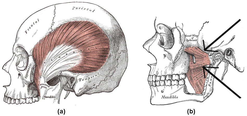
Location of the temporalis muscle and the lateral pterygoid: (a) Highlighted in orange, the temporalis muscle is a broad muscle extending from zygomatic bone. (b) Arrows indicate the location of the lateral pterygoid, highlighted with the medial pterygoid in orange.
KEY MOVEMENTS
Elevation of the Jaw: Produced by the masseter, temporalis and medial pterygoid.
Depression of the Jaw: Produced by the lateral pterygoid, assisted by the digastric, mylohyoid and geniohyoid muscles found in the neck.
Protraction of the Jaw: Produced by the lateral pterygoid.
Retraction of the Jaw: Produced by the temporalis.
Lateral Movement of the Jaw: Produced by the lateral and medial pterygoid.
Neck Muscles
Cervical muscles are those associated with the front of the neck; vertebral muscles are associated with the vertebral column. Outline the neck muscles and their movements. Numerous muscles contribute to the processes of speaking and swallowing. These muscles can be divided into suprahyoid and ingrahyoid groups based on their locations relative to the hyoid bone. The hyoid bone, located beneath the mandible, acts as a key attachment point for muscles involved in speaking and swallowing. Numerous muscles contribute to both the stabilization and fine movements of the head and neck.
Key Terms
Suprahyoid muscles: A group of muscles located above the hyoid bone, responsible for its elevation which widens the esophagus.
Hyoid bone: shaped bone that sits below the mandible
and in front of the esophagus, facilitating the wide range of movements associated with speaking and swallowing.Infrahyoid muscles: A group of muscles located below the hyoid bone, responsible for its depression which narrows the esophagus.
Muscles of the neck play important roles in mastication (chewing), swallowing, speaking and supporting and moving the head. All muscles found in the neck are paired, meaning they exist to both the left and right side of the spine.
Muscles Involved in Swallowing and Speaking
Located to the anterior of the neck, these muscles are split into two based on their location relative to the hyoid bone. The U-shaped hyoid bone sits below the mandible and in front of the esophagus, providing a level of protection and facilitating the wide range of muscle activity required for speaking and swallowing.
Suprahyoid Muscles

Suprahyoid and infrahyoid muscles of the neck: Suprahyoid and infrahyoid muscle groups are named based on their location relative to the hyoid bone. The hyoid bone sits below the mandible and in front of the esophagus, providing a level of protection but also facilitating the wide range of muscle activity required for speaking and swallowing.
The four suprahyoid muscles found above the hyoid bone act in concert to elevate the hyoid bone, assisting with swallowing by widening the esophagus.
- Stylohyoid: The most superior of the suprahyoid muscles, the stylohyoid originates from the skull and attaches to the hyoid bone.
- Digastric: The digastric muscle is split into two parts that are connected by a tendon attached to the hyoid bone. The anterior section originates from the mandible and the posterior section from the skull.
- Mylohyoid: The mylohyoid is a broad flat muscle which forms the floor of the oral cavity. It originates from the mandible and attaches to the hyoid bone.
- Geniohyoid: The deepest of the suprahyoid muscles, the geniohyoid muscle originates from the mandible and attaches to the hyoid bone.
Infrahyoid Muscles
The four infrahyoid muscles found below the hyoid bone act in concert to depress the hyoid bone during swallowing and speaking, compressing the esophagus.
- Sternohyoid: A superficial muscle which originates from the sternum and attaches onto the hyoid bone.
- Omohyoid: Located laterally to the sternohyoid, the omohyoid muscle is split in two parts attached by a tendon. The inferior region originates from the scapula, joins the superior region, and attaches to the hyoid bone.
- Sternothyroid: Sitting deeper than the sternohyoid, the sternothyroid originates from the sternum and attaches to the thyroid cartilage associated with the hyoid bone.
- Thyrohyoid: A short continuation of the sternothyroid muscle, the thyrohyoid originates from the thyroid cartilage and attaches to the hyoid bone.
Muscles of the Back and Neck
Muscles of the back and neck: Muscles of the back and neck play an important role in maintaining posture and the movement of the head and neck.
The muscles of the back and neck are responsible for maintaining posture and facilitating the movement of the head and neck. They are divided into three layers.
Superficial Layer
Two muscles in the superficial layer are responsible for the rotation of the head.
- Splenius Capitis: A thick rectangular muscle, the most superior of the neck muscles.
- Attachments: Originates from the upper spine and attaches to the skull.
- Actions: Rotates and extends the head and neck.
- Splenius Cervicis: A small triangular-shaped muscle located immediately below the splenius capitis.
- Attachments: Originates from the spine and attaches several vertebrae higher.
- Actions: Rotate and extend the head and neck.
Intermediate Layer
Three columnar muscles in the intermediate layer are responsible for flexion and extension of the neck as well as posture maintenance. All three originate from a common tendon associated with the pelvis and can be divided into thoracic, cervicis, and capitis regions.
- Iliocostalis: The most laterally located of the three intermediate muscles.
- Attachments: Originates from the common tendon and attaches to the ribs and lower neck.
- Actions: Extends and controls abduction and adduction of the spine and neck.
- Longissimus: Located between the iliocostalis and spinalis muscles, this is the largest of the intermediate layer muscles.
- Attachments: Originates from the common tendon and attaches to the lower ribs, the spine, and the skull.
- Actions: Extends and controls abduction and adduction of the spine and neck.
- Spinalis: The most medially located and smallest of the three intermediate layer muscles.
- Attachments: Originates from the common tendon and attaches to the upper spine and skull.
- Actions: Extends and flexes to control abduction and adduction of the spine and neck.
Deep Layer
Two muscles in the deep layer are responsible for the maintenance of posture and rotation of the neck.
- Semispinalis: The semispinalis is the most superficial of the deep muscles.
- Attachments: A broad origin on the upper regions of the spine, with each origin attaching several vertebrae higher or to the skull.
- Actions: Extends and rotates the head and maintains posture.
- Multifidus: The multifidus is located underneath the semispinalis muscle and is key in maintaining posture.
- Attachments: A broad origin up the length of the spine, with each origin attaching several vertebrae higher.
- Actions: Maintains posture through the spine.
Other Muscles That Act on the Neck
Several other muscles act on the head and neck. Below are three with a larger impact.
- Trapezius: The trapezius is the most superficial muscle of the back and forms a broad flat triangle.
- Attachments: The trapezius originates from the skull and spine of the upper back and neck. It attaches to the clavicle and scapula.
- Actions: The superior region supports the arm and elevates and rotates the scapula. It controls adduction, abduction and rotation of the head, the intermediate region retracts the scapula, and the inferior region rotates and depresses the scapula.
- Sternocleidomastoid: A thick rectangular muscle that is responsible for many movements within the neck.
- Attachments: Dual-headed, the sternocleidomastoid originates from the clavicle and the sternum and attaches to the mandible.
- Actions: Abduction, adduction, extension, flexion, and rotation of the neck depending on intra and inter-muscle contractions.
- Platysma: A broadsheet of muscle arising from the fascia covering the pectorals.
- Attachments: Originates from the fascia covering the pectorals and attaches to various locations within the mandible and dermis of the face and neck.
- Actions: Depresses the mandible and angles the lip and mouth, wrinkling the skin upon the neck flexing.
KEY MOVEMENTS
The extension (tilting head backwards): Produced by the semispinalis, splenus capitis, longissimus, trapezius (superior fibers), and sternocleidomastoid (posterior fibers).
Flexion (tilting head forwards): Produced by the sternocleidomastoid (anterior fibers).
Abduction (tilting head towards shoulder): Produced by the sternocleidomastoid, longissimus, splenius capitis, semispinalis, and trapezius (superior fibers)
Adduction (returning head to midline): Produced by the sternocleidomastoid, longissimus, splenius capitis, semispinalis, and trapezius (superior fibers)
Rotation (rotation head to left or right): Produced by the sternocleidomastoid, longissimus, splenius capitis, semispinalis, and trapezius (superior fibers)
Anterior Muscles Trunk
The anterior muscles of the torso (trunk) are those on the front of the body, including the muscles of the chest, abdomen, and pelvis.
Key Points
- The intercostal muscles form the chest wall and function in respiration.
- The diaphragm is a sheet-like muscle that extends underneath the rib cage and aids in respiration by physically moving the lungs.
- The obliques are abdominal muscles that assist during bending and twisting of the torso.
- The rectus abdominis are the muscles often referred to as the “six-pack abs” and are involved in numerous aspects of trunk stabilization and bending.
Key Terms
- diaphragm: The key muscle in the control of respiration.
- abdominal wall: A layer of muscle and fascia that protects and encloses the abdominal cavity, allowing for its compression as well as torso movement.
- linea alba: A tough, fibrous line running down the midline of the abdomen, formed from the aponeuroses of the abdominal muscles.
- intercostal: Muscles forming the chest wall, which aid in respiration.
The anterior muscles of the trunk (torso) are associated with the front of the body, include chest and abdominal muscles. Chest muscles function in respiration while abdominal muscles function in torso movement and in the maintenance of balance and
posture.
Muscles of the Chest
Pectoral Muscles
Pectoral muscles lie in the chest and exert force through the shoulder to move the upper arm.
- Pectoralis Major: The pectoralis major is a fan-shaped muscle covering the chest and comprised of clavicular and sternocostal regions.
- Attachments: The clavicular region originates from the clavicle and the sternocostal region originates from the sternum and the fascia of the oblique muscles of the abdomen. Both attach to the humerus.
- Actions: Adducts and rotates the upper arm.
- Pectoralis Minor: The smaller pectoralis minor muscle lies beneath the pectoralis major.
- Attachments: The pectoralis minor originates from the third to fifth ribs and attaches to the scapula.
- Actions: Supports and depresses the scapula.
- Serratus Anterior: The serratus anterior is located in the lateral wall of the chest.
- Attachments: The muscle is formed of several strips originating from the second to eighth ribs, each of which attaches to the scapula.
- Actions: Supports the scapula, allowing for elevation of the upper arm.
Intercostal Muscles
Intercostal muscles of the anterior trunk: Deep muscles of the chest and front of the arm, with the boundaries of the axilla. The intercostals are muscles between the ribs that form the chest cavity wall.
Lying below the pectoral muscles, the intercostal muscles form the chest wall and play a key role in respiration. All intercostal muscles originate on the lower border of a rib and attach to the upper border of the rib below.
- External Intercostals: The external intercostals are the most superficial of the intercostal muscles. They are continuous with the external oblique muscle of the abdomen.
- Actions: Elevate the ribs.
- Internal Intercostals: Lying below the external intercostals, the internal intercostals are continuous with the internal oblique muscle of the abdomen.
- Actions: Elevate or depress the ribs.
- Innermost Intercostals: The deepest lying of the intercostals, these muscles are similar in structure to the internal intercostals.
- Actions: Elevate or depress the ribs.
Other Muscles
Diaphragm: The diaphragm is a large, flat, sheet-like muscle that extends horizontally underneath the rib cage.
Functionally, the diaphragm separates the thoracic cavity, containing the lungs and heart and enclosed by the rib cage from the abdominal cavity, which contains the digestive organs. The diaphragm’s position allows it to aid in respiration. When it contracts, it physically moves the lungs and deforms the volume of the thoracic cavity.
- Attachments: The diaphragm has several points of origin along the sternum, the lower ribs, and lower vertebrae. The muscle fibers combine into a central tendon, which ascends and attaches to the surface of the pericardium.
- Actions: Contracts, flattening and increasing the volume of the thoracic cavity. Relaxes and returns to original shape, reducing the volume of the thoracic cavity.
Muscles of the Abdomen
The skeletal muscles of the abdomen form part of the abdominal wall, which holds and protects the gastrointestinal system. Five muscles form the abdominal wall, divided into vertical and flat groups. The flat muscles act to flex, laterally flex, and rotate the trunk. The fibers run in different directions and cross each other, strengthening the abdominal wall. The vertical muscles aid in compressing the abdominal cavity, stabilizing the pelvis, and depressing the ribs when a person is walking. Toward the midline, the muscles form aponeuroses, which merge into the linea alba.
Location of the external obliques: Highlighted in orange, the external obliques lie inferior to the pectoral muscles
- External Oblique: The external oblique is the largest and most superficial of the flat muscles.
- Attachments: Originates from the lower ribs and attaches to the pelvis, forming an aponeurosis toward the midline and linea alba.
- Internal Oblique: Lying deep to the external oblique, the internal oblique is smaller and thinner. Its fibers run perpendicular to the external oblique, improving the strength of the abdominal wall.
- Attachments: Originates from the pelvis and thoracolumbar fascia, running through the back. Attaches to the lower ribs and forms an aponeurosis toward the midline and linea alba.
- Transversus Abdominis: The deepest of the flat muscles, the transversus abdominis consists of transversely running fibers.
- Attachments: Originates from the lower ribs, thoracolumbar fascia, and pelvis, forming an aponeurosis toward the midline and linea alba.
- Rectus Abdominis: A long vertical muscle that covers the abdomen, lying below the flat muscles. It is split through the midline by the linea alba formed from the aponeuroses of the abdominal muscles and separated by horizontal tendinous intersections which give rise to the six-pack.
- Attachments: Originates from the pubis and attaches to the lower edge of the rib cage and sternum.
- Pyramidalis: Lying superficial to the rectus abdomini,s the pyramidalis is a small, triangular vertical muscle.
- Attachments: Originates from the pubis and attaches to the linea alba.
Posterior Muscles
Muscles of the posterior portion of the trunk include muscles of the back, suboccipital region, and perineum region.
Key Points
- The back is characterized by numerous muscle groups which allow movement of the shoulder, head, and neck, as well as aid in respiration and maintain posture and balance.
- The superficial muscles of the back are responsible for movement of the shoulder.
- The intermediate muscles of the back assist in the movement of the rib cage during respiration.
- The intrinsic back muscles facilitate movement of the head and neck and are fundamental in maintaining posture and balance.
The posterior or back muscles perform a wide range of functions, including movement of the shoulder, head, and neck and assisting in respiration, posture, and balance. Posterior muscles are split into three groups depending on their physiological location.
Superficial Posterior Muscles
Location of the latissimus dorsi muscle: Highlighted in orange, the latissimus dorsi is a muscle of the posterior torso.
The superficial posterior muscles are associated with movement of the shoulder. As the name suggests, they are the most superficially located of the muscles covering the intermediate and intrinsic layers.
- Trapezius: The trapezius is the most superficial muscle of the back and forms a broad flat triangle.
- Attachments: The trapezius originates from the skull and spine of the upper back and neck. It attaches to the clavicle and scapula.
- Actions: The superior region supports the arm and elevates and rotates the scapula, the intermediate region retracts the scapula, and the inferior region rotates and depresses the scapula.
- Latissimus Dorsi: The latissimus dorsi originates from the lower back and covers a wide area.
- Attachments: The latissimus dorsi originates from the lower spine and ribs and the upper pelvis and fascia of the deep trunk muscles. The muscle converges into a tendon attaching to the humerus.
- Actions: Extends, adducts, and medially rotates the upper arm.
- Levator Scapulae: A small, strap-like muscle that joins the neck to the scapula.
- Attachments: Originates from the side of the spine in the neck and attaches to the scapula.
- Actions: Elevates the scapula.
- Rhomboid Major: Sits inferiorly to the levator scapulae.
- Attachments: Originates from the spine in the upper back and attaches to the scapula inferior to the levator scapulae attachment.
- Actions: Retracts and rotates the scapula.
- Rhomboid Minor: Sits between the levator scapulae and rhomboid major, with which it is paired in action and function, this retracts and rotates the scapula.
Intermediate Posterior Muscles
The intermediate muscles of the posterior contribute to movements of the ribcage during respiration.
Serratus Posterior Superior – The serratus posterior superior is a thin, rectangular-shaped muscle lying below the rhomboid muscles.
- Attachments: Originates from the lower spine and attaches to ribs 2 through 5.
- Actions: Elevates ribs 2 through 5.
Serratus Posterior Inferior: The serratus posterior inferior is a broad muscle lying beneath the latissimus dorsi.
- Attachments: Originates from the spine and attaches to ribs 9 through 12.
- Actions: Depresses ribs 9 through 12.
Intrinsic Posterior Muscles
The intrinsic muscles of the posterior are responsible for maintaining posture and facilitating the movement of the head and neck. They are divided into three layers.
Superficial Layer
Location of the splenius muscle.: The splenius capitis is highlighted in orange, with the splenius cervicis directly below
Two muscles in the superficial layer are responsible for the rotation of the head.
- Splenius Capitis: This thick rectangular muscle is the most superior of the next muscles.
- Attachments: Originates from the upper spine and attaches to the skull.
- Actions: Rotates and extends the head and neck.
- Splenius Cervicis: A small triangular-shaped muscle located immediately below the splenius capitis.
- Attachments: Originates from the spine and attaches several vertebrae higher.
- Actions: Rotates and extends the head and neck.
Intermediate Layer
Three columnar muscles in the intermediate layer are responsible for flexing and extending the neck as well as maintaining posture. All three originate from a common tendon associated with the pelvis.
- Iliocostalis: The most laterally located of the three intermediate muscles.
- Attachments: Originates from the common tendon and attaches to the ribs and lower neck.
- Actions: Extends and controls abduction and adduction of the spine and neck.
- Longissimus: Located between the iliocostalis and spinal muscles, it is the largest of the intermediate layer muscles.
- Attachments: Originates from the common tendon and attaches to the lower ribs, spine, and skull.
- Actions: Extends and controls abduction and adduction of the spine and neck.
- Spinalis: The most medially located and smallest of the three intermediate layer muscles.
- Attachments: Originates from the common tendon and attaches to the upper spine and skull.
- Actions: Extends, flexes, and controls abduction and adduction of the spine and neck.
Deep Layer
Two muscles in the deep layer are responsible for the maintenance of posture and rotation of the neck.
- Semispinalis: The semispinalis is the most superficial of the deep muscles.
- Attachments: A broad origin on the upper regions of the spine, with each origin attaching several vertebrae higher or to the skull.
- Actions: Extends and rotates the head and maintains posture.
- Multifidus: The multifidus is located underneath the semispinalis muscle, and is key in maintaining posture.
- Attachments: A broad origin up the length of the spine, with each origin attaching several vertebrae higher.
- Actions: Maintains posture through the spine.
Muscles of the Upper Limb
Muscles of the Humerus that Act on the Forearm
Humerus that act on the forearm are primarily involved in flexion and extension. Diagram the movements of the humerus muscles that act on the forearm
Key Points
- Muscles of both the upper arm and forearm control movement of the forearm.
- The biceps brachii flex the forearm and work with the supinator of the forearm to rotate it so the palm faces upward.
- The triceps brachii extend the forearm.
- The pronator teres and quadratus control pronation, or rotation of the forearm so that the palm faces downward.
Key Terms
Pronator Teres: A muscle of the anterior compartment of the forearm that controls pronation.
Supinator: A muscle of the posterior compartment of the forearm that controls supination.
Pronator Quadraturs: A muscle of the anterior compartment of the forearm that controls pronation.
Brachioradialis: A muscle of the posterior compartment of the forearm that flexes the forearm.
Biceps Brachii: A muscle of the anterior compartment of the upper arm that flexes the forearm.
Triceps Brachii: A muscle of the posterior compartment of the upper arm that extends the forearm.
The humerus is a long bone in the arm that runs from the shoulder to the elbow. Anatomically, it interacts with the scapula to form the shoulder joint and the radius and ulna of the lower arm to form the elbow joint. Forearm rotation is controlled by two joints: the proximal radioulnar joint which exists immediately below the elbow, and the distal radioulnar joint located immediately before the wrist.
Upper Arm
There are four muscles in the upper arm split into an anterior and posterior compartment.
Anterior Compartment
Superficial muscles of the chest and upper arm: The biceps brachii is located in the anterior compartment of the upper arm and flexes and supinates the forearm at the elbow.
Three muscles are located in the anterior compartment of the upper arm.
- Biceps Brachii: The biceps brachii is a two-headed muscle. Although the majority of the muscle mass is located anteriorly to the humerus, it has no attachment to the bone itself.
- Coracobrachialis: The coracobrachialis lies within the two heads of the biceps brachii.
- Attachments: Originates from the scapula and attaches to the humerus.
- Action: Flexing of the arm at the shoulder, and weak adduction.
- Brachialis: The brachialis muscle lies within the distal region of the biceps brachii.
- Attachments: Originates from the humerus and attaches to the ulna.
- Action: Flexing of the arm at the elbow.
Posterior Compartment
The posterior compartment of the upper arm contains only one muscle.
- Triceps Brachii: The triceps brachii is a three-headed muscle.
- Attachments: The long head originates from the scapula, the lateral head from the proximal region of the humerus, and the medial head from the distal region of the humerus. All three converge into one tendon which attaches to the ulna.
- Action: Extension of the arm at the elbow.
Forearm
Superficial muscles of the posterior forearm: The anconeus, located in the superficial region of the posterior forearm compartment, moves the ulna during pronation and extends the forearm at the elbow.
As with the upper arm, the forearm is split into anterior and posterior compartments. Only those responsible for the movement of the forearm are discussed below; the muscles responsible for the movement of the hand and wrist are discussed in a later section.
Anterior
The anterior compartment of the forearm is split into superficial, intermediate, and deep regions.
- Pronator Teres: A rectangular muscle located in the superficial region of the anterior compartment.
- Attachments: The pronator teres have two origins, one on the proximal end of the humerus and one of the distal end of the ulna. It attaches to the mid-region of the radius.
- Action: Pronates the forearm.
- Pronator Quadratus: A square-shaped muscle located adjacent to the wrist in the deep region of the anterior compartment.
- Attachments: Originates from the ulna and attaches to the radius.
- Action: Pronates the forearm.
Posterior
The posterior compartment of the forearm is split into superficial and deep regions.
- Anconeus: The anconeus is located in the superficial region of the forearm posterior compartment and is blended with the triceps brachii.
- Attachments: Originates from the humerus and attaches to the ulna.
- Action: Moves the ulna during pronation and extends the forearm at the elbow.
- Brachioradialis: The brachioradialis is located in the superficial region of the forearm posterior compartment,
- Attachments: Originates from the humerus and attaches to the distal end of the radius.
- Action: Flexes the forearm at the elbow.
- Supinator: The supinator is located in the deep region of the forearm posterior compartment.
- Attachments: The supinator has two heads: one originating from the humerus, the other from the ulna. Together they attach to the radius.
- Action: Supinates the forearm.
KEY MOVEMENTS
The extension (forearm away from the upper arm): Produced by the triceps brachii and anconeus of the forearm.
Flexion (forearm towards the upper arm): Produced by the brachialis, biceps brachii, and brachioradialis of the forearm.
Pronation (rotation of the forearm so the palm faces downwards): Produced by the pronator quadratus and pronator teres of the forearm.
Supination(rotation of the forearm so the palm faces upwards): Produced by the supinator of the forearm and biceps brachii.
Muscles of the Wrist and Hand
Muscles in the forearm move the wrists, and hand movement is caused by both forearm and hand muscles.
Key Points
- Movements of the wrist include abduction, adduction, extension, and flexion.
- Movements of the fingers and thumb include abduction, adduction, extension, and flexion.
- Rotation of the thumb and little finger allows for opposition.
- Muscles of the forearm that act on the wrist and hand are referred to as extrinsic muscles, or external to the hand.
- Muscles within the wrist and hand are referred to as intrinsic muscles.
Key Terms
Palmaris Longus: A long muscle originating near the elbow and passing through into the wrist, attaching to the base of the hand.
Flexor Digitorum Superficialis: A key muscle controlling wrist and finger flex.
Flexor Carpi Ulnaris: A long muscle originating near the elbow and passing through into the wrist, attaching to one of the carpal bones in the wrist.
Flexor Carpi Radialis: A long muscle originating near the elbow and passing through into the wrist, attaching to the base of the digits (fingers).
Flexor Digitorum Profundus: A long muscle originating near the elbow and passing through into the wrist, flexing the wrist and the most distant regions of the fingers.
Pronator Teres: A rectangular muscle that pronates the forearm.
Flexor Pollicis Longus: A long, deep muscle responsible for flexing the thumb.
Pronator quadratus: A square-shaped muscle located adjacent to the wrist.
Muscles associated with the wrist include those of the forearm and hand that move the wrist and digits. The wrist and hand exhibit a remarkable range of movement, key for grasping and interaction with objects. These muscles can generate highly variable force, from the strong grip required when lifting a heavy object to the delicate movements required to write.
Muscles and tendons of the forearm and hand: The extrinsic muscles of the forearm are responsible for the movement of the wrist and fingers. Often providing the more forceful movements required.
Muscles of the forearm that act on the wrist and hand are referred to as extrinsic muscles, or external to the hand. Those located within the hand are referred to as intrinsic.
Muscles of the Forearm
As with the upper arm, the forearm is split into the anterior and posterior compartments. Each contains many more muscles than described below due to the requirement for more complex movements in the wrist and hand.
Anterior
The anterior compartment of the forearm is split into superficial, intermediate, and deep layers. These muscles are generally responsible for flexing of the wrist and fingers and pronation of the forearm.
Superficial Layer
Three muscles are located in the superficial layer of the anterior compartment of the forearm.
- Flexor Carpi Ulnaris: A long muscle originating near the elbow and passing through into the wrist.
- Attachments: Originates from the humerus and ulna and attaches to one of the carpal bones in the wrist.
- Actions: Flexion and adduction at the wrist.
- Palmaris Longus: A long muscle originating near the elbow and passing through into the wrist.
- Attachments: Originates from the humerus and attaches to the base of the hand.
- Actions: Flexion at the wrist.
- Flexor Carpi Radialis: A long muscle originating near the elbow and passing through into the wrist.
- Attachments: Originates from the humerus and attaches to the base of the digits.
- Actions: Flexion and abduction at the wrist.
- Pronator Teres: A rectangular muscle.
- Attachments: The pronator teres has two origins, one on the proximal end of the humerus and one of the distal end of the ulna. It attaches to the mid region of the radius.
- Actions: Pronates the forearm.
Intermediate Layer
There is just one muscle in the intermediate layer of the anterior compartment of the forearm.
- Flexor Digitorum Superficialis: Lying below the superficial region, the flexor digitorum superficialis is a key muscle controlling wrist and finger flex.
- Attachments: Originates from the humerus and the radius, splitting into four tendons at the wrist which travel through the carpal tunnel and attach to the fingers.
- Actions: Flexes fingers and wrist.
Deep Layer
There are three muscles in the deep layer of the anterior compartment of the forearm.
- Flexor Digitorum Profundus: A long muscle originating near the elbow and passing through into the wrist, lying adjacent to the flexor pollicis longus.
- Attachments: Originates from the ulna, splitting into four tendons at the wrist which travel through the carpal tunnel and attach distally to the fingers.
- Actions: Flexes the wrist and the most distal regions of the fingers.
- Flexor Pollicis Longus: A long muscle originating near the elbow and passing through into the wrist, lying adjacent to the flexor digitorum profundus.
- Attachments: Originates from the radius and attaches to the base of the thumb.
- Actions: Flexes the thumb.
- Pronator quadratus: A square-shaped muscle located adjacent to the wrist.
- Attachments: Originates from the ulna and attaches to the radius.
- Actions: Pronates the forearm.
Posterior
The posterior compartment of the forearm is split into superficial and deep regions. The muscles are generally responsible for the extension of the wrist and fingers.
Superficial Layer
The superficial layer of the posterior forearm contains seven muscles.
- Aconeus: The anconeus is located in the superficial region of the forearm posterior compartment and is blended with the triceps brachii.
- Attachments: Originates from the humerus and attaches to the ulna.
- Actions: Moves the ulna during pronation and extends the forearm at the elbow.
- Brachioradialis: The brachioradialis is located in the superficial region of the forearm posterior compartment.
- Attachments: Originates from the humerus and attaches to the distal end of the radius.
- Actions: Flexes the forearm at the elbow.
- Extensor Carpi Radialis Longus and Brevis: A pair of muscles located on the side of the forearm, allowing them to control extension and abduction of the wrist.
- Attachments: Both originate from the humerus and attach to the base of the hand.
- Actions: Extend and abduct the wrist.
- Extensor Digitorum: The extensor digitorum is the main extensor of the fingers.
- Attachments: Originates from the humerus, splitting into four tendons at the wrist which travel through the carpal tunnel and attach to the digits.
- Actions: Extends fingers.
- Extensor Digiti Minimi: Originates from the extensor digitorum. In some people, these muscles cannot be individually defined.
- Attachments: Originates from the humerus and attaches to the little finger.
- Actions: Extends the little finger, and contributes to extension at the wrist.
- Extensor Carpi Ulnaris: Located on the other side of the forearm to the extensor carpi radialis longus and brevis, it performs a similar role.
- Attachments: Originates from the humerus and attaches to the base of the hand.
- Actions: Extension and adduction of the wrist.
Deep Layer
There are four muscles in the deep layer of the posterior compartment of the forearm.
- Supinator: The supinator is located in the deep region of the forearm posterior compartment.
- Attachments: The supinator has two heads: one originates from the humerus, the other from the ulna. Together they attach to the radius.
- Actions: Supinates the forearm.
- Abductor Pollicis Longus: The abductor pollicis longus is situated immediately distal to the supinator muscle.
- Attachments: Originates from the radius and ulna attaching to the base of the thumb.
- Actions: Abducts the thumb.
- Extensor Pollicis Brevis: The extensor pollicis brevis is located below the abductor pollicis longus.
- Attachments: Originates from radius and attaches to the base of the thumb.
- Actions: Extends the thumb.
- Extensor Indicis Proprius: This muscle allows the index finger to be independent of the other fingers during extension.
- Attachments: Originates from the ulna and attaches to the index finger.
- Actions: Extends the index finger.
Muscles of the Hand
The extrinsic muscles of the hand are responsible for the larger scale, stronger movements of the wrist and hand. The intrinsic muscles produce finer, more controlled movements and play important roles in maintaining grip.
Thenar Muscles
The thenar muscles are three short muscles located at the base of the thumb and responsible for its fine movement.
- Opponens Pollicis: The opponent pollicis is the largest and deepest-lying of the thenar muscles.
- Attachments: Originates from the wrist and attaches to the thumb.
- Actions: Rotates the thumb towards the palm, producing opposition and improving grip.
- Abductor Pollicis Brevis : Located anteriorly to the opponens pollicis and proximal to the flexor pollicis Brevis.
- Attachments: Originates from the wrist and attaches to the thumb.
- Actions: Abducts the thumb.
- Flexor Pollicis Brevis: The smallest and most distal of the thenar muscles.
- Attachments: Originates from the wrist and attaches to the thumb.
- Actions: Flexes the thumb.
Hypothenar Muscles
The hypothenar muscles are located at the base of the little finger. Their naming, function, and organization are similar to those of the thenar muscles.
- Opponens Digiti Minimi: The opponens digit minimi is the deepest-lying of the hypothenar muscles.
- Attachments: Originates from the wrist and attaches to the little finger.
- Actions: Rotates little finger towards the palm, producing opposition and improving grip.
- Abductor Digiti Minimi: The most superficial of the hypothenar muscles.
- Attachments: Originates from the wrist and attaches to the little finger.
- Actions: Abducts the little finger.
- Flexor Digiti Minimi Brevis: Located laterally to the digiti minimi.
- Attachments: Originates from the wrist and attaches to the little finger.
- Actions: Flexes little finger.
Lubricants
These are four lumbricals in the hand, each associated with an individual finger.
- Attachments: Originates from a tendon of attached to the flexor digitorum profundus of the forearm, each attaching to an individual finger
- Actions: Flexes and extends the fingers.
Interossei
The interossei muscles are located between the fingers; they can be split into two groups.
- Dorsal Interossei: Located superficially on the dorsal side of the hand, there are four dorsal interossei muscles.
- Attachments: Originates from the base of the finger, each attaching after the first finger joint.
- Actions: Abducts the fingers.
- Palmar Interossei: Located on the anterior side of the hand, there are three palmar interossei, with the index finger controlled by the extensor indicis proprius.
- Attachments: Originates from the base of the finger, each attaching after the first finger joint.
- Actions: Adducts the fingers.
Other Muscles
One other muscle in the hand is not easily grouped with the above categories.
- Palmaris Brevis: The palmaris brevis is a small superficial muscle found in the palm.
- Attachments: Originates from the fascia of the palm and attaches to the dermis.
- Actions: Wrinkles the skin and deepens the curvature of the palm improving grip.
Muscles of the Shoulder
Muscles of the shoulder include those that attach to the bones of the shoulder to move and stabilize the joint.
Key Points
- The shoulder exhibits a wide range of movement, which makes it susceptible to dislocation and injury.
- The trapezius muscles rotate the scapulae upward.
- The rhomboid major and the rhomboid minor press the scapula against the thoracic wall, retracting the scapula towards the spine.
- The deltoid is a complex muscle that forms the rounded edge of the shoulder and participates in many articulations of the shoulder joint.
- The rotator cuff are the muscles that stabilize movement of the shoulder.
- The pectoralis minor and pectoralis major are large muscles of the chest that participate in many movements, including flexion of the humerus.
Key Terms
pectoralis major: A large, fan-shaped muscle of the chest.
rotator cuff: A set of four smaller muscles in the shoulder responsible for rotating the humerus (upper arm bone).
trapezius: A large vertebrate skeletal muscle divided into an ascending, descending, and transverse portion, attaching the neck and central spine to the outer extremity of the scapula. It functions in scapular elevation, adduction, and depression.
deltoid: The deltoid muscle, a triangular muscle on the human shoulder.
The shoulder or glenohumeral joint is a ball and socket joint formed between the humerus and scapula. Due to the shallowness of the socket and relatively loose connections, the shoulder joint allows for a wide range of motion; however, this wide range makes the joint unstable and thus more prone to dislocation and injury than other joints.
Two other joints make up the shoulder; the acromioclavicular joint of the clavicle and scapula, which allows the arm to be raised above the head, and the sternoclavicular joint of the clavicle and sternum, which plays an important role in facilitating movement of the upper arm and connecting it to the rest of the skeleton.
Muscles that act on the shoulder can be classified as extrinsic, intrinsic, pectoral, or upper arm. Upper arm muscles will be discussed in a later section since they primarily promote forearm movement.
Extrinsic Shoulder Muscles
Extrinsic muscles of the shoulder originate from the trunk and attach to the bones of the shoulder. They can be further subdivided into superficial and deep layers.
Superficial
Location of the trapezius muscle: Highlighted in orange, the trapezius is a large, broad muscle of the back that acts on the shoulder.
As suggested by the name, superficial muscles lie on the surface. There are two superficial extrinsic muscles.
- Trapezius: The trapezius is the most superficial muscle of the back and forms a broad flat triangle.
- Attachments: The trapezius originates from the skull and spine of the upper back and neck. It attaches to the clavicle and scapula.
- Actions: The superior region supports the arm and elevates and rotates the scapula, the intermediate region retracts the scapula, and the inferior region rotates and depresses the scapula.
- Latissimus Dorsi: The latissimus dorsi originates from the lower back and covers a wide area.
- Attachments: The latissimus dorsi originates from the lower spine and ribs and the upper pelvis and fascia of the deep trunk muscles. The muscle converges into a tendon attaching to the humerus.
- Actions: Extends, adducts, and medially rotates the upper arm.
Deep
Three deep muscles lie below the superficial muscles of the shoulder.
- Levator Scapulae: A small, strap-like muscle that joins the neck to the scapula.
- Attachments: Originates from the side of the spine in the neck and attaches to the scapula.
- Actions: Elevates the scapula.
- Rhomboid Major: Sits inferiorly to the levator scapulae.
- Attachments: Originates from the spine in the upper back and attaches to the scapula in an inferior position to the levator scapulae attachment.
- Actions: Retracts and rotates the scapula.
- Rhomboid Minor: Sits between the levator scapulae and rhomboid major, with which it is paired in action and function. It retracts and rotates the scapula.
Intrinsic
Location of the deltoid muscles: Highlighted in orange, the deltoids cover the rounding of the shoulder joint.
Intrinsic muscles originate from the scapula or clavicle and attach to the humerus. There are six intrinsic muscles, four of which form the rotator cuff.
- Deltoid: The deltoid muscle is a triangular muscle that covers the shoulder. The action of the muscle is complex, with the components acting in opposing and separate ways during the course of a contraction.
- Attachments: The deltoid muscle originates from the scapula and clavicle and attaches to the lateral surface of the humerus.
- Actions: The anterior region assists the pectoralis major during transverse flexion of the shoulder and acts weakly in strict transverse flexion. The lateral region assists in shoulder flexion when the shoulder is rotating, although it also assists the transverse abduction of the shoulder. The posterior region is the hyperextension of the shoulder, contributing to transverse
extension.
- Teres Major: The teres major is a thick flattened muscle connecting the lower scapula with the humerus.
- Attachments: Originates from the posterior of the scapula and attaches to the humerus.
- Actions: Adducts the shoulder and assists in rotation of the arm.
Rotator Cuff
A rotator cuff is a group of four muscles that pull the ball of the humerus into the shallow socket of the scapula, adding required stability. The rotator cuff complex is composed of the supraspinatus, infraspinatus, subscapularis, and teres minor all of which originate from the scapula and connect to the humerus. The supraspinatus is involved in the abduction of the arm in association with the deltoid, while the other muscles facilitate rotation of the arm.
Muscles of the rotator cuff: Muscles of the rotator cuff and presented with the triceps brachii.
Pectoral
Pectoral muscles lie in the chest and exert force through the shoulder to move the upper arm. Three pectoral muscles interact with the shoulder.
- Pectoralis Major: The pectoralis major is a large, fan-shaped muscle covering the chest. It is comprised of clavicular and sternocostal regions.
- Attachments: The clavicular region originates from the clavicle and the sternocostal region originates from the sternum and the fascia of the oblique muscles of the abdomen. Both attach to the humerus.
- Actions: Adducts and rotates the upper arm.
- Pectoralis Minor: The pectoralis minor muscle is smaller and lies beneath the pectoralis major.
- Attachments: The pectoralis minor originates from the third to fifth ribs and attaches to the scapula.
- Actions: Supports and depresses the scapula.
- Serratus Anterior: The serratus anterior is located in the lateral wall of the chest.
- Attachments: The muscle is formed of several strips originating from the second to eight ribs, each of which attaches to the scapula.
- Actions: Supports the scapula allowing for elevation of the upper arm.
KEY MOVEMENTS
The extension (upper limb backward behind back): Produced by the posterior deltoid, latissimus dorsi, and teres major.
Flexion (upper limb forwards past chest): Produced by the biceps brachii (both heads), pectoralis major, anterior deltoid, and coracobrachialis.
Abduction (upper limb away from the trunk, spreading arms wide): Produced by the supraspinatus and deltoid. Past 90 degrees, the scapula needs to be rotated by the trapezius and serratus anterior to achieve abduction.
Adduction (upper limb towards the trunk, bringing arms down to side): Produced by contraction of the pectoralis major, latissimus dorsi, and teres major.
Medial Rotation (rotation of arm inwards to cover abdomen): Produced by contraction of the subscapularis, pectoralis major, latissimus dorsi, teres major, and anterior deltoid.
Lateral Rotation (rotation of arm outwards away from the abdomen): Produced by contraction of the infraspinatus and teres minor
Muscles of the Lower Limb
Muscles that Cause Movement at the Hip Joint
The four main groups of hip muscles are gluteal, adductor, iliopsoas, and lateral rotator, defined by the type of movement they mediate.
Key Points
- The gluteus maximus extends the hip, while the gluteus medius and minimus are involved in hip rotation and abduction (moving hip out from the midline).
- The adductor group (adductor brevis, longus, and magnus along with petineus and gracilis) moves the femur towards the midline from an abducted position.
- The iliopsoas group of muscles (iliacus and psoas major) is responsible for hip flexion.
- The lateral rotator group of muscles (externus and internus obturators, the piriformis, the superior and inferior gemelli, and the quadratus femoris) turns the anterior surface of the femur outward. This motion is aided by the gluteus maximus and the adductor magnus.
Key Terms
adductor group: The adductor brevis, adductor longus, adductor magnus, pectineus, and gracilis.
lateral rotator group: The externus and internus obturators, the piriformis, the superior and inferior gemelli, and the quadratus femoris.
gluteal group: The gluteus maximus, gluteus medius, gluteus minimus, and tensor fasciae latae.
iliopsoas group: The iliacus and psoas major.
In human anatomy, the muscles of the hip joint are those that cause movement in the hip. Hip joint muscles are divided into four groups according to their orientation and function. Movement at the hip is similar to that of the shoulder joint, but due to increased weight-bearing requirements, the range of potential movements is reduced.
Gluteal Group
Key muscles of the hip: The gluteus maximus can be seen at the top, cut away to expose the underlying muscles.
Muscles in the gluteal group are superficially located and act mainly to abduct and extend the thigh at the hip.
- Gluteus Maximus: The gluteus maximus is the largest of the gluteal muscles and gives structure to the buttocks.
- Attachments: Originates from the posterior of the pelvis and coccyx (tailbone) and attaches to the femur.
- Actions: Extends of the thigh and assists with rotation. Is only used when the generation of force is required (e.g. when climbing).
- Gluteus Medius: The fan-shaped gluteus medius muscle lies between the gluteus maximus and minimus and performs a similar function to the gluteus minimus.
- Attachments: Originates from the posterior of the pelvis and attaches to the femur.
- Actions: Abducts and medially rotates the thigh and fixes the pelvis during walking.
- Gluteus Minimus: The gluteus minimus is the deepest and smallest of the superficial gluteal muscles and performs a similar function to the gluteus medius.
- Attachments: Originates from the pelvis and attaches to the femur.
- Actions: Abducts and medially rotates the thigh and fixes the pelvis during walking.
Lateral Rotator Group
The muscles of the lateral rotator group are deeply located and as the name suggests, act to laterally rotate the thigh at the hip. All of the lateral rotator group muscles originate from the pelvis and attach to the femur.
- Piriformis: The piriformis is the most superior of the lateral rotator group muscles.
- Actions: Lateral rotation and abduction of the thigh at the hip.
- Obturator Internus: The obturator internus lines the internal wall of the pelvis.
- Actions: Lateral rotation and abduction of the thigh at the hip.
- Gemelli: The gemelli are two (superior and inferior) narrow and triangular muscles, separated by the obturator internus tendon.
- Actions: Lateral rotation and abduction of the thigh at the hip.
- Quadratus Femoris: The quadratus femoris is a flat, square-shaped muscle (actually composed of four distinct muscles). It is the most inferior of the lateral rotator group muscles, located below the gemelli and obturator internus.
- Actions: Lateral rotation of the thigh at the hip, plays a major role in the extension of the lower leg at the knee as well.
Adductor Group
The five muscles of the adductor group are responsible for the adduction of the thigh, although several have additional functions.
- Adductor Longus: The adductor longus is a large, flat muscle covering the adductor magnus and adductor brevis.
- Attachments: Originates from the pubis and broadly attaches to the femur.
- Actions: Adduction and medial rotation of the thigh.
- Adductor Magnus: The adductor magnus is the largest and most posterior of the adductor group muscles.
- Attachments: Originates from the pubis and attaches to the femur.
- Actions: Adducts, flexes, and extends the thigh.
- Adductor Brevis: The adductor brevis is a short muscle lying underneath the adductor longus.
- Attachments: Originates from the pubis and attaches to the femur.
- Actions: Adduction of the thigh.
- Obturator Externus: This is one of the smaller muscles of the medial thigh, and it is located most superiorly.
- Attachments: Originates from the pubis and attaches to the femur.
- Actions: Laterally rotates the thigh.
- Gracilis: The gracilis is the most superficial and medial of the adductor group muscles. Crossing both the hip and knee joints, it can induce movement at both the hip and knee.
- Attachments: Originates from the pubis and attaches to the tibia.
- Actions: Adduction of the thigh at the hip, and flexing of the thigh at the knee.
(a) Adductor Group Muscles and (b) Key muscles associated with movement at the hip: The deep-lying adductor group muscles originate from the pubis and attach to the length of the femur. The iliacus and psoas major comprise the iliopsoas group and can be seen at the pelvis and lower spine.
Other Muscles
There are several other muscles that induce movement around the hip joint.
- Psoas Major: The psoas major is located deep in the back near the midline immediately adjacent to the spine. The iliacus and psoas major comprise the iliopsoas group.
- Attachments: Originates from the base of the spine, combining with the iliacus to attach to the femur.
- Actions: Flexing of the thigh at the hip joint.
- Iliacus: The iliacus muscle is a large, fan-shaped muscle which lines the interior of the pelvis. The iliacus and psoas major comprise the iliopsoas group.
- Attachments: Originates from the pelvis and the base of the spine, combining with the psoas major to attach to the femur.
- Actions: Flexing of the thigh at the hip joint.
- Sartorius: The sartorius is a long thin muscle in the thigh, the longest muscle in the body.
- Attachments: Originates from the pelvis and attaches to the tibia.
- Actions: Flexing, abducting, and rotation of the thigh at the hip joint.
- Pectineus: The pectineus muscle is a large flat muscle found in the thigh.
- Attachments: Originates from the pelvis and attaches to the femur.
- Actions: Adduction and flexing at the thigh at the hip joint.
- Biceps Femoris: A similar muscle to the biceps brachii in the upper arm, also double-headed. Two synergistic muscles are associated with the biceps femoris, the semitendinosus, and the semimembranosus.
- Attachments: Originates from the pelvis and femur and attaches to the fibula.
- Actions: Extends and laterally rotates at the hip. The main action is flexing of the lower leg at the knee.
KEY MOVEMENTS
The extension (bringing thigh behind the body) Produced by the gluteus maximus, adductor Magnus, and biceps femoris. Flexion (extending thigh to front of the body): Produced by the gracilis, psoas major, iliacus, and pectineus.
Abduction (moving thigh laterally away from pelvis): Produced by the gluteus medius and minimus, obturator externus, gemelli, and sartorius.
Adduction (returning thigh to midline): Produced by the adductor group of muscles.
Rotation (rotation of the thigh around the hip joint): Produced by the lateral rotator group of muscles and the biceps femoris, sartorius, and gluteus medius and minimus.
Muscles that Cause Movement at the Knee Joint
Three sets of muscles (popliteus, quadriceps and hamstrings) allow for movement, balance, and stability at the knee joint.
Key Points
- At full extension, the tibia and femur “lock” into position, providing stability in the leg and improving load-bearing capacity. The popliteus muscle at the back of the leg unlocks the knee by rotating the femur on the tibia, allowing flexion of the joint.
- The quadriceps femoris muscle group (rectus femoris, vastus lateralis, vastus medius, and vastus intermedius) crosses the knee via the patella and acts to extend the leg.
- The hamstring group muscles (semitendinosus, semimembranosus, and biceps femoris) flex the knee and extend the hip.
Key Terms
hamstring group: A group of three muscles found in the posterior region of the thigh, responsible for the flexing of the lower leg at the knee.
quadriceps femoris: A group of four muscles found in the anterior region of the thigh, responsible for extension of the lower leg at the knee.
popliteus: A muscle located behind the knee which “unlocks” the fully extended knee joint allowing for flexion.
The knee joint allows for movement of the lower leg relative to the thigh across the knee joint. The knee joint is in fact comprised of two joints: the tibiofemoral joint between the femur and tibia, which is the weight-bearing knee joint, and the patellofemoral joint, which joins the patella (kneecap) with the femur.
The tibiofemoral joint is relatively weak and easily damaged, so it relies on muscles and ligaments to ensure stability. When the knee is fully extended the femur rotates slightly on the tibia to lock the joint into place, allowing for efficient load-bearing.
The patella is the attachment point for the quadriceps femoris muscle and is attached by a ligament to the tibia. This increases the leverage afforded to the quadriceps femoris muscle, thus increasing its efficiency when extending the lower leg. The patella additionally protects the knee joint from damage.
The patellofemoral has two key functions: increasing leverage of the quadriceps tendon to improve muscle stability and protecting the knee joint from damage.
Muscles that generate movement across the knee are mainly located in the thigh and can be split into anterior and posterior compartments. The popliteus muscle, located in the lower leg, is responsible for “unlocking” the knee joint after extension.
Anterior Muscles of the Thigh
- Sartorius: The sartorius, a thin muscle in the thigh, is the body’s longest muscle. There are four muscles in the anterior region of the thigh. The pectineus and iliopsoas muscles are responsible for movement at the hip and are discussed elsewhere.
-
- Attachments: Originates from the pelvis and attaches to the tibia.
- Actions: Flexing of the lower leg at the knee joint.
- Quadriceps Femoris: The quadriceps femoris is actually composed of four muscles that comprise the front of the thigh: three deep-lying vastus muscles (lateralis, intermedius, and medialis) and the rectus femoris which covers them. All four muscles are the key extensors of the lower leg at the knee joint and also stabilize and protect the patella.
- Attachments: The vastus lateralis, intermedius, and medialis originate from the femur and attach to the patella. The rectus femoris originates from the pelvis and attaches to the patella.
- Actions: Extends the lower leg at the knee joint and stabilizes the patella. The rectus femoris additionally facilitates rotation at the hip.
Posterior Muscles of the Thigh
There are three muscles in the posterior compartment of the thigh: the biceps femoris and two synergistic muscles (the semitendinosus and semimembranosus). These muscles are sometimes termed the hamstring group. The posterior region of the thigh displays similarity with the anterior region of the upper arm in both structure and function.
- Biceps Femoris: A similar muscle to the biceps brachii in the upper arm and also double-headed. Two synergistic muscles are associated with the biceps femoris, the semitendinosus and the semimembranosus.
- Attachments: Originates from the pelvis and femur and attaches to the fibula.
- Actions: Extends and laterally rotates at the hip, main action is flexing of the lower leg at the knee.
Other Muscles
- Popliteus: The popliteus is located behind the knee joint and acts to “unlock” the knee by rotating the femur on the tibia allowing for the lower leg to be flexed.
- Attachments: Originates from the posterior of the tibia and attaches to the femur.
- Actions: Laterally rotates the femur on the tibia “unlocking” the knee joint so that flexion can occur.
KEY MOVEMENTS
- Extension: Produced by the sartorius and quadriceps femoris group of muscles.
- Flexion: Produced by the biceps femoris, semitendinosus, and semimembranosus muscles. The popliteus muscle facilitates this movement by unlocking the fully extended knee joint.
- Rotation: The knee joint allows for slight rotation when flexed, which is produced by the biceps femoris, semitendinosus, semimembranosus, gracilis, and sartorius.
(a) Posterior muscles of the thigh and (b) posterior region of the lower leg: The biceps femoris and synergistic semitendinosus and the semimembranosus muscles are responsible for the flexing of the lower leg at the knee. Posterior view of muscles of the lower leg, the popliteus can be seen at the top located behind the knee.
Muscles that Cause Movement at the Ankle
Muscles of the leg insert into ankle and foot bones to facilitate ankle movement.
Key Points
- The ankle consists of two joints that permit dorsiflexion, plantarflexion, inversion, and eversion of the foot.
- Strong ligaments hold the ankle joint in place, although it is susceptible to damage.
- Muscles controlling movement at the ankle are found in the leg and can be split into anterior, posterior, and lateral compartments.
Key Terms
plantarflexion: Movement of the foot downwards away from the lower leg.
eversion: Tilting of the foot so the sole faces away from the midline.
inversion: Tilting of the foot so the sole faces into the midline.
dorsiflexion: Movement of the foot upwards towards the lower leg.
Movement at the ankle is controlled by two joints. The ankle or talocrural joint is formed from the tibia and fibula of the lower leg and talus of the foot. Functionally, it acts as a hinge, allowing dorsiflexion (pulling the foot upwards towards the lower leg) and plantarflexion (pulling the foot downwards away from the lower leg). Eversion (tilting of the sole of the foot away from the midline) and inversion (tilting of the sole of the foot inwards towards the midline) is controlled by the subtalar joint formed between the talus and calcaneus bones of the foot.
The ankle joint is held in place by numerous strong ligaments that can be easily damaged when excessive force is placed on the ankle, particularly during strenuous inversion and eversion. Movement at the ankle is key for the maintenance of posture and balance but is most important in locomotion. Variation in muscle activation can control the movement of the ankle joint, allowing the foot to generate graduated force.
Muscles that generate movement at the ankle are generally found in the lower leg and can be split into three categories.
Anterior Compartment
Three muscles in the anterior compartment of the leg act to dorsiflex and invert the foot at the ankle joint.
- Tibialis Anterior: The tibialis anterior muscle is located alongside the lateral surface of the tibia and is the strongest dorsiflexor of the foot.
- Attachments: Originates from the lateral surface of the tibia and attaches to the base of the big toe.
- Actions: Dorsiflexion and inversion of the foot.
- Extensor Digitorum Longus: The extensor digitorum longus is a deep-lying extrinsic muscle that runs the length of the tibia.
- Attachments: Originates from the tibia and transitions into a tendon, passes into the foot, split into four, and attaches to the toes.
- Actions: Extension of the toes and dorsiflexion of the foot.
- Extensor Hallucis Longus: The extensor hallucis longus is a deep-lying extrinsic muscle beneath the extensor digitorum longus.
- Attachments: Originates from the fibula and attaches to the big toe.
- Actions: Extension of the big toe, and dorsiflexion of the foot.
(a) Anterior Compartment of the Leg and (b) Posterior Compartment of the leg: Anterior view of leg showing the muscles and tendons involved in ankle movement. : Posterior view of leg showing muscles and tendons involved in ankle movement.
Posterior Compartment
Several muscles are located in the posterior compartment of the leg, typically grouped into superficial and basal groups. The majority of these muscles work to plantarflex the foot at the ankle.
Superficial Muscles
The superficial muscles give rise to the characteristic shape of the lower leg.
- Gastrocnemius: The gastrocnemius, a two-headed muscle, is the most superficial of the muscles in the posterior compartment.
- Attachments: Both heads originate from the femur. The fibers converge to form the calcaneal tendon which attaches to the heel.
- Actions: Plantarflexes the foot, can also flex the lower leg at the knee but is not key in this movement.
- Plantaris: The plantaris is a small muscle lying between the gastrocnemius and soleus. It is absent in 10% of people.
- Attachments: Originates from the femur and attaches to the heel via the calcaneal tendon.
- Actions: Plantarflexes the foot, can also flex the lower leg at the knee but is not key in this movement.
- Soleus: The soleus is a large flat muscle that is the deepest lying of the superficial muscles.
- Attachments: Originates from the tibia and fibula and attaches to the heel via the calcaneal tendon.
- Actions: Plantarflexes the foot.
Deep Muscles
- Tibialis Posterior: The tibialis posterior is the deepest lying of the muscles in the posterior compartment.
- Attachments: Originates from the tibia and fibula and attaches to the plantar surfaces of the toes.
- Actions: Inverts and plantarflexes the foot, maintains the arch of the foot.
Lateral Compartment
Two muscles found in the lateral compartment function to control the eversion of the foot. Physiologically, there is a preference for the foot to invert, so these muscles also prevent excessive inversion.
- Fibularis Longus: The fibularis longus is the longer and more superficial of the two muscles.
- Attachments: Originates from the fibula and tibia. The fibers converge into a tendon that passes under the foot and attaches to the medial side of the foot.
- Actions: Eversion and plantarflexion of the foot.
- Fibularis Brevis: The fibularis brevis muscles is the deeper and shorter of the two muscles.
- Attachments: Originates from the lateral surface of the fibula and attaches to the little toe.
- Actions: Eversion of the foot.
KEY MOVEMENTS
- Eversion of the Foot (tilting of the sole of the foot away from the midline): Performed by the fibularis brevis and fibularis longus.
- Inversion of the Foot (tilting of the sole of the foot inwards towards the midline): Performed by the tibialis posterior and tibialis anterior.
- Dorsiflexion of the Foot (pulling the foot upwards towards the leg): Performed by the tibialis anterior, extensor hallucis longus and extensor digitorum longus.
- Plantarflexion of the Foot (pulling the foot downwards away from the lower leg): Performed by the gastrocnemius, plantaris, soleus, and fibularis longus.
Muscles that Cause Movement at the Foot
Movement of the foot and toes requires the action of many muscles.
Key Points
- The hallux or large toe is extended by the extensor hallucis brevis on the top of the foot.
- The flexor hallucis brevis and abductor hallucis flex and abduct the big toe. The adductor hallucis adducts the big toe.
- The remaining toes are flexed by the flexor digitorum longus, lumbricals, flexor digitorum brevis, and quadratus plantae.
- The little toe is also controlled by the flexor digiti minimi and abductor digiti minimi.
- The toes (other than the big toe) are extended by the extensor digitorum brevis.
- The dorsal and plantar interossei are muscles between the metatarsals that help maintain the foot’s arch. They also aid in flexion and extension.
Key Terms
plantar: The sole of the foot.
dorsal: The top surface of foot.
The foot plays a fundamental role in maintaining upright walking posture and balance, so it must provide a strong base while allowing for small, accurate movements. The foot can be split into dorsal and plantar compartments, the latter of which contains significantly more muscles.
Dorsal Compartment
Although many extrinsic muscles attach within the dorsal compartment, there are only three intrinsic muscles that act on the foot and two extrinsic muscles which act on the foot rather than the ankle.
- Extensor Digitorum Longus: The extensor digitorum longus is a deep-lying extrinsic muscle that lies runs down the length of the tibia.
- Attachments: Originates from the tibia and transitions into a tendon, passes into the foot, splits into four, and attaches to the toes.
- Actions: Extension of the toes.
- Extensor Digitorum Brevis: The extensor digitorum brevis muscle is a deep-lying intrinsic muscle lying beneath the tendon of the extensor digitorum longus.
- Attachments: Originates from the heel and attaches to the toes.
- Actions: Extension of the toes.
- Extensor Hallucis Longus: The extensor hallucis longus is a deep-lying extrinsic muscle lying beneath the extensor digitorum longus.
- Attachments: Originates from the fibula and attaches to the big toe.
- Actions: Extension of the big toe.
- Extensor Hallucis Brevis: The extensor hallucis brevis muscle is an intrinsic muscle of the foot lying between the extensor digitorum longus and lateral to extensor hallucis longus.
- Attachments: Originates from the heel and attaches to the big toe.
- Actions: Extension of the big toe.
- Dorsal Interossei: There are four dorsal interossei located between the metatarsals. Each arises from two metatarsals.
- Attachments: Originates from the sides of metatarsals one to five (big toe to little toe). The first muscle attaches to the medial side of the phalanx of the second toe. The second to fourth interossei attach to the lateral sides of the phalanxes of toes two to four.
- Actions: Abduct and flexes digits two to four.
Plantar Compartment
The muscles of the plantar compartment play a key role in stabilizing the arch of the foot and controlling the digits, providing the necessary strength and fine movements to maintain balance and posture and promote walking.
Plantar View of Foot: The muscles, tendons, and ligaments of the sole of the foot with flexor digitorum brevis shown in red.
- Abductor Hallucis: The abductor hallucis muscle is located on the medial side of the sole.
- Attachments: Originates from the heel and attaches to the base of the big toe.
- Actions: Abducts and flexes the big toe.
- Flexor Digitorum Brevis: The flexor digitorum brevis muscle is located laterally to the abductor hallucis and lies in the middle of the sole of the foot.
- Attachments: Originates from the heel and attaches to all the toes excluding the little toe.
- Actions: Flexes the toes at the proximal interphalangeal (between the phalanges preoxmales and phalanges mediae) joints.
- Abductor Digiti Minimi: The abductor digit minimi muscle is located on the lateral side of the foot.
- Attachments: Originates from the heel and attaches to the little toe.
- Actions: Abducts and flexes the little toe.
- Quadratus Plantae: The quadratus Plantae muscle is located superior to the flexor digitorum longus tendons.
- Attachments: Originates from the heel and attaches to the tendons of flexor digitorum longus.
- Actions: Assists flexor digitorum longus in flexing the lateral four toes.
- Lumbricals: The lumbricals are four small skeletal muscles, accessory to the tendons of the flexor digitorum longus and numbered from the medial side of the foot.
- Attachments: Originates from the tendons of flexor digitorum longus. Attaches to the toes.
- Actions: Flexes at the metatarsophalangeal (between the metatarsals and phalanges proximal) joints, while extending the interphalangeal joints (between the phalanges proximal and phalanges mediae).
- Flexor Hallucis Brevis: The flexor hallucis brevis muscle is located on the medial side of the foot.
- Attachments: Originates from the plantar surfaces of the cuboid and lateral cuneiform bones in the sole of the foot and attaches to the big toe.
- Actions: Flexes the big toe.
- Adductor Hallucis: The adductor hallucis muscle is located laterally to the flexor hallucis Brevis.
- Attachments: The adductor hallucis originates from the bases of the first four metatarsals, and also from the plantar ligaments before attaching to the big toe.
- Actions: Adduct the big toe and maintains the arch of the foot.
- Plantar Interossei: The three plantar interossei lie beneath rather than between the metatarsal bones. Each is connected with one metatarsal bone.
- Attachments: Originates from the medial side of metatarsals three to five. Attaches to the medial sides of the phalanges of digits three to five.
- Actions: Adducts and flexes digits three to five.
- Flexor Digiti Minimi Brevis: Similar to the interossei in structure, the flexor digiti minimi brevis muscle is located on the lateral side of the foot underneath the metatarsal of the little toe.
- Attachments: Originates from the base of the fifth metatarsal and attaches to the base of the phalanx of the little toe
- Actions: Flexes the little toe.
References