Calcaneus fractures are rare but potentially debilitating injuries. The calcaneus is one of seven tarsal bones and is part of the hind-foot which includes the calcaneus and the talus. The hindfoot articulates with the tibia and fibula creating the ankle joint. The subtalar or calcaneotalar joint accounts for at least some foot and ankle dorsal/plantar flexion. Calcaneal anatomy is demonstrated in Figure 1. Historically a burst fracture of the calcaneus was coined a “Lovers Fracture” as the injury would occur as a suitor would jump off a lover’s balcony to avoid detection. [rx][rx][rx]
Causes of Calcaneus Fractures
Calcaneal fractures are often attributed to shearing stress adjoined with compressive forces combined with a rotary direction (Soeur, 1975[rx]). These forces are typically linked to injuries in which an individual falls from a height, involvement in an automobile accident, or muscular stress where the resulting forces can lead to the trauma of fracture. Overlooked aspects of what can lead to a calcaneal fracture are the roles of osteoporosis and diabetes.
Unfortunately, the prevention of falls and automobile accidents is limited and applies to unique circumstances that should be avoided. The risk of muscular stress fractures can be reduced through stretching and weight-bearing exercise, such as strength training. In addition, footwear can influence forces that may cause a calcaneal fracture and can prevent them as well. A 2012 study conducted by Salzler[rx] showed that the increasing trend toward minimalist footwear or running barefoot can lead to a variety of stress fractures including that of the calcaneus.

Symptoms of Calcaneus Fractures
The most common symptom is pain over the heel area, especially when the heel is palpated or squeezed. Patients usually have a history of recent trauma to the area or fall from a height. Other symptoms include: inability to bear weight over the involved foot, limited mobility of the foot, and limping. Upon inspection, the examiner may notice swelling, redness, and hematomas. A hematoma extending to the sole of the foot is called “Mondor Sign”, and is pathognomonic for calcaneal fracture.[rx][rx] The heel may also become widened with associated edema due to the displacement of lateral calcaneal border. Soft tissue involvement should be evaluated because of the association with serious complications (see below).[rx][rxrx]
Diagnosis ofCalcaneus Fractures
A traumatic event will almost invariably precede the presentation of calcaneal injury.
-
Patients will present with diffuse pain, edema, and ecchymosis at the affected fracture site.
-
The patient is not likely able to bear weight.
-
Plantar ecchymosis extending through the plantar arch of the foot should raise suspicion significantly.
-
There may be associated disability of the Achilles tendon, also raising the suspicion of a calcaneus injury.
Evaluation of a potential calcaneus fracture should include the following:
-
Complete neurovascular examination as well as evaluation of all lower extremity tendon function. Loss of ipsilateral dorsalis pedis or posterior tibial pulse compared to contralateral limb should raise suspicion of arterial injury and prompt further investigation with angiography or Doppler scanning.
-
Initial bony evaluation with AP, lateral, and oblique plain films of the foot and ankle is needed. A Harris View may be obtained which demonstrates the calcaneus in an axial orientation.
-
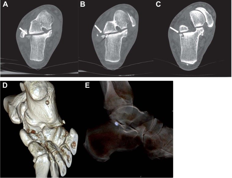
Noncontrast computed tomography remains the gold standard for traumatic calcaneal injuries. CT scan is used for preoperative planning, classification of fracture severity, and in instances where the index of suspicion for a calcaneal fracture is high despite negative initial plain radiographs.
-
Mondors Sign is a hematoma identified on CT that extends along the sole and is considered pathognomic for calcaneal fracture.
-
Stress fractures such as those seen in runners would be best evaluated with a bone scan or MRI.
-
Bohler’s Angle may be depressed on plain radiographs. Defined as the angle between two lines drawn on plain film. The first line is between highest point on the tuberosity and the highest point of posterior facet and the second is the highest point on the anterior process and the highest point on the posterior facet. Normal angle is between 20-40 degrees.
-
The Critical Angle of Gissane may be increased. Defined as the angle between two lines drawn on plain film. The first along the anterior downward slope of the calcaneus and the second along the superior upward slope. A normal angle is 130-145 degrees.
-
Normal Bohlers and Gissane angles do not rule out a fracture.
-
Abnormalities of either of these findings should prompt a CT scan for further classification and evaluation of the fracture.
Calcaneal fractures can be classified into two general categories.[rx][rx]
-
Extraarticular fractures account for 25 % of calcaneal fractures. These typically are avulsion injuries of either the calcaneal tuberosity from the Achilles tendon, the anterior process from the bifurcate ligament, or the sustenaculum tali.
-
Intraarticular Fractures account for the remaining 75%. The talus acts as a hammer or wedge compressing the calcaneus at the angle of Gissane causing the fracture.
There are two main classification systems of extraarticular fractures.
Essex-Lopresti:
-
Joint depression type with a single verticle fracture line through the angle of Gissane separating the anterior and posterior portions of the calcaneus.
-
Tongue type which has the same verticle fracture line as a depression type with another horizontal fracture line running posteriorly, creating a superior posterior fragment.
Sanders Classification: Based on reconstituted CT findings.
-
Type I fractures: 1 nondisplaced or minimally displaced bony fragment
-
Type II fractures: 2 bony fragments involving the posterior facet. Subdivided into types A, B, and C depending on the medial or lateral location of the fracture line.
-
Type III fractures: 3 bony fragments including an additional depressed middle fragment. Subdivided into types AB, AC, and BC, depending on the position and location of the fracture lines.
-
Type IV fractures: 4 comminuted bony fragments.
Treatment of Calcaneus Fractures
Emergent treatment includes
-
Aggressive wound care and antibiotics as needed for contaminated wounds.
-
Analgesics.
-
ICE and elevation.
-
Immobilization with splinting.
-
All patients who are candidates for outpatient treatment are nonweight bearing at discharge.
Open fractures require more urgent surgical treatment and wound care.
Closed fracture reduction can be delayed.
-
All surgical treatment is aimed at restoration of heel height and width (i.e., reconstructing the anatomy to reapproximate Bohler and Gissane angles), repair and realignment of the subtalar joint, and returning the mechanical axis of the hindfoot to functionality.
-
Most extraarticular fractures are treated conservatively with 10-12 weeks of casting.
-
Calcaneal tuberosity avulsion, displaced sustenaculum tali, and large substantial calcaneal body fractures may require operative management.
-
Some intraarticular injuries may be treated in a closed fashion depending upon severity. Many are treated with either open surgical reduction and internal fixation, percutaneous pinning, or sometimes arthrodesis.
-
Nondisplaced Sanders type I fractures may be treated in a conservative, closed fashion.
About the author