Category Archive Health A – Z

By

Treatment of Osteoarthritis of Knee Exercise, Complications

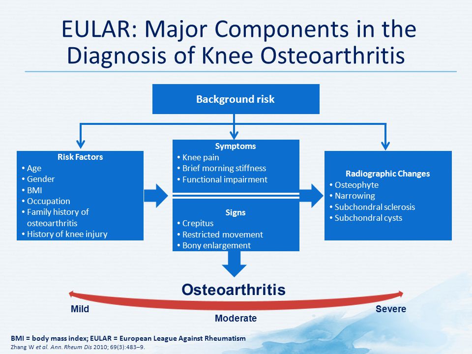
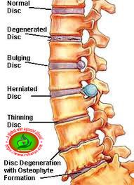
Treatment of Osteoarthritis of Knee (OA) a common disease of an aged population and one of the leading causes of disability. The incidence of knee OA is rising by increasing average age of the general population. Age, weight, trauma to joint due to repetitive movements, in particular, squatting and kneeling are common risk factors of knee OA. Several factors including cytokines, leptin, and mechanical forces are pathogenic factors of knee OA. In patients with knee pain attribution of pain to knee OA should be considered with caution. Since a proportion of knee OA is asymptomatic and in a number of patients identification of knee OA is not possible due to the low sensitivity of radiographic examination. In this review data presented in regard to prevalence, pathogenesis, risk factors.

Treatment of Osteoarthritis (OA) of Knee

Treatment for knee osteoarthritis can be broken down into non-surgical and surgical management. Initial treatment begins with non-surgical modalities and moves to surgical treatment once the non-surgical methods are no longer effective. A wide range of non-surgical modalities is available for the treatment of knee osteoarthritis. These interventions do not alter the underlying disease process, but they may substantially diminish pain and disability.

The non-pharmacological approach includes

  • Education –Encourage patients to participate in self-management programmes (such as those conducted by the Arthritis Foundation in the United States and Arthritis Care in the United Kingdom), and provide resources for social support and instruction on coping skills.
  • Weight loss – Encourage overweight patients with osteoarthritis of the hip and knee to lose weight through a combination of diet and exercise.
  • Exercise  – increases aerobic capacity, muscle strength, and endurance and also facilitates weight loss. All people capable of exercise should be encouraged to take part in a low impact aerobic exercise programme (walking, cycling, or swimming or another aquatic exercise). Exercises to strengthen the quadriceps lead to reductions in pain and improvements in function.
  • Physical therapy –  consists of several strategies to facilitate the resolution of symptoms and improve functional deficits, including a range of motion exercise, muscle strengthening, muscle stretching, and soft tissue mobilization.
  • Knee braces and orthotics – For those with the instability of the knee and varus misalignment, valgus bracing and orthotics shift the load away from the medial compartment and, in doing so, may provide relief of pain and improvement in function. Though some research has shown that heel wedges can reduce medial compartment loads, there is no evidence that, used alone, they improve knee symptoms. Appropriate supportive footwear should be worn by people who have osteoarthritis of the knee and hip.
  • Activity modification
  • Weight loss
  • Knee Bracing

The first-line treatment for all patients with symptomatic knee osteoarthritis includes patient education and physical therapy. A combination of supervised exercises and a home exercise program have been shown to have the best results. These benefits are lost after 6 months if the exercises are stopped. The American Academy of Orthopedic Surgeons (AAOS) recommends this treatment.

Treatment of Osteoarthritis of Knee

www.rxharun.com

Medicine of Osteoarthritis (OA) of Knee

The goal of treatment is to decrease pain and increase mobility.

  • Analgesics Paracetamol (up to 4 g/day) is the oral analgesic of choice for mild to moderate pain in osteoarthritis. NSAIDs should be added or substituted in patients who respond inadequately and are sometimes the first choice because of greater efficacy and patients’ preference. There are, however, certain disadvantages of routinely using NSAIDs—for example, all NSAIDs (non-selective and COX 2 selective) are associated with potential toxicity, particularly in elderly people. COX 2 selective inhibitors have also been associated with an increased risk for cardiovascular disease. Rofecoxib, a COX 2 selective inhibitor, was recently withdrawn because of such concerns. In people with an increased gastrointestinal risk, nonselective NSAIDs plus a gastroprotective agent or a selective COX 2 inhibitor should be used. Opioid analgesics are useful alternatives in patients in whom NSAIDs are contraindicated, ineffective, or poorly tolerated. Topical formulations of NSAIDs and capsaicin may be helpful.
  • Antidepressants –  A Drugs that block pain messages from your brain and boost the effects of endorphins (your body’s natural painkillers).
  • Nonsteroidal anti-inflammatory drugs (NSAIDs) Common pain remedies such as aspirin, acetaminophen, ibuprofen, and naproxen can offer short-term relief. All are available in low doses without a prescription. Other medications, including muscle relaxants and anti-seizure medications, treat aspects such as muscle spasms and damaged nerves.
  • Corticosteroid injections – Your doctor will inject a steroid such as prednisone into your thumb joints. Steroids make inflammation go down. However, because of side effects, they are used sparingly
  • Anesthetics – Used with precision, an injection of a “nerve block” can stop the pain for a time.
  • Muscle Relaxants – These medications provide relief from spinal muscle spasms.
  • Neuropathic Agents –  Drugs(pregabalin & gabapentin) that address neuropathic—or nerve-related—pain. This includes burning, numbness, and tingling.
  • Opioids  – Also known as narcotics, these medications are intense pain relievers that should only be used under a doctor’s careful supervision.
  • Topical Medications: These prescription-strength creams, gels, ointments, patches, and sprays help relieve pain and inflammation through the skin.
  • Calcium & vitamin D3 – to improve bones health and healing fracture.
  • Glucosamaine & diacerine – can be used to tightening the loose tension and regenerate cartilage or inhabit the further degeneration of cartilage.
  • Muscle relaxants – such as cyclobenzaprine might be prescribed to relieve the discomfort associated with muscle spasms. However, these medicines might cause confusion in older people. Depending on the level of pain, prescription pain medicines might be used in the initial period of treatment.
  • Over-the-counter – non-narcotic pain relievers and anti-inflammatory medications are usually the first choices of therapy for arthritis of the knee. Acetaminophen is a simple, over-the-counter pain reliever that can be effective in reducing arthritis pain that is following
  • Glucosamine compounds –  in particular, have attracted a great deal of attention, mostly in the lay press. Possibly as a function of this publicity, osteoarthritis is the leading medical condition for which people use alternative therapies.w6 Glucosamine and chondroitin seem to have the same benefit as placebo,and there is controversy over whether they also have structure modifying benefits.
  • COX-2 inhibitors
  • Glucosamine and chondroitin sulfate
  • Corticosteroid injections
  • Hyaluronic acid (HA)

Intra-articular Knee Injection Treatments

  • Viscosupplementation with hyaluronic acid (HA) – Injection into the knee with HA (similar to the main component in cartilage) has been reported to provide temporary pain relief for up to three months. Evidence to date on the use of HA has been contradictory, and recommendations regarding its use remain inconclusive[, ]Currently, the American Academy of Orthopaedic Surgeons does not recommend using hyaluronic acid for patients with symptomatic end-stage osteoarthritis (OA) of the knee. There are no existing data that any of the HA injections will cause regression of osteophytes, subchondral bone remodeling, or regeneration of cartilage and meniscus in patients with substantial, irreversible bone and cartilage damage. Further investigations are required to determine whether high-molecular-weight and cross-linked preparations of HA have superior efficacy compared with other HA preparations or other currently available treatments. In addition, studies involving long-term outcomes of efficacy, safety, and economic cost-benefit analyses are needed. Because of the paucity of data supporting the effectiveness of HA injections to justify their cost, careful patient selection and decreasing the use of HA among patients with end-stage knee OA may represent a substantial cost reduction without negatively affecting the quality of health care.
  • Intra-articular corticosteroids – Intra-articular injections of corticosteroids have long been used to try to relieve symptoms from knee OA, but studies addressing their efficacy have been contradictory. The American Academy of Orthopaedic Surgeons guidelines for nonoperative treatment options for patients with OA of the knee does not recommend for or against the use of intra-articular corticosteroids into the knee. Furthermore, a recent Cochrane systematic review concluded that clinically important benefits of one to six weeks remain unclear because of the overall quality of the studies, the heterogeneity between trials, and the presence of small-study effects.
  • Intra-articular hyaluronan – Though the meta-analyses reviewing the efficacy of intra-articular hyaluronan are not in complete agreement, mainly because of variation in study methods, most suggest that the effect size is relatively small and the placebo response is significant.
Biologics: Biologic injections include cell-based therapies and platelet-rich plasma (PRP).
  • Cell-based therapies – Cell-based therapies for knee OA are in development stages. A recent systematic review suggested that intra-articular cellular injections for OA and focal cartilage defects in the human knee had positive results and seemed safe. However, improvement in patient symptoms was modest and a placebo effect could not be disregarded. The overall quality of the literature was poor; therefore, accurate assessment and optimization of these therapies will require further research. Most of the studies analyzed reported on the use of autologous cellular therapies. Bone marrow-derived cells were the source chosen more often, followed by adipose-derived cells and blood stem cells.
  • Platelet-rich plasma – Multiple studies and systematic reviews have reported on the use of intra-articular PRP for the treatment of knee OA. Initial observations support an inference that PRP appears to be safe. Although some transient pain or swelling has been reported after its use, these symptoms typically resolve within two to three days, and no long-term side effects have been reported. Use of PRP, especially a lower leukocyte concentration known as leukocyte-poor PRP, showed improved results compared with HA and placebo, showing beneficial effects of amelioration in pain and improvement in function about two months after application and lasting up to a year. On the basis of the current evidence, although PRP injections have been demonstrated to more effectively reduce pain and improve overall physical function compared with control studies, the quality of evidence is lacking, and further research is required to establish the efficacy of using PRP as a treatment option.
  • Glucose Amaine with  Chondroitin Sulphate – Glucosamine and chondroitin have been widely promoted as a treatment for OA. Glucosamine, an amino sugar, is thought to promote the formation and repair of cartilage. Chondroitin, a carbohydrate, is a cartilage component that is thought to promote water retention and elasticity and to inhibit the enzymes that break down cartilage. Both compounds are manufactured by the body. Glucosamine supplements are derived from shellfish shells; chondroitin supplements are generally made from cow cartilage.
  • Duloxetine – This antidepressant, which was evaluated for OA for the first time in these guidelines, may help with chronic pain. However, side effects like nausea, dry mouth, fatigue, constipation, and increased sweating may keep some people from taking it. Duloxetine is appropriate for people with OA of the knee and other joints. Whether people with knee-only OA and coexisting health conditions should take it is uncertain.
  • Capsaicin  – This analgesic, which is derived from chili peppers, is better than a placebo at reducing pain, but it can cause side effects such as a burning sensation or skin redness. It’s appropriate for knee-only OA in people without existing health conditions, but uncertain in people with multi-joint OA and those with health conditions.
  • Opioid and narcotic analgesics – A review of 18 randomized controlled trials showed a significant reduction in pain and some improvement in physical function with the use of these strong pain relievers. However, these benefits were countered by significant side effects, including nausea, constipation, dizziness, sleepiness, and vomiting. Overall, about a quarter of patients treated with opioids—and particularly with strong drugs like oxycodone and oxymorphone—dropped out of studies because of side effects. The recommendation on both oral and patch forms of opioid pain relievers is “uncertain.”
  • Corticosteroid injections  – Injecting corticosteroid compounds directly into affected joints can be useful for decreasing pain in the short term, although the effect tends to wear off after a few weeks. To maintain relief, you may need to have corticosteroid injections in the same joint every few months or use another treatment.
  • Hyaluronic acid injections   – Hyaluronic acid is meant to supplement a natural substance that gives joint fluid its viscosity. Most of the studies conducted have been in patients with knee OA. Although the study outcomes differ, some do find the treatment relieves pain. Hyaluronic acid injections seem to reach their peak benefit 8 weeks after the shot is given, and the effect lasts for about 24 weeks. Side effects were minimal, including temporary pain and swelling at the injection site. Because of inconsistent study results, hyaluronic acid injections are “not appropriate” for people with OA of the hip and other joints, and “uncertain” for those with knee-only OA.
  • Risedronate (Actonel)  – This bisphosphonate drug is better known for treating osteoporosis, but it may also reduce cartilage degeneration. More studies are needed to determine whether risedronate helps with symptoms, function, or OA disease progression. For now, the drug is deemed “not appropriate.”
  • Bracing – Knee braces are available for treatment of medial compartmental osteoarthritis (arthritis on the inside of the knee joint). These braces work by unloading the medial (inside) portion of the knee. These braces need to be custom made and therefore can be expensive.
  • Duloxetine  – Normally used as an antidepressant, this medication is also approved to treat chronic pain, including osteoarthritis pain.
  • Disease-modifying anti-rheumatic drugs (DMARDs) – are used to slow the progression of rheumatoid arthritis. Drugs like methotrexate, sulfasalazine, and hydroxychloroquine are commonly prescribed. In addition, biologic DMARDs like etanercept and adalimumab may reduce the body’s overactive immune response. Because there are many different drugs today for rheumatoid arthritis, a rheumatology specialist is often required to effectively manage medications.
  • Glucosamine and chondroitin sulfate substances found naturally in joint cartilage, can be taken as dietary supplements. Although patient reports indicate that these supplements may relieve pain, there is no evidence to support the use of glucosamine and chondroitin sulfate to decrease or reverse the progression of arthritis.
  • Avocado-soybean unsaponifiables – This nutritional supplement — a mixture of avocado and soybean oils — is widely used in Europe to treat knee and hip osteoarthritis. It acts as an anti-inflammatory, and some studies have shown it may slow down or even prevent joint damage.
  • Alternative therapies – Many alternative forms of therapy are unproven but may be helpful to try, provided you find a qualified practitioner and keep your doctor informed of your decision. Alternative therapies to treat pain include the use of acupuncture and magnetic pulse therapy.

Strengthening Exercises

Knee extension

Straightening the knee against resistance will strengthen the quadriceps muscles at the front of the thigh. This can be achieved in a number of ways, including using ankle weights and resistance machines. One of the easiest ways involves using a resistance band

  • The patient sits on a chair with one end of the band tied around their ankle and the other end around one of the chair legs.
  • The band should be taut when the knee is bent at 90 degrees. The patient straightens the knee before slowly bending it and returning the foot back to the floor.
  • Perform 10-15 repetitions and repeat this 2-3 times.

Knee flexion

Still using the resistance band:

  • The patient lies on the floor with the band tied around one ankle and the other end attached to something sturdy, close to the floor. The band should be taught when the knee is straight
  • The patient bends the knee to bring the heel towards the buttock as far as possible, and then slowly straightens the knee back again.
  • Perform 10-15 repetitions and repeat this 2-3 times.

Calf raise

Calf raises are good for strengthening the Gastrocnemius muscle which is one of the two main calf muscles, but the only one which crosses the knee joint.

  • The patient stands with the feet should width apart and close to a wall or chair which can be held for balance if required.
  • The patient rises up on to the toes, keeping the knees straight, before slowly lowering the heels back to the floor.
  • Perform 10-15 repetitions and repeat this 2-3 times.
  • This exercise can be progressed by performing on one leg only.

Squats

Squats are really good exercises for strengthening all the main muscle groups of the legs and buttocks. They can start off as very shallow movements and progress until the knees reach a 90 degree angle at which point weights can be added.

  • The patient stands with the feet shoulder width apart and back straight.
  • The knees are then bent as if trying to sit on a chair.
  • The back should remain straight and the knees should not move forwards past the toes.
  • Perform 10-15 repetitions and repeat this 2-3 times.

Acupuncture – uses fine needles to stimulate specific body areas to relieve pain or temporarily numb an area. Although it is used in many parts of the world and evidence suggests that it can help ease the pain of arthritis, there are few scientific studies of its effectiveness. Be sure your acupuncturist is certified, and do not hesitate to ask about his or her sterilization practices.

Magnetic pulse therapy – is painless and works by applying a pulsed signal to the knee, which is placed in an electromagnetic field. Like many alternative therapies, magnetic pulse therapy has yet to be proven.

Nonsurgical Treatment

As with other arthritic conditions, initial treatment of arthritis of the knee is nonsurgical. Your doctor may recommend a range of treatment options.

Lifestyle modificationsSome changes in your daily life can protect your knee joint and slow the progress of arthritis.

  • Minimize activities that aggravate the condition, such as climbing stairs.
  • Switching from high impact activities (like jogging or tennis) to lower impact activities (like swimming or cycling) will put less stress on your knee.
  • Losing weight can reduce stress on the knee joint, resulting in less pain and increased function.
  • Physical therapy Specific exercises can help increase the range of motion and flexibility, as well as help strengthen the muscles in your leg. Your doctor or a physical therapist can help develop an individualized exercise program that meets your needs and lifestyle.
  • Assistive devices Using devices such as a cane, wearing shock-absorbing shoes or inserts, or wearing a brace or knee sleeve can be helpful. A brace assists with stability and function and may be especially helpful if the arthritis is centered on one side of the knee. There are two types of braces that are often used for knee arthritis: An “unloader” brace shifts weight away from the affected portion of the knee, while a “support” brace helps support the entire knee load.
  • Occupational therapy An occupational therapist can help you discover ways to do everyday tasks or do your job without putting extra stress on your already painful joint. For instance, a toothbrush with a large grip could make brushing your teeth easier if you have finger osteoarthritis. A bench in your shower could help relieve the pain of standing if you have knee osteoarthritis.
  • Other remedies – Applying heat or ice, using pain-relieving ointments or creams, or wearing elastic bandages to provide support to the knee may provide some relief from pain.
  • Aerobic exercise – programs may make OA patients feel better, help reduces the joint pain, and make it easier for them to perform daily tasks. Exercise programs under medical supervision should be balanced with rest and joint care.[] Aerobic programs truly border both clinical (rehabilitation) and home programs. Regardless of the setting, this program type was found to be effective for reducing pain in the hip and knee.[] Patients are typically recommended to exercise between 50% and 70% target heart rate for a minimum of 30 min, 3 times a week, for overall weight management, health benefits, and a reduction in pain which was noted after a 6-month program.[
  • Hydrotherapy (balneotherapy) – involves the use of water in any form or at any temperature (steam, liquid, ice) for the purpose of healing. In aquatic physical therapy or hydrotherapy, exercise activities are carried out in heated pools by a variety of providers.[] Hydrotherapy/balneotherapy and aquatic therapy displayed positive results when conducted for testing a subject’s strength and flexibility. The results emphasized the role of these therapies in aiding normal walking and relieving joint pain.[] The sessions typically are run from 6 to 48 weeks for the duration of 60 min and are conducted in a shallow pool with water temperatures ranging from 29°C to 34°C.[]

Surgical Treatment

Your doctor may recommend surgery if your pain from arthritis causes disability and is not relieved with nonsurgical treatment. As with all surgeries, there are some risks and possible complications with different knee procedures. Your doctor will discuss the possible complications with you before your operation.

Arthroscopy – During arthroscopy, doctors use small incisions and thin instruments to diagnose and treat joint problems. Arthroscopic surgery is not often used to treat arthritis of the knee. In cases where osteoarthritis is accompanied by a degenerative meniscal tear, arthroscopic surgery may be recommended to treat the torn meniscus.

Cartilage grafting – Normal, healthy cartilage tissue may be taken from another part of the knee or from a tissue bank to fill a hole in the articular cartilage. This procedure is typically considered only for younger patients who have small areas of cartilage damage.

Synovectomy – The joint lining damaged by rheumatoid arthritis is removed to reduce pain and swelling.

Osteotomy – In a knee osteotomy, either the tibia (shinbone) or femur (thighbone) is cut and then reshaped to relieve pressure on the knee joint. Knee osteotomy is used when you have early-stage osteoarthritis that has damaged just one side of the knee joint. By shifting your weight off the damaged side of the joint, an osteotomy can relieve pain and significantly improve function in your arthritic knee.

Total or partial knee replacement (arthroplasty) Your doctor will remove the damaged cartilage and bone, and then position new metal or plastic joint surfaces to restore the function of your knee.

(Left) A partial knee replacement is an option when damage is limited to just one part of the knee. (Right) A total knee replacement prosthesis.

Surgical Treatment Options

  • Osteotomy
  • Unicompartmental knee arthroplasty (UKA)
  • Total knee arthroplasty (TKA)

A high tibial osteotomy (HTO) may be indicated for unicompartmental knee osteoarthritis associated with malalignment. Typically an HTO is done for varus deformities where the medial compartment of the knee is worn and arthritic. The ideal patient for an HTO would be a young, active patient in whom arthroplasty would fail due to excessive component wear. An HTO preserves the actual knee joint, including the cruciate ligaments, and allows the patient to return to high-impact activities once healed. It does require additional healing time compared to an arthroplasty, is more prone to complications, depends on bone and fracture healing, is less reliable for pain relief, and ultimately does not replace cartilage that is already lost or repair any remaining cartilage. An osteotomy will delay the need for an arthroplasty for up to 10 years.

Indications for HTO

  • Young (less than 50 years old), active patient
  • Healthy patient with good vascular status
  • Non-obese patients
  • Pain and disability interfering with daily life
  • Only one knee compartment is affected
  • Compliant patient who will be able to follow postoperative protocol

Contraindications for HTO

  • Inflammatory arthritis
  • Obese patients
  • Knee flexion contracture greater than 15 degrees
  • Knee flexion less than 90 degrees
  • If the procedure will need greater than 20 degrees of deformity correction
  • Patellofemoral arthritis
  • Ligamentous instability

A UKA also is indicated in unicompartmental knee osteoarthritis. It is an alternative to an HTO and a TKA. It is indicated for older patients, typically 60 years or older, and relatively thin patients; although, with newer surgical techniques the indications are being pushed.

Indications for UKA

  • Older (60 years or older), lower demand patients
  • Relatively thin patients

Contraindications for UKA

  • Inflammatory arthritis
  • ACL deficiency
  • Fixed varus deformity greater than 10 degrees
  • Fixed valgus deformity greater than 5 degrees
  • Arc of motion less than 90 degrees
  • Flexion contracture greater than 10 degrees
  • Arthritis in more than one compartment
  • Younger, higher activity patients or heavy laborers
  • Patellofemoral arthritis

A TKA is the surgical treatment option for patients failing conservative management and those with osteoarthritis in more than one compartment. It is regarded as a valuable intervention for patients who have severe daily pain along with radiographic evidence of knee osteoarthritis.

Indications for TKA

  • Symptomatic knee OA in more than one compartment
  • Failed non-surgical treatment options

Contraindications for TKA

Absolute

  • Active or latent knee infection
  • Presence of active infection elsewhere in the body
  • Incompetent quadriceps muscle or extensor mechanism

Relative

  • Neuropathic arthropathy
  • Poor soft tissue coverage
  • Morbid obesity
  • Noncompliance due to major psychiatric disorder or alcohol or drug abuse
  • Insufficient bone stock for reconstruction
  • Poor health or presence of comorbidities that make the patient an unsuitable candidate for major surgery and anesthesia
  • Patient’s poor motivation or unrealistic expectations
  • Severe peripheral vascular disease

Advantages of UKA vs TKA

  • Faster rehabilitation and quicker recovery
  • Less blood loss
  • Less morbidity
  • Less expensive
  • Preservation of normal kinematics
  • Smaller incision
  • Less postsurgical pain and shorter hospital stay

Advantages of UKA vs HTO

  • Faster rehabilitation and quicker recovery
  • Improved cosmesis
  • Higher initial success rate
  • Fewer short-term complications
  • Lasts longer
  • Easier to convert to TKA

Natural Relief From Arthritis Pain 

Arthritis Pain

Osteoarthritis of Knee

Arthritis is a painful and degenerative condition marked by inflammation in the joints that causes stiffness and pain. Osteoarthritis, the most common type of arthritis, gets worse with age and is caused by wear and tear over the years.

Doctors traditionally treat arthritis with anti-inflammatory medications and painkillers. However, some medications cause side effects, and a natural approach to pain relief is becoming more popular. Remember to consult your doctor before trying these natural remedies.

Connect With Others Who Have Arthritis
“You do feel as if you are on your own, but with being part of the group you know you are not and it is very helpful to get thoughts and ideas from others who are suffering the same pain as you. “

Lose Weight

Your weight can make a big impact on the amount of pain you experience from arthritis.

Extra weight puts more pressure on your joints—, especially your knees, hips, and feet. Reducing the stress on your joints by losing weight will improve your mobility, decrease pain, and prevent future damage to your joints.

Get More Exercise

There are more benefits to exercise than just weight loss. Regular movement helps to maintain flexibility in your joints. Weight-bearing exercises like running and walking can be damaging. Instead, try low-impact exercises like water aerobics or swimming to flex your joints without adding further stress.

Use Hot and Cold Therapy

Simple hot and cold treatments can make a world of difference when it comes to arthritis pain. Long, warm showers or baths—especially in the morning—help ease stiffness in your joints. Use an electric blanket or heating pad at night to keep your joints loose and use moist heating pads.

Cold treatments are best for relieving joint pain. Wrap a gel ice pack or a bag of frozen vegetables in a towel and apply it to painful joints for quick relief

Use Meditation to Cope With Pain

Meditation and relaxation techniques may be able to help you reduce pain from arthritis by reducing stress and enabling you to cope with it better. According to the National Institutes of Health (NIH), studies have found that the practice of mindfulness meditation is helpful for some people with painful joints. Researchers also found that those with depression and arthritis benefitted the most from meditation.

Include the Right Fatty Acids in Your Diet

Everyone needs omega-3 fatty acids in their diets for optimum health. However, these fats may also help your arthritis. Fish oil supplements, which are high in omega-3s, may help reduce joint stiffness and pain.

Another fatty acid that can help is gamma-linolenic acid or GLA. It’s found in the seeds of certain plants like evening primrose, borage, hemp, and black currants. You can also buy the oils of the seeds as a supplement. However, be sure to check with your doctor before taking them.

Turmeric to Dishes

Turmeric, the yellow spice common in Indian dishes, contains a chemical called curcumin that may be able to reduce arthritis pain. The secret is its anti-inflammatory properties.

The NIH reports that turmeric given to lab rats reduced inflammation in their joints. Research on humans is scarce, but it can’t hurt to add this tasty spice to your dinners.

Massage

According to the Arthritis Foundation, regular massaging of arthritic joints can help reduce pain and stiffness and improve your range of motion. Work with a physical therapist to learn self-massage, or schedule appointments with a massage therapist regularly

Your massage therapist should be experienced with working on people who have arthritis. Check with your doctor for a recommendation.

Consider Herbal Supplements

There are many kinds of herbal supplements on the market that claim to be able to reduce joint pain. Some of the herbs touted for arthritis pain include boswellia, bromelain, devil’s claw, ginkgo, stinging nettle, and thunder god vine.

Always talk to your doctor before trying a new supplement to avoid side effects and dangerous drug interactions.

Complications

Complications associated with non-surgical treatment are largely associated with NSAID use.

Common Adverse Effects of NSAID Use

  • Stomach pain and heartburn
  • Stomach ulcers
  • A tendency to bleed, especially while taking aspirin
  • Kidney problems

Common Adverse Effects of Intra-Articular Corticosteroid Injection

  • Pain and swelling (cortisone flare)
  • Skin discoloration at the site of injection
  • Elevated blood sugar
  • Infection
  • Allergic reaction

Common Adverse Effects of Intra-Articular HA Injection

  • Injection site pain
  • Muscle pain
  • Trouble walking
  • Fever
  • Chills
  • A headache

Complications Associated with HTO

  • Recurrence of deformity
  • Loss of posterior tibial slope
  • Patella Baja
  • Compartment syndrome
  • Peroneal nerve palsy
  • Malunion or nonunion
  • Infection
  • Persistent pain
  • Blood clot

Complications Associated with UKA

  • Stress fracture of the tibia
  • Tibial component collapse
  • Infection
  • Osteolysis
  • Persistent pain
  • Neurovascular injury
  • Blood clot

Complications Associated with TKA

  • Infection
  • Instability
  • Osteolysis
  • Neurovascular injury
  • Fracture
  • Extensor mechanism rupture
  • Patellar maltracking
  • Patellar clunk syndrome
  • Stiffness
  • Peroneal nerve palsy
  • Wound complications
  • Heterotopic ossification
  • Blood clot

References

Treatment of Osteoarthritis of Knee

ByRx Harun

Osteoarthritis of Knee – Symptoms, Diagnosis, Treatment

Osteoarthritis of Knee (OA) which is also known as osteoarthrosis or degenerative joint disease (DJD), is a progressive disorder of the joints caused by gradual loss of cartilage and resulting in the development of bony spurs and cysts at the margins of the joints. The name osteoarthritis comes from three Greek words meaning bone, joint, and inflammation.

Osteoarthritis is a multifactorial process in which mechanical factors have a central role and is characterized by changes in structure and function of the whole joint. There is no cure, and current therapeutic strategies are primarily aimed at reducing pain and improving joint function. We searched Medline for relevant articles (1966 to January 2006) and the Cochrane library (to first quarter of 2006) and consulted experts in rheumatology to produce a narrative review with an update on management for primary care doctors. We concentrated on osteoarthritis of the knee as this is associated with the greatest public health burden.

Osteoarthritis (OA) a common disease of an aged population and one of the leading causes of disability. The incidence of knee OA is rising by increasing average age of the general population. Age, weight, trauma to joint due to repetitive movements, in particular, squatting and kneeling are common risk factors of knee OA. Several factors including cytokines, leptin, and mechanical forces are pathogenic factors of knee OA. In patients with knee pain attribution of pain to knee OA should be considered with caution. Since a proportion of knee OA is asymptomatic and in a number of patients identification of knee OA is not possible due to the low sensitivity of radiographic examination. In this review data presented in regard to prevalence, pathogenesis, risk factors.


osteoarthritis of knee

Epidemiology /Etiology of Osteoarthritis of knee

Knee osteoarthritis is classified as either primary or secondary, depending on its cause. Primary knee osteoarthritis is the result of articular cartilage degeneration without any known reason. This is typically thought of as degeneration due to age as well as wear and tear. Secondary knee osteoarthritis is the result of articular cartilage degeneration due to a known reason.

Osteoarthritis is the most prevalent form of arthritis and occurs especially in the knee joint. It affects nearly 6% of all adults, but more women are affected than men.“According to a number of published reports, anywhere from 6% to over 13% of men, but between 7% and 19% of women, over 45 years of age are affected, resulting in a 45% less risk of incidence in men (Coleman, et al).

Possible Causes of Secondary Knee OA

  • Posttraumatic
  • Postsurgical
  • Congenital or malformation of the limb
  • Malposition (Varus/Valgus)
  • Scoliosis
  • Rickets
  • Hemochromatosis
  • Chondrocalcinosis
  • Ochronosis
  • Wilson disease
  • Gout
  • Pseudogout
  • Acromegaly
  • Avascular necrosis
  • Rheumatoid arthritis
  • Infectious arthritis
  • Psoriatic arthritis
  • Hemophilia
  • Paget disease
  • Sickle cell disease

Causes of Osteoarthritis (OA) of Knee

Articular cartilage increased the water content

  • alterations in proteoglycans eventual decrease in the number of proteoglycans
  • collagen abnormalities organization and orientation are lost
  • binding of proteoglycans to hyaluronic acid

Synovium and capsule

An early phase of OA

  • mild inflammatory changes in synovium

The middle phase of OA

  • moderate inflammatory changes of the synovium
  • synovium becomes hypervascular

Late phases of OA

  • synovium becomes increasingly thick and vascular

Bones

Subchondral bone attempts to remodel

  • forming lytic lesion with sclerotic edges (different than bone cysts in RA)
  • bone cysts form in late stages

Cell biology

Proteolytic enzymes matrix metalloproteases (MMPs)

Responsible for cartilage matrix digestion

  • examples
  •  stromelysin
  •  plasmin
  • aggrecanase-1 (ADAM-TS-4)

Tissue inhibitors of MMPS (TIMPs)

  • control MMP activity preventing excessive degradation
  • the imbalance between MMPs and TIMPs has been demonstrated in OA tissues

Inflammatory cytokines

Secreted by synoviocytes and increase MMP synthesis, examples

  •  IL-1
  •  IL-6
  • TNF-alpha

Genetics >Inheritance

  • in-mendelian

Genes potentially linked to OA

  • vitamin D receptor
  • estrogen receptor 1
  • inflammatory cytokines
  •  IL-1
  • leads to the catabolic effect
  •  IL-4
  •  matrilin-3
  • BMP-2, BMP-5

Risk Factor

Age is not the only factor that plays a role in the evolution of OA. Other risk factors are

  • Obesity
  • Joint hypermobility or instability
  • Sports stress with high impact loading
  • Repetitive knee bending or heavy weight lifting
  • Specific occupations
  • Peripheral neuropathy
  • Injury to the joint
  • History of immobilization
  • Family history

Modifiable

  • Articular trauma
  • Occupation – prolonged standing and repetitive knee bending
  • Muscle weakness or imbalance
  • Weight
  • Health – metabolic syndrome

Non-modifiable

  • Gender – females more common than males
  • Age
  • Genetics
  • Race
 How does a normal knee work?

Your knee joint is where your thigh bone (femur) and your shin bone (tibia) meet. It allows the bones to move freely but within limits.

Your knee is the largest joint in the body and also one of the most complicated. It needs to be strong enough to take our weight and must lock into position so we can stand upright. But it also has to act as a hinge so we can walk, and it must withstand extreme stresses, twists, and turns, such as when we run or play sports.

Osteoarthritis formation

When your knee has osteoarthritis its surfaces become damaged and it doesn’t move as well as it should do. The following happens:

  • The cartilage becomes rough and thin – this can happen over the main surface of your knee joint and in the cartilage underneath your kneecap.
  • The bone underneath the cartilage reacts by growing thicker and becoming broader.
  • All the tissues in your joint become more active than normal as if your body is trying to repair the damage.
  • The bone at the edge of your joint grows outwards, forming bony spurs called osteophytes.
  • The synovium may swell and produce extra fluid, causing the joint to swell – this is called an effusion or sometimes water on the knee.
  • The capsule and ligaments slowly thicken and contract.

These changes in and around your joint are partly the result of the inflammatory process and partly an attempt by your body to repair the damage. In many cases, your body’s repairs are quite successful and the changes inside your joint won’t cause much pain or, if there is the pain, it’ll be mild and may come and go.

However, in other cases, the repair doesn’t work as well and your knee is damaged. This leads to instability and more weight being put onto other parts of the joint. This can cause symptoms to become gradually worse and more persistent over time.

Risk factors

osteoarthritis of knee -follow chart

Risk Factors can be Classified into  Primary & secondary

Primary

A number of studies have shown that there is a greater prevalence of the disease among siblings and especially identical twins, indicating a hereditary basis. Although a single factor is not generally sufficient to cause the disease, about half of the variation in susceptibility has been assigned to genetic factors

As early human ancestors evolved into bipeds, changes occurred in the pelvis, hip joint, and spine which increased the risk of osteoarthritis. Additionally, genetic variations that increase the risk were likely not selected against because usually problems only occur after reproductive success.

The development of osteoarthritis is correlated with a history of previous joint injury and with obesity, especially with respect to knees. Since the correlation with obesity has been observed not only for knees but also for non-weight bearing joints and the loss of body fat is more closely related to symptom relief than the loss of body weight, it has been suggested that there may be a metabolic link to body fat as opposed to just mechanical loading.

Changes in sex hormone levels may play a role in the development of osteoarthritis as it is more prevalent among post-menopausal women than among men of the same age. A study of mice found natural female hormones to be protective while injections of the male hormone dihydrotestosterone reduced protection.

Secondary

Secondary osteoarthritis (due to an old injury with fracture) of the ankle in a woman of 82 years old

This type of osteoarthritis is caused by other factors but the resulting pathology is the same as for primary osteoarthritis:

  • Older age. The risk of osteoarthritis increases with age.
  • Sex. Women are more likely to develop osteoarthritis, though it isn’t clear why.
  • Obesity. Carrying extra body weight contributes to osteoarthritis in several ways, and the more you weigh, the greater your risk. Increased weight puts added stress on weight-bearing joints, such as your hips and knees. In addition, fat tissue produces proteins that may cause harmful inflammation in and around your joints.
  • Joint injuries. Injuries, such as those that occur when playing sports or from an accident, may increase the risk of osteoarthritis. Even injuries that occurred many years ago and seemingly healed can increase your risk of osteoarthritis.
  • Certain occupations. If your job includes tasks that place repetitive stress on a particular joint, that joint may eventually develop osteoarthritis.
  • Genetics. Some people inherit a tendency to develop osteoarthritis.
  • Bone deformities. Some people are born with malformed joints or defective cartilage, which can increase the risk of osteoarthritis.
  • Alkaptonuria
  • Congenital disorders of joints
  • Diabetes doubles the risk of having a joint replacement due to osteoarthritis and people with diabetes have joint replacements at a younger age than those without diabetes.
  • Ehlers-Danlos Syndrome
  • Hemochromatosis and Wilson’s disease
  • Inflammatory diseases (such as Perthes’ disease), (Lyme disease), and all chronic forms of arthritis (e.g., costochondritis, gout, and rheumatoid arthritis). In gout, uric acid crystals cause the cartilage to degenerate at a faster pace.
  • Injury to joints or ligaments (such as the ACL), as a result of an accident or orthopedic operations.
  • Ligamentous deterioration or instability may be a factor.
  • Marfan syndrome
  • Joint infection

The symptom of Osteoarthritis (OA) of Knee

The main symptoms of osteoarthritis of the knee are:

  • Pain (particularly when you’re moving your knee or at the end of the day – this usually gets better when you rest)
  • Stiffness (especially after rest – this usually eases after a minute or so as you get moving)
  • Crepitus, a creaking, crunching, grinding sensation when you move the joint
  • Hard swellings (caused by osteophytes)
  • Soft swellings (caused by extra fluid in the joint).
  • Loss of flexibility – You may not be able to move your joint through its full range of motion.
  • Grating sensation – You may hear or feel a grating sensation when you use the joint.
  • Bone spurs – These extra bits of bone, which feel like hard lumps, may form around the affected joint.

Other symptoms can include:

  • your knee giving way because your muscles have become weak or the joint structure is less stable
  • your knee not moving as freely or as far as normal
  • your knees becoming bent and bowed
  • the muscles around your joint looking thin or wasted.

It’s unusual, but some people have pain in their knee that wakes them up at night. This generally only happens with severe osteoarthritis.

You’ll probably find that your pain will vary and that you have good days and bad days, sometimes depending on how active you’ve been but sometimes for no clear reason.

Some people find that changes in the weather (especially damp weather and low pressure) make their pain and stiffness worse. This may be because nerve fibers in the capsule of their knee are sensitive to changes in atmospheric pressure.

Diagnosis of Osteoarthritis (OA) of Knee

Physical Examination

osteoarthritis of knee -diagnosis

During the physical examination, your doctor will look for:

Grading Knee

osteoarthritis of knee -diagnosis/Grade+of+Osteoathritis

For the grading of osteoarthritis in the knee, the most reliable systems are the International Knee Documentation Committee (IKDC) system and the Ahlbäck system, which have been found to be superior to for example systems by Kellgren-Lawrence, Fairbank, Brandt, and Jäger-Wirth. These findings refer to posteroanterior weight-bearing projection radiographs made with the knee in 45° of flexion.

Ahlbäck classification
Grade Findings
I Joint space narrowing, with or without subchondral sclerosis. Joint space narrowing is defined by this system as a joint space less than 3 mm, or less than half of the space in the other compartment, or less than half of the space of the homologous compartment of the other knee.
II Obliteration of the joint space
III Bone defect/loss <5 mm
IV Bone defect/loss between 5 and 10 mm
V Bone defect/loss >10 mm, often with subluxation and arthritis of the other compartment

For the patellofemoral joint, a classification by Merchant 1974 uses a 45° “skyline” view of the patella

Stage Description
1 (mild) Patellofemoral joint space > 3mm
2 (moderate Joint space < 3 mm but no bony contact
3 (severe) Bony surfaces in contact over less than one-quarter of the joint surface
4 (very severe) Bony contact throughout the entire joint surface

Goldberg recommends the following as a common approach to the examination of all joints

  • Make sure the area is well exposed—no clothing covering either side. Patient gowns come in handy
  • Carefully inspect the joint or joints in question. Are there signs of inflammation or injury (swelling, redness, warmth)? Deformity? Because many joints are symmetrical, compare it with the opposite side
  • Understand normal functional anatomy. What does this joint normally do?
  • Observe the joint while the patient attempts to perform the normal activity. What can’t the patient do? What specifically limits him/her? Was there a discrete event (eg, trauma) that caused this? If so, what was the mechanism of injury?
  • Palpate the joint in question. Is there warmth? Point tenderness? If so, over what anatomic structures?
  • Assess the range of motion, both active (patient moves it) and passive (you move it). If the active range of motion is limited, determine causes of pain
  • Perform strength and neurovascular assessments
  • Perform specific provocative maneuvers related to pathology occurring in that joint (Goldbergpresents some for each joint)
  • In the setting of acute injury and pain, it is often very difficult to assess a joint because the patient “protects” the affected area, limiting movement and thus your examination. It helps to examine the unaffected side first. This will help to set the patient at ease and will help the physician to gain a sense of the patient’s normal range of motion.

Lab Diagnostic

Laboratory tests and x-rays are often used in addition to these criteria.

Osteoarthritis of the hand can often be diagnosed on the basis of these criteria alone, and laboratory tests and x-rays may be unnecessary. But in some cases it needed.

A normal erythrocyte sedimentation rate (ESR)

The presence of bony outgrowths (osteophytes) on x-rays

The presence of joint space narrowing on x-rays, indicating a loss of cartilage

osteoarthritis of knee -Osteoathritis

Rx

www.rxharun.com

Laboratory tests 

The number of characteristics associated with knee pain varies depending on whether a diagnosis is being made using clinical criteria only, using clinical and radiographic criteria, or using clinical and laboratory criteria, as follows

Clinical: Knee pain for most days of the prior month, in addition to at least 3 of the following:
  • crepitus on active joint motion
  • morning stiffness less than 30 minutes’ duration
  • age older than 50 years
  • bony enlargement of the knee on examination
  • bony tenderness of the knee on examination
  • no palpable warmth.
Clinical plus radiographic: Knee pain for most days of the prior month, plus radiographic evidence of osteophytes on joint margins in addition to 1 of the following:
  • crepitus on active motion
  • morning stiffness less than 30 minutes’ duration
  • age older than 50 years.
Clinical plus laboratory: Knee pain for most days of the prior month, in addition to at least 5 of the following:
  • crepitus on active joint motion
  • morning stiffness less than 30 minutes’ duration
  • age older than 50 years
  • bony tenderness to palpation
  • bony enlargement
  • no palpable warmth
  • erythrocyte sedimentation rate below 40 mm/h
  • a rheumatoid factor less than 1:40
  • synovial fluid consistent with OA (white blood cell count < 2000/μL).

Laboratory tests may be recommended to help diagnose OA by ruling out conditions with similar symptoms.

Imaging tests —

 X-rays are often helpful for tracking the status of OA over time, but x-rays may appear normal during the early stages.

Other types of imaging tests, such as ultrasound and magnetic resonance imaging (MRI), may be used to detect damage to cartilage, ligaments, and tendons, which cannot be known by the following an investigation

Skin

  • scars
  • trauma
  • erythema
  • Swelling
  • Muscle atrophy
  • normal quadriceps circumference >10 cm (VMO), 15 cm (quadriceps)
  • Asymmetry

Gait

  • antalgia
  • stride length
  • muscle weakness

Standing limb alignment

  • neutral, varus, valgus
  • Joint line tenderness

Tenderness over soft tissue structures

  • pes anserine bursae
  • patellar tendon
  • iliotibial band

Point of maximal tenderness

  • Effusion
  • patella balloting
  • milking

Active and passive

flexion/extension normal range

  • 10° extension (recurvatum) to 130° flexion

Rotation varies with flexion

  • in full extension, there is ma inimal rotation
  • at 90° flexion, 45° ER and 30° IR

Abduction/adduction

  • in full extension, essentially 0°
  • at 30° flexion, a few degrees of passive motion possible

Sensation

  • medial thigh – obturator
  • anterior thigh – femoral
  • posterolateral calf – sciatic
  • dorsal foot – peroneal
  • plantar foot – tibial

Motor

  • thigh adduction – obturator
  • knee extension – femoral
  • knee flexion – sciatic
  • toe extension – peroneal
  • toe flexion – tibial

Vascular

Pulses

  • popliteal
  • dorsalis pedis
  • posterior tibial

Ankle-brachial index

  • ABI < 0.9 is abnormal
  • Large hemarthrosis
  • Quadriceps avoidance gait (does not actively extend knee)

Pivot shift

  • extension to flexion: reduces at 20-30° of flexion
  • patient must be completely relaxed (easier to elicit under anesthesia)
  • mimics the actual giving way event

KT-1000

  • useful to quantify anterior laxity
  • measured with knee in slight flexion and 10-30° externally rotation

PCL Injury

Posterior sag sign

patient lies supine with hips and knees flexed to 90°, examiner supports ankles and observes for a posterior shift of the tibia as compared to the uninvolved knee

Posterior drawer (at 90° flexion)

With the knee at 90° of flexion, a posteriorly directed force is applied to the proximal tibia and posterior tibial translation is quantified

  • the medial tibial plateau of a normal knee at rest is ~1 cm anterior to the medial femoral condyle

Most accurate maneuver for diagnosing PCL injury

Quadriceps active test

  • attempt to extend a knee flexed at 90° to elicit quadriceps contraction
  • positive if anterior reduction of the tibia occurs relative to the femur

MCL Injury

Valgus instability = medial opening

  • 30° only – isolated MCL
  • 0° and 30° – combined MCL and ACL and/or PCL

Classification

  • Grade I: 0-5 mm opening
  • Grade II: 6-10 mm opening
  • Grade III: 11-15 mm opening

Anterior Drawer with tibia in external rotation

  • grade III MCL tears often associated with ACL and posteriomedial corner tears
  • pthe ostive test will indicate associated ligamentous injury

LCL Injury

Varus instability = lateral opening

  • 30° only – isolated LCL
  • 0° and 30° – combined LCL and ACL and/or PCL
  • Varus opening and increased external tibial rotatory instability at 30° – combined LCL and posterolateral corner

PLC Injury

Gait

Varus thrust or hyperextension thrust

Varus stress test

  • varus laxity at 0° indicates both LCL & cruciate (ACL or PCL) injury
  • varus laxity at 30° indicates LCL injury.

Dial test

  • > 10° ER asymmetry at 30° only consistent with isolated PLC injury
  • > 10° ER asymmetry at 30° & 90° consistent with PLC and PCL injury

Reversed pivot shift test

  • with the knee positioned at 90°, ER and valgus forces are applied to tibia
  • as the knee is extended, the tibia reduces with a palpable clunk
    • tibia reduces from a posterior subluxed position at ~20° of flexion to a reduced position in full extension (reduction force from IT band transitioning from a flexor to an extensor of the knee)

External rotation recurvatum test

  • positive when the leg falls into ER and recurvatum when the lower extremity is suspended by the toes in a supine patient
  • Peroneal nerve assessment injury present with altered sensation to foot dorsum and weak ankle dorsiflexion

Meniscus Injury

  • Joint line tenderness
  • Effusion

McMurray’s test

  • flex the knee and place a hand on the medial side of knee externally rotate the leg and bring the knee into extension
  • a palpable pop or click is a positive test and can correlate with a medial meniscus tear

Patella Pathology

Large Hemarthrosis

  • The absence of swelling supports ligamentous laxity and habitual dislocation mechanism
  • Medial-sided tenderness (over MPFL)
  • Increase in passive patellar translation
  • measured in quadrants of translation (midline of the patella is considered “0”) and should be compared to the contralateral side
  • normal motion is <2 quadrants of patellar translation
  • lateral translation of medial border of patella to lateral edge of trochlear groove is considered “2” quadrants and is an abnormal amount of translation
  • Patellar apprehension
  • Increased Q angle

J sign

  • excessive lateral translation in extension which “pops” into groove as the patella engages the trochlea early in flexion
  • associated with patella alta

[stextbox id=’custom’]

American College of Rheumatology criteria for the diagnosis of knee osteoarthritis

Using history and clinical examination*
   Pain in the knee and three of the following
1-Age >50 years
2- Morning stiffness <30 minutes
3-Crepitus on active motions
4-Bony tenderness
5-Bony enlargement
6-No palpable warmth of synovium
Using history and clinical examination and radiographic findings
   Pain in the knee and one of the following
1-Age >50 years
2- Morning stiffness < 30 minutes
3-Crepitus on active motions and osteophyte
Using history and clinical examination and laboratory findings
   Pain in the knee and 5 of the following
1- Age >50 years
2- Morning stiffness <30 minutes
3-Crepitus on active motions
4-Bony enlargement
5-No palpable warmth of synovium
6-ESR <40 mm/h
7-Rhgeumatoid Factor <1/40
8-Synovial fluid signs of osteoarthritis

*Reference: Altman, R, et al.: Arthritis Rheum 29:1039, 1986.

[/stextbox]

Histopathology

Cartilage Changes in Aging

  • Water content – decreased
  • Collagen – same
  • Proteoglycan content – decreased
  • Proteoglycan synthesis – same
  • Chondrocyte size – increased
  • Chondrocyte number – decreased
  • Modulus of elasticity – increased

Cartilage Changes in OA

  • Water content – increased
  • Collagen – disorganized
  • Proteoglycan content – decreased
  • Proteoglycan synthesis – increased
  • Chondrocyte size – same
  • Chondrocyte number – same
  • Modulus of elasticity – decreased

Matrix Metalloproteases

Responsible for cartilage matrix degradation

  • Stromelysin
  • Plasmin
  • Aggrecanase-1 (ADAMTS-4)
  • Collagenase
  • Gelatinase

Tissue inhibitors of MMPs

Control MMP activity preventing excess degradation

  • TIMP-1
  • TIMP-2
  • Alpha-2-macroglobulin

History and Physical

Patients typically present to their healthcare provider with the chief complaint of knee pain. It is essential to obtain a detailed history of their symptoms. Pay careful attention to the history as knee pain can be referred from the lumbar spine or the hip joint. It is equally important to obtain a detailed medical and surgical history to identify any risk factors associated with secondary knee OA.

The history of the present illness should include the following:

  • Onset of symptoms
  • The specific location of pain
  • Duration of pain and symptoms
  • Characteristics of the pain
  • Alleviating and aggravating factors
  • Any radiation of pain
  • Specific timing of symptoms
  • Severity of symptoms
  • The patient’s functional activity

Clinical Symptoms of Knee OA

Knee pain

  • Typically of gradual onset
  • Worse with prolonged activity
  • Worse with repetitive bending or stairs
  • Worse with inactivity
  • Worsening over time
  • Better with rest
  • Better with ice or anti-inflammatory medication
  • Knee stiffness
  • Knee swelling
  • Decreased ambulatory capacity

Physical examination of the knee should begin with a visual inspection. With the patient standing, look for periarticular erythema and swelling, quadriceps muscle atrophy, and varus or valgus deformities. Observe gait for signs of pain or abnormal motion of the knee joint that can be indicative of ligamentous instability. Inspect the surrounding skin for the presence and location of any scars from previous surgical procedures, overlying evidence of trauma, or any soft tissue lesions.

Range of motion (ROM) testing is a very important aspect of the knee exam. Active and passive ROM with regard to flexion and extension should be assessed and documented.

Palpation along the bony and soft tissue structures is an essential part of any knee exam. The palpatory exam can be broken down into the medial, midline, and lateral structures of the knee.

Areas of focus for the medial aspect of the knee

  • Vastus medialis obliquus
  • Superomedial pole patella
  • A medial facet of the patella
  • Origin of the medial collateral ligament (MCL)
  • Midsubstance of the MCL
  • Broad insertion of the MCL
  • Medial joint line
  • Medial meniscus
  • Pes anserine tendons and bursa

Areas of focus for the midline of the knee

  • Quadricep tendon
  • Suprapatellar pouch
  • Superior pole patella
  • Patellar mobility
  • Prepatellar bursa
  • Patellar tendon
  • Tibial tubercle

Areas of focus for the lateral aspect of the knee

  • Iliotibial band
  • Lateral facet patella
  • Lateral collateral ligament (LCL)
  • Lateral joint line
  • Lateral meniscus
  • Gerdy’s tubercle

A thorough neurovascular exam should be performed and documented. It is important to assess the strength of the quadriceps and hamstring muscles as these often times will become atrophied in the presence of knee pain. A sensory exam of the femoral, peroneal, and tibial nerve should be assessed as there may be concomitant neurogenic symptoms associated. Palpation of a popliteal, dorsal pedis, and posterior tibial pulse is important as any abnormalities may raise the concern for vascular problems.

Other knee tests may be performed, depending on clinical suspicion based on history.

Special knee tests

  • Patella apprehension – patellar instability
  • J-sign – patellar maltracking
  • Patella compression/grind – chondromalacia or patellofemoral arthritis
  • Medial McMurray – a medial meniscus tear
  • Lateral McMurray – lateral meniscus tear
  • Thessaly test – a meniscus tear
  • Lachman – anterior cruciate ligament (ACL) injury
  • Anterior drawer – ACL injury
  • Pivot shift – ACL injury
  • Posterior drawer – posterior cruciate ligament (PCL) injury
  • Posterior sag – PCL injury
  • Quadriceps active test – PCL injury
  • Valgus stress test – MCL injury
  • Varus stress test – LCL injury

Differential Diagnosis

Any potential cause of local or diffuse knee pain should be considered in the differential diagnosis of knee osteoarthritis.

  • Hip arthritis
  • Low back pain
  • Spinal stenosis
  • Patellofemoral syndrome
  • Meniscal tear
  • Pes anserine bursitis
  • Infections arthritis
  • Gout
  • Pseudogout
  • Iliotibial band syndrome
  • Collateral or cruciate ligament injury

Radiographic Findings of OA

  • Joint space narrowing
  • Osteophyte formation
  • Subchondral sclerosis
  • Subchondral cysts

[stextbox id=’info’]

Radiographic and MRI findings in knee osteoarthritis

1- Radiographic findings
  Osteophytes
  Joint space narrowing
  Subchondral sclerosis
  Subchondral cysts
2- MRI findings in knee osteoarthritis
  Cartilage abnormalities,
  Osteophytes,
  Bone edema,
  Subarticular cysts,
  Bone attrition,
  Meniscal tears,
  Ligament abnormalities
  Synovial thickening,
  Joint effusion
  Intra-articular loose bodies
  Periarticular cysts


[/stextbox]

Treatment of Osteoarthritis (OA) of Knee

Treatment for knee osteoarthritis can be broken down into non-surgical and surgical management. Initial treatment begins with non-surgical modalities and moves to surgical treatment once the non-surgical methods are no longer effective. A wide range of non-surgical modalities is available for the treatment of knee osteoarthritis. These interventions do not alter the underlying disease process, but they may substantially diminish pain and disability.

The non-pharmacological approach includes

  • Education –Encourage patients to participate in self-management programmes (such as those conducted by the Arthritis Foundation in the United States and Arthritis Care in the United Kingdom), and provide resources for social support and instruction on coping skills.
  • Weight loss – Encourage overweight patients with osteoarthritis of the hip and knee to lose weight through a combination of diet and exercise.
  • Exercise  – increases aerobic capacity, muscle strength, and endurance and also facilitates weight loss. All people capable of exercise should be encouraged to take part in a low impact aerobic exercise programme (walking, cycling, or swimming or another aquatic exercise). Exercises to strengthen the quadriceps lead to reductions in pain and improvements in function.
  • Physical therapy –  consists of several strategies to facilitate the resolution of symptoms and improve functional deficits, including a range of motion exercise, muscle strengthening, muscle stretching, and soft tissue mobilization.
  • Knee braces and orthotics – For those with the instability of the knee and varus misalignment, valgus bracing and orthotics shift the load away from the medial compartment and, in doing so, may provide relief of pain and improvement in function. Though some research has shown that heel wedges can reduce medial compartment loads, there is no evidence that, used alone, they improve knee symptoms. Appropriate supportive footwear should be worn by people who have osteoarthritis of the knee and hip.
  • The first-line treatment for all patients with symptomatic knee osteoarthritis includes patient education and physical therapy. A combination of supervised exercises and a home exercise program have been shown to have the best results. These benefits are lost after 6 months if the exercises are stopped. The American Academy of Orthopedic Surgeons (AAOS) recommends this treatment.
  • Activity modification
  • Weight loss
  • Knee Bracing
Osteoarthritis of Knee

www.rxharun.com

Medicine of Osteoarthritis (OA) of Knee

The goal of treatment is to decrease pain and increase mobility.

  • Analgesics Paracetamol (up to 4 g/day) is the oral analgesic of choice for mild to moderate pain in osteoarthritis. NSAIDs should be added or substituted in patients who respond inadequately and are sometimes the first choice because of greater efficacy and patients’ preference. There are, however, certain disadvantages of routinely using NSAIDs—for example, all NSAIDs (non-selective and COX 2 selective) are associated with potential toxicity, particularly in elderly people. COX 2 selective inhibitors have also been associated with an increased risk for cardiovascular disease. Rofecoxib, a COX 2 selective inhibitor, was recently withdrawn because of such concerns. In people with an increased gastrointestinal risk, nonselective NSAIDs plus a gastroprotective agent or a selective COX 2 inhibitor should be used. Opioid analgesics are useful alternatives in patients in whom NSAIDs are contraindicated, ineffective, or poorly tolerated. Topical formulations of NSAIDs and capsaicin may be helpful.
  • Antidepressants –  A Drugs that block pain messages from your brain and boost the effects of endorphins (your body’s natural painkillers).
  • Nonsteroidal anti-inflammatory drugs (NSAIDs) Common pain remedies such as aspirin, acetaminophen, ibuprofen, and naproxen can offer short-term relief. All are available in low doses without a prescription. Other medications, including muscle relaxants and anti-seizure medications, treat aspects such as muscle spasms and damaged nerves.
  • Corticosteroid injections – Your doctor will inject a steroid such as prednisone into your thumb joints. Steroids make inflammation go down. However, because of side effects, they are used sparingly
  • Anesthetics – Used with precision, an injection of a “nerve block” can stop the pain for a time.
  • Muscle Relaxants – These medications provide relief from spinal muscle spasms.
  • Neuropathic Agents –  Drugs(pregabalin & gabapentin) that address neuropathic—or nerve-related—pain. This includes burning, numbness, and tingling.
  • Opioids  – Also known as narcotics, these medications are intense pain relievers that should only be used under a doctor’s careful supervision.
  • Topical Medications: These prescription-strength creams, gels, ointments, patches, and sprays help relieve pain and inflammation through the skin.
  • Calcium & vitamin D3 – to improve bones health and healing fracture.
  • Glucosamaine & diacerine – can be used to tightening the loose tension and regenerate cartilage or inhabit the further degeneration of cartilage.
  • Muscle relaxants – such as cyclobenzaprine might be prescribed to relieve the discomfort associated with muscle spasms. However, these medicines might cause confusion in older people. Depending on the level of pain, prescription pain medicines might be used in the initial period of treatment.
  • Over-the-counter – non-narcotic pain relievers and anti-inflammatory medications are usually the first choices of therapy for arthritis of the knee. Acetaminophen is a simple, over-the-counter pain reliever that can be effective in reducing arthritis pain that is following
  • Glucosamine compounds –  in particular, have attracted a great deal of attention, mostly in the lay press. Possibly as a function of this publicity, osteoarthritis is the leading medical condition for which people use alternative therapies.w6 Glucosamine and chondroitin seem to have the same benefit as placebo,and there is controversy over whether they also have structure modifying benefits.
  • COX-2 inhibitors
  • Glucosamine and chondroitin sulfate
  • Corticosteroid injections
  • Hyaluronic acid (HA)

Intra-articular Knee Injection Treatments

  • Viscosupplementation with hyaluronic acid (HA) – Injection into the knee with HA (similar to the main component in cartilage) has been reported to provide temporary pain relief for up to three months. Evidence to date on the use of HA has been contradictory, and recommendations regarding its use remain inconclusive[, Currently, the American Academy of Orthopaedic Surgeons does not recommend using hyaluronic acid for patients with symptomatic end-stage osteoarthritis (OA) of the knee. There are no existing data that any of the HA injections will cause regression of osteophytes, subchondral bone remodeling, or regeneration of cartilage and meniscus in patients with substantial, irreversible bone and cartilage damage. Further investigations are required to determine whether high-molecular-weight and cross-linked preparations of HA have superior efficacy compared with other HA preparations or other currently available treatments. In addition, studies involving long-term outcomes of efficacy, safety, and economic cost-benefit analyses are needed. Because of the paucity of data supporting the effectiveness of HA injections to justify their cost, careful patient selection and decreasing the use of HA among patients with end-stage knee OA may represent a substantial cost reduction without negatively affecting the quality of health care.
  • Intra-articular corticosteroids – Intra-articular injections of corticosteroids have long been used to try to relieve symptoms from knee OA, but studies addressing their efficacy have been contradictory. The American Academy of Orthopaedic Surgeons guidelines for nonoperative treatment options for patients with OA of the knee does not recommend for or against the use of intra-articular corticosteroids into the knee. Furthermore, a recent Cochrane systematic review concluded that clinically important benefits of one to six weeks remain unclear because of the overall quality of the studies, the heterogeneity between trials, and the presence of small-study effects.
  • Intra-articular hyaluronan – Though the meta-analyses reviewing the efficacy of intra-articular hyaluronan are not in complete agreement, mainly because of variation in study methods, most suggest that the effect size is relatively small and the placebo response is significant.

Biologics: Biologic injections include cell-based therapies and platelet-rich plasma (PRP).

  • Cell-based therapies – Cell-based therapies for knee OA are in development stages. A recent systematic review suggested that intra-articular cellular injections for OA and focal cartilage defects in the human knee had positive results and seemed safe. However, improvement in patient symptoms was modest and a placebo effect could not be disregarded. The overall quality of the literature was poor; therefore, accurate assessment and optimization of these therapies will require further research. Most of the studies analyzed reported on the use of autologous cellular therapies. Bone marrow-derived cells were the source chosen more often, followed by adipose-derived cells and blood stem cells.
  • Platelet-rich plasma – Multiple studies and systematic reviews have reported on the use of intra-articular PRP for the treatment of knee OA. Initial observations support an inference that PRP appears to be safe. Although some transient pain or swelling has been reported after its use, these symptoms typically resolve within two to three days, and no long-term side effects have been reported. Use of PRP, especially a lower leukocyte concentration known as leukocyte-poor PRP, showed improved results compared with HA and placebo, showing beneficial effects of amelioration in pain and improvement in function about two months after application and lasting up to a year. On the basis of the current evidence, although PRP injections have been demonstrated to more effectively reduce pain and improve overall physical function compared with control studies, the quality of evidence is lacking, and further research is required to establish the efficacy of using PRP as a treatment option.
  • Glucose Amaine with  Chondroitin Sulphate – Glucosamine and chondroitin have been widely promoted as a treatment for OA. Glucosamine, an amino sugar, is thought to promote the formation and repair of cartilage. Chondroitin, a carbohydrate, is a cartilage component that is thought to promote water retention and elasticity and to inhibit the enzymes that break down cartilage. Both compounds are manufactured by the body. Glucosamine supplements are derived from shellfish shells; chondroitin supplements are generally made from cow cartilage.
  • Duloxetine – This antidepressant, which was evaluated for OA for the first time in these guidelines, may help with chronic pain. However, side effects like nausea, dry mouth, fatigue, constipation, and increased sweating may keep some people from taking it. Duloxetine is appropriate for people with OA of the knee and other joints. Whether people with knee-only OA and coexisting health conditions should take it is uncertain.
  • Capsaicin  – This analgesic, which is derived from chili peppers, is better than a placebo at reducing pain, but it can cause side effects such as a burning sensation or skin redness. It’s appropriate for knee-only OA in people without existing health conditions, but uncertain in people with multi-joint OA and those with health conditions.
  • Opioid and narcotic analgesics – A review of 18 randomized controlled trials showed a significant reduction in pain and some improvement in physical function with the use of these strong pain relievers. However, these benefits were countered by significant side effects, including nausea, constipation, dizziness, sleepiness, and vomiting. Overall, about a quarter of patients treated with opioids—and particularly with strong drugs like oxycodone and oxymorphone—dropped out of studies because of side effects. The recommendation on both oral and patch forms of opioid pain relievers is “uncertain.”
  • Corticosteroid injections  – Injecting corticosteroid compounds directly into affected joints can be useful for decreasing pain in the short term, although the effect tends to wear off after a few weeks. To maintain relief, you may need to have corticosteroid injections in the same joint every few months or use another treatment.
  • Hyaluronic acid injections   – Hyaluronic acid is meant to supplement a natural substance that gives joint fluid its viscosity. Most of the studies conducted have been in patients with knee OA. Although the study outcomes differ, some do find the treatment relieves pain. Hyaluronic acid injections seem to reach their peak benefit 8 weeks after the shot is given, and the effect lasts for about 24 weeks. Side effects were minimal, including temporary pain and swelling at the injection site. Because of inconsistent study results, hyaluronic acid injections are “not appropriate” for people with OA of the hip and other joints, and “uncertain” for those with knee-only OA.
  • Risedronate (Actonel)  – This bisphosphonate drug is better known for treating osteoporosis, but it may also reduce cartilage degeneration. More studies are needed to determine whether risedronate helps with symptoms, function, or OA disease progression. For now, the drug is deemed “not appropriate.”
  • Bracing – Knee braces are available for treatment of medial compartmental osteoarthritis (arthritis on the inside of the knee joint). These braces work by unloading the medial (inside) portion of the knee. These braces need to be custom made and therefore can be expensive.
  • Duloxetine  – Normally used as an antidepressant, this medication is also approved to treat chronic pain, including osteoarthritis pain.
  • Disease-modifying anti-rheumatic drugs (DMARDs) – are used to slow the progression of rheumatoid arthritis. Drugs like methotrexate, sulfasalazine, and hydroxychloroquine are commonly prescribed. In addition, biologic DMARDs like etanercept and adalimumab may reduce the body’s overactive immune response. Because there are many different drugs today for rheumatoid arthritis, a rheumatology specialist is often required to effectively manage medications.
  • Glucosamine and chondroitin sulfate substances found naturally in joint cartilage, can be taken as dietary supplements. Although patient reports indicate that these supplements may relieve pain, there is no evidence to support the use of glucosamine and chondroitin sulfate to decrease or reverse the progression of arthritis.
  • Avocado-soybean unsaponifiables – This nutritional supplement — a mixture of avocado and soybean oils — is widely used in Europe to treat knee and hip osteoarthritis. It acts as an anti-inflammatory, and some studies have shown it may slow down or even prevent joint damage.
  • Alternative therapies – Many alternative forms of therapy are unproven but may be helpful to try, provided you find a qualified practitioner and keep your doctor informed of your decision. Alternative therapies to treat pain include the use of acupuncture and magnetic pulse therapy.

Strengthening Exercises

Knee extension

Straightening the knee against resistance will strengthen the quadriceps muscles at the front of the thigh. This can be achieved in a number of ways, including using ankle weights and resistance machines. One of the easiest ways involves using a resistance band

  • The patient sits on a chair with one end of the band tied around their ankle and the other end around one of the chair legs.
  • The band should be taut when the knee is bent at 90 degrees. The patient straightens the knee before slowly bending it and returning the foot back to the floor.
  • Perform 10-15 repetitions and repeat this 2-3 times.

Knee flexion

Still using the resistance band:

  • The patient lies on the floor with the band tied around one ankle and the other end attached to something sturdy, close to the floor. The band should be taught when the knee is straight
  • The patient bends the knee to bring the heel towards the buttock as far as possible, and then slowly straightens the knee back again.
  • Perform 10-15 repetitions and repeat this 2-3 times.

Calf raise

Calf raises are good for strengthening the Gastrocnemius muscle which is one of the two main calf muscles, but the only one which crosses the knee joint.

  • The patient stands with the feet should width apart and close to a wall or chair which can be held for balance if required.
  • The patient rises up on to the toes, keeping the knees straight, before slowly lowering the heels back to the floor.
  • Perform 10-15 repetitions and repeat this 2-3 times.
  • This exercise can be progressed by performing on one leg only.

Squats

Squats are really good exercises for strengthening all the main muscle groups of the legs and buttocks. They can start off as very shallow movements and progress until the knees reach a 90 degree angle at which point weights can be added.

  • The patient stands with the feet shoulder width apart and back straight.
  • The knees are then bent as if trying to sit on a chair.
  • The back should remain straight and the knees should not move forwards past the toes.
  • Perform 10-15 repetitions and repeat this 2-3 times.

Acupuncture – uses fine needles to stimulate specific body areas to relieve pain or temporarily numb an area. Although it is used in many parts of the world and evidence suggests that it can help ease the pain of arthritis, there are few scientific studies of its effectiveness. Be sure your acupuncturist is certified, and do not hesitate to ask about his or her sterilization practices.

Magnetic pulse therapy – is painless and works by applying a pulsed signal to the knee, which is placed in an electromagnetic field. Like many alternative therapies, magnetic pulse therapy has yet to be proven.

Nonsurgical Treatment

As with other arthritic conditions, initial treatment of arthritis of the knee is nonsurgical. Your doctor may recommend a range of treatment options.

Lifestyle modificationsSome changes in your daily life can protect your knee joint and slow the progress of arthritis.

  • Minimize activities that aggravate the condition, such as climbing stairs.
  • Switching from high impact activities (like jogging or tennis) to lower impact activities (like swimming or cycling) will put less stress on your knee.
  • Losing weight can reduce stress on the knee joint, resulting in less pain and increased function.
  • Physical therapy Specific exercises can help increase the range of motion and flexibility, as well as help strengthen the muscles in your leg. Your doctor or a physical therapist can help develop an individualized exercise program that meets your needs and lifestyle.
  • Assistive devices Using devices such as a cane, wearing shock-absorbing shoes or inserts, or wearing a brace or knee sleeve can be helpful. A brace assists with stability and function and may be especially helpful if the arthritis is centered on one side of the knee. There are two types of braces that are often used for knee arthritis: An “unloader” brace shifts weight away from the affected portion of the knee, while a “support” brace helps support the entire knee load.
  • Occupational therapy An occupational therapist can help you discover ways to do everyday tasks or do your job without putting extra stress on your already painful joint. For instance, a toothbrush with a large grip could make brushing your teeth easier if you have finger osteoarthritis. A bench in your shower could help relieve the pain of standing if you have knee osteoarthritis.
  • Other remedies – Applying heat or ice, using pain-relieving ointments or creams, or wearing elastic bandages to provide support to the knee may provide some relief from pain.
  • Aerobic exercise – programs may make OA patients feel better, help reduces the joint pain, and make it easier for them to perform daily tasks. Exercise programs under medical supervision should be balanced with rest and joint care.[] Aerobic programs truly border both clinical (rehabilitation) and home programs. Regardless of the setting, this program type was found to be effective for reducing pain in the hip and knee.[] Patients are typically recommended to exercise between 50% and 70% target heart rate for a minimum of 30 min, 3 times a week, for overall weight management, health benefits, and a reduction in pain which was noted after a 6-month program.[
  • Hydrotherapy (balneotherapy) – involves the use of water in any form or at any temperature (steam, liquid, ice) for the purpose of healing. In aquatic physical therapy or hydrotherapy, exercise activities are carried out in heated pools by a variety of providers.[] Hydrotherapy/balneotherapy and aquatic therapy displayed positive results when conducted for testing a subject’s strength and flexibility. The results emphasized the role of these therapies in aiding normal walking and relieving joint pain.[] The sessions typically are run from 6 to 48 weeks for the duration of 60 min and are conducted in a shallow pool with water temperatures ranging from 29°C to 34°C.[]

Surgical Treatment

Your doctor may recommend surgery if your pain from arthritis causes disability and is not relieved with nonsurgical treatment. As with all surgeries, there are some risks and possible complications with different knee procedures. Your doctor will discuss the possible complications with you before your operation.

Arthroscopy – During arthroscopy, doctors use small incisions and thin instruments to diagnose and treat joint problems. Arthroscopic surgery is not often used to treat arthritis of the knee. In cases where osteoarthritis is accompanied by a degenerative meniscal tear, arthroscopic surgery may be recommended to treat the torn meniscus.

Cartilage grafting – Normal, healthy cartilage tissue may be taken from another part of the knee or from a tissue bank to fill a hole in the articular cartilage. This procedure is typically considered only for younger patients who have small areas of cartilage damage.

Synovectomy – The joint lining damaged by rheumatoid arthritis is removed to reduce pain and swelling.

Osteotomy – In a knee osteotomy, either the tibia (shinbone) or femur (thighbone) is cut and then reshaped to relieve pressure on the knee joint. Knee osteotomy is used when you have early-stage osteoarthritis that has damaged just one side of the knee joint. By shifting your weight off the damaged side of the joint, an osteotomy can relieve pain and significantly improve function in your arthritic knee.

Total or partial knee replacement (arthroplasty) Your doctor will remove the damaged cartilage and bone, and then position new metal or plastic joint surfaces to restore the function of your knee.

(Left) A partial knee replacement is an option when damage is limited to just one part of the knee. (Right) A total knee replacement prosthesis.

Surgical Treatment Options

  • Osteotomy
  • Unicompartmental knee arthroplasty (UKA)
  • Total knee arthroplasty (TKA)

A high tibial osteotomy (HTO) may be indicated for unicompartmental knee osteoarthritis associated with malalignment. Typically an HTO is done for varus deformities where the medial compartment of the knee is worn and arthritic. The ideal patient for an HTO would be a young, active patient in whom arthroplasty would fail due to excessive component wear. An HTO preserves the actual knee joint, including the cruciate ligaments, and allows the patient to return to high-impact activities once healed. It does require additional healing time compared to an arthroplasty, is more prone to complications, depends on bone and fracture healing, is less reliable for pain relief, and ultimately does not replace cartilage that is already lost or repair any remaining cartilage. An osteotomy will delay the need for an arthroplasty for up to 10 years.

Indications for HTO

  • Young (less than 50 years old), active patient
  • Healthy patient with good vascular status
  • Non-obese patients
  • Pain and disability interfering with daily life
  • Only one knee compartment is affected
  • Compliant patient who will be able to follow postoperative protocol

Contraindications for HTO

  • Inflammatory arthritis
  • Obese patients
  • Knee flexion contracture greater than 15 degrees
  • Knee flexion less than 90 degrees
  • If the procedure will need greater than 20 degrees of deformity correction
  • Patellofemoral arthritis
  • Ligamentous instability

A UKA also is indicated in unicompartmental knee osteoarthritis. It is an alternative to an HTO and a TKA. It is indicated for older patients, typically 60 years or older, and relatively thin patients; although, with newer surgical techniques the indications are being pushed.

Indications for UKA

  • Older (60 years or older), lower demand patients
  • Relatively thin patients

Contraindications for UKA

  • Inflammatory arthritis
  • ACL deficiency
  • Fixed varus deformity greater than 10 degrees
  • Fixed valgus deformity greater than 5 degrees
  • Arc of motion less than 90 degrees
  • Flexion contracture greater than 10 degrees
  • Arthritis in more than one compartment
  • Younger, higher activity patients or heavy laborers
  • Patellofemoral arthritis

A TKA is the surgical treatment option for patients failing conservative management and those with osteoarthritis in more than one compartment. It is regarded as a valuable intervention for patients who have severe daily pain along with radiographic evidence of knee osteoarthritis.

Indications for TKA

  • Symptomatic knee OA in more than one compartment
  • Failed non-surgical treatment options

Contraindications for TKA

Absolute

  • Active or latent knee infection
  • Presence of active infection elsewhere in the body
  • Incompetent quadriceps muscle or extensor mechanism

Relative

  • Neuropathic arthropathy
  • Poor soft tissue coverage
  • Morbid obesity
  • Noncompliance due to major psychiatric disorder or alcohol or drug abuse
  • Insufficient bone stock for reconstruction
  • Poor health or presence of comorbidities that make the patient an unsuitable candidate for major surgery and anesthesia
  • Patient’s poor motivation or unrealistic expectations
  • Severe peripheral vascular disease

Advantages of UKA vs TKA

  • Faster rehabilitation and quicker recovery
  • Less blood loss
  • Less morbidity
  • Less expensive
  • Preservation of normal kinematics
  • Smaller incision
  • Less postsurgical pain and shorter hospital stay

Advantages of UKA vs HTO

  • Faster rehabilitation and quicker recovery
  • Improved cosmesis
  • Higher initial success rate
  • Fewer short-term complications
  • Lasts longer
  • Easier to convert to TKA

Natural Relief From Arthritis Pain 

Arthritis Pain

Osteoarthritis of Knee

Arthritis is a painful and degenerative condition marked by inflammation in the joints that causes stiffness and pain. Osteoarthritis, the most common type of arthritis, gets worse with age and is caused by wear and tear over the years.

Doctors traditionally treat arthritis with anti-inflammatory medications and painkillers. However, some medications cause side effects, and a natural approach to pain relief is becoming more popular. Remember to consult your doctor before trying these natural remedies.

Connect With Others Who Have Arthritis
“You do feel as if you are on your own, but with being part of the group you know you are not and it is very helpful to get thoughts and ideas from others who are suffering the same pain as you. “

Lose Weight

Your weight can make a big impact on the amount of pain you experience from arthritis.

Extra weight puts more pressure on your joints—, especially your knees, hips, and feet. Reducing the stress on your joints by losing weight will improve your mobility, decrease pain, and prevent future damage to your joints.

Get More Exercise

There are more benefits to exercise than just weight loss. Regular movement helps to maintain flexibility in your joints. Weight-bearing exercises like running and walking can be damaging. Instead, try low-impact exercises like water aerobics or swimming to flex your joints without adding further stress.

Use Hot and Cold Therapy

Simple hot and cold treatments can make a world of difference when it comes to arthritis pain. Long, warm showers or baths—especially in the morning—help ease stiffness in your joints. Use an electric blanket or heating pad at night to keep your joints loose and use moist heating pads.

Cold treatments are best for relieving joint pain. Wrap a gel ice pack or a bag of frozen vegetables in a towel and apply it to painful joints for quick relief

Use Meditation to Cope With Pain

Meditation and relaxation techniques may be able to help you reduce pain from arthritis by reducing stress and enabling you to cope with it better. According to the National Institutes of Health (NIH), studies have found that the practice of mindfulness meditation is helpful for some people with painful joints. Researchers also found that those with depression and arthritis benefitted the most from meditation.

Include the Right Fatty Acids in Your Diet

Everyone needs omega-3 fatty acids in their diets for optimum health. However, these fats may also help your arthritis. Fish oil supplements, which are high in omega-3s, may help reduce joint stiffness and pain.

Another fatty acid that can help is gamma-linolenic acid or GLA. It’s found in the seeds of certain plants like evening primrose, borage, hemp, and black currants. You can also buy the oils of the seeds as a supplement. However, be sure to check with your doctor before taking them.

Turmeric to Dishes

Turmeric, the yellow spice common in Indian dishes, contains a chemical called curcumin that may be able to reduce arthritis pain. The secret is its anti-inflammatory properties.

The NIH reports that turmeric given to lab rats reduced inflammation in their joints. Research on humans is scarce, but it can’t hurt to add this tasty spice to your dinners.

Massage

According to the Arthritis Foundation, regular massaging of arthritic joints can help reduce pain and stiffness and improve your range of motion. Work with a physical therapist to learn self-massage, or schedule appointments with a massage therapist regularly

Your massage therapist should be experienced with working on people who have arthritis. Check with your doctor for a recommendation.

Consider Herbal Supplements

There are many kinds of herbal supplements on the market that claim to be able to reduce joint pain. Some of the herbs touted for arthritis pain include boswellia, bromelain, devil’s claw, ginkgo, stinging nettle, and thunder god vine.

Always talk to your doctor before trying a new supplement to avoid side effects and dangerous drug interactions.

Complications

Complications associated with non-surgical treatment are largely associated with NSAID use.

Common Adverse Effects of NSAID Use

  • Stomach pain and heartburn
  • Stomach ulcers
  • A tendency to bleed, especially while taking aspirin
  • Kidney problems

Common Adverse Effects of Intra-Articular Corticosteroid Injection

  • Pain and swelling (cortisone flare)
  • Skin discoloration at the site of injection
  • Elevated blood sugar
  • Infection
  • Allergic reaction

Common Adverse Effects of Intra-Articular HA Injection

  • Injection site pain
  • Muscle pain
  • Trouble walking
  • Fever
  • Chills
  • Headache

Complications Associated with HTO

  • Recurrence of deformity
  • Loss of posterior tibial slope
  • Patella baja
  • Compartment syndrome
  • Peroneal nerve palsy
  • Malunion or nonunion
  • Infection
  • Persistent pain
  • Blood clot

Complications Associated with UKA

  • Stress fracture of the tibia
  • Tibial component collapse
  • Infection
  • Osteolysis
  • Persistent pain
  • Neurovascular injury
  • Blood clot

Complications Associated with TKA

  • Infection
  • Instability
  • Osteolysis
  • Neurovascular injury
  • Fracture
  • Extensor mechanism rupture
  • Patellar maltracking
  • Patellar clunk syndrome
  • Stiffness
  • Peroneal nerve palsy
  • Wound complications
  • Heterotopic ossification
  • Blood clot

References

Osteoarthritis of Knee

By

Causes of Spondylolisthesis Diagnosis Grading

Causes of Spondylolisthesis is a condition in which one vertebra slips forward over the one below it. It is a slipping of the vertebra that occurs, in most cases, at the base of the spine. Spondylolysis, which is a defect or fracture of one or both wing-shaped parts of a vertebra, can result in vertebrae slipping backward, forward, or over a bone below

Spondylolisthesis most commonly occurs in the lower lumbar spine but can also occur in the cervical spine and rarely, except for trauma, in the thoracic spine. Degenerative spondylolisthesis predominately occurs in adults and is more common in females than males with increased risk in the obese.  Isthmic spondylolisthesis is more common in the adolescent and young adult population but may go unrecognized until symptoms develop in adulthood. There is a higher prevalence of isthmic spondylolisthesis in males. Dysplastic spondylolisthesis is more common in the pediatric population with females more commonly affected than males. Current estimates for prevalence are 6-7% for isthmic spondylolisthesis by the age of 18 years and up to 18% of adult patients undergoing MRI of the lumbar spine. Grade I spondylolisthesis accounts for 75% of all cases.  Spondylolisthesis most commonly occurs at the L5-S1 level with anterior translation of the L5 vertebral body on the S1 vertebral body.  The L4-5 level is the second most common location for spondylolisthesis.

Spondylolisthesis is where a bone in the spine (vertebra) slips out of position, either forwards or backward.

Symptoms of Spondylolisthesis

It’s most common in the lower back (lumbar spine), but it can also occur in the mid to upper back (thoracic spine) or the neck (cervical spine). Spondylolisthesis isn’t the same as a slipped disc, which is where one of the spinal discs between the vertebrae ruptures.

The word spondylolisthesis comes from the Greek words spondylosis, which means “spine” or “vertebra,” and listhesis, which means “to slip or slide

Classification / Types of Spondylolisthesis

Spondylolisthesis can be categorized by cause, location, and severity.

1.  By causes

  • Degenerative  – spondylolisthesis (a.k.a. type 3) is a disease of the older adult that develops as a result of facet arthritis and joint remodeling. Joint arthritis, and ligamentum flavum weakness may result in slippage of a vertebra. Degenerative forms are more likely to occur in women, persons older than fifty, and African Americans.
  • Traumatic – spondylolisthesis is rare and results from acute fractures in the neural arch, other than the pars.
  • Dysplastic – spondylolisthesis (a.k.a. type 1) results from congenital abnormalities of the upper sacral facets or inferior facets of the fifth lumbar vertebra, and accounts for 14% to 21% of all spondylolisthesis.
  • Isthmic – spondylolisthesis (a.k.a. type 2) is caused by a defect in the pars interarticularis but it can also be seen with an elongated par.
  • Pathologic – spondylolisthesis (a.k.a. type 5) is caused by either infection or a malignancy.
  • Post-surgical/iatrogenic – spondylolisthesis (a.k.a. type 6) is caused by complications after surgery.

2.  By location

Causes of Spondylolisthesis

Spondylolisthesis location includes which vertebrae are involved, and may also specify which parts of the vertebrae are affected.

Isthmic spondylolisthesis is where there is a defect in the pars interarticularis. It is the most common form of spondylolisthesis; also called spondylolytic spondylolisthesis, it occurs with a reported prevalence of 5–7 percent in the US population. A slip or fracture of the intravertebral joint is usually acquired between the ages of 6 and 16 years but remains unnoticed until adulthood. Roughly 90 percent of these isthmic slips are low-grade (less than 50 percent slip) and 10 percent are high-grade (greater than 50 percent slip). It is divided into three subtypes

  • A: pars fatigue fracture
  • B: pars elongation due to multiple healed stress fx
  • C: pars acute fracture

Overall Types 0f spondylolisthesis

There are different types of spondylolisthesis. The more common types include.

  • Congenital spondylolisthesis — Congenital means “present at birth.” Congenital spondylolisthesis is the result of abnormal bone formation. In this case, the abnormal arrangement of the vertebrae puts them at greater risk for slipping.
  • Isthmic spondylolisthesis — This type occurs as a result of spondylolysis, a condition that leads to small stress fractures (breaks) in the vertebrae. In some cases, the fractures weaken the bone so much that it slips out of place.
  • Degenerative spondylolisthesis — This is the most common form of the disorder. With aging, the discs — the cushions between the vertebral bones — lose water, becoming less spongy and less able to resist movement by the vertebrae.

Less common forms of spondylolisthesis include

  • Traumatic spondylolisthesis, in which an injury leads to a spinal fracture or slippage
  • Pathological spondylolisthesis, which results when the spine is weakened by disease — such as osteoporosis — an infection, or tumor
  • Post-surgical spondylolisthesis, which refers to slippage that occurs or becomes worse after spinal surgery
  • Isthmic – spondylolisthesis refers to a defect within the pars interarticular is usually from repetitive microtrauma and accounts for the vast majority of cases in children and adolescents.
  • Degenerative – spondylolisthesis is the most common form of spondylolisthesis seen in adults. It is due to chronic degenerative changes at the posterior elements resulting in the incompetence of the surrounding ligamentous structures, leading to elongation and slippage.
  • Traumatic – spondylolisthesis can occur following a high-energy injury flexion/extension that causes a fracture-dislocation at the posterior elements.
  • Dysplastic – spondylolisthesis which is a result of an abnormal formation of the posterior elements resulting in this subsequent instability.

Grading/Types of Spondylolisthesis

  • Type I – This is also called dysplastic or congenital spondylolisthesis. Congenital means that you’re born with it. Type I spondylolisthesis, then, is a defect in the articular processes of the vertebra (the part that’s supposed to control the movement of the vertebra; your facet joints are made of the articular processes). It usually happens in the area where the lumbar spine and the sacrum come together, the L5-S1 area. The defect allows the L5 vertebra (your last vertebra in your lumbar spine) to slip forward over the sacrum.
  • Type II – Also called isthmic, this is the most common kind of spondylolisthesis. With type II spondylolisthesis, there’s a problem with the pars interarticularis, a particular region of your vertebra. (You can see an illustration of this in Anatomy of Spondylolisthesis). Isthmic spondylolisthesis is divided further:
  • Type II A – Gymnasts, weightlifters, and football linemen are especially prone to this kind of spondylolisthesis. It’s caused by multiple micro-fractures on the pars interarticularis—micro-fractures that occur because of hyperextension (overarching) and overuse. The pars fractures completely in type II-A.
  • Type II B – This type is also caused by micro-fractures. The difference, though, is that the pars don’t fracture completely. Instead, new bones grow as the pars heal, causing it to stretch. Longer pars can then cause the vertebra to slide forward.
  • Type II C – Like type II A, this type has a complete fracture. However, it’s caused by trauma. The impact in a car accident could fracture your pars, for example.

A pars fracture can lead to a mobile piece of bone – the detached inferior articular process can actually move around. This bone fragment, also called a Gill fragment, can pinch the exiting nerve root, so the bone may need to be removed. Problems with the pars interarticular are can also be called spondylolysis. The word looks like spondylolisthesis, and they are somewhat related. Micro-fractures in the pars interarticularis—the kind that gymnasts, football linemen, and weightlifters are prone to—are a form of spondylolysis. The fractures are called spondylolysis; if the vertebra slips forward because it’s not being held in place properly, it’s called spondylolisthesis.

  • Type III – Aging can also cause spondylolisthesis. As you grow older, the parts of your spine can degenerate; they can wear out. Usually, your intervertebral discs change first. The older you get, the less water and proteoglycan content the discs have—and less fluid makes them less able to handle movement and shock. Less fluid can also cause the disc to thin, and a thinner disc brings the facet joints closer together. Without the disc acting as the cushion, the facets can’t control the spine’s movement as well, and they become hypermobile.
  • Type IV – Similar to type II C, type IV involves a fracture. However, it’s a fracture of any other part of the vertebra other than the pars interarticular is. Your facet joints, for example, can fracture, separating the front part of your vertebra from the back part.
  • Type V –  Tumors on the vertebra can also cause spondylolisthesis because they weaken the bones and can cause fractures that split your vertebra, leading to instability and a potential slip.
  • Type VI – You have this type of spondylolisthesis if surgery caused your vertebra to slip forward. It’s also known as iatrogenic spondylolisthesis, and it’s caused by a weakening of the pars, often as a result of a laminectomy (a typical back surgery, but type VI spondylolisthesis isn’t a typical result of the surgery).

Retrolisthesis

Retrolistheses are found most prominently in the cervical spine and lumbar region but can also be seen in the thoracic area.

Causes of Spondylolisthesis

Causes of Spondylolisthesis

There are five main types of spondylolisthesis, each with a different cause. Spondylolisthesis can be caused by

  • a birth defect in part of the vertebra – this can cause it to slip forward (dysplastic spondylolisthesis)
  • repetitive trauma to the spine – this results in a defect developing in the vertebra, which can cause it to slip; this is known as isthmic spondylolisthesis and is more common in athletes such as gymnasts and weightlifters
  • the joints of the vertebrae becoming worn and arthritic – this is known as degenerative spondylolisthesis and is more common in older people
  • a sudden injury or trauma to the spine – such as a fracture, which can result in the vertebra slipping forward (traumatic spondylolisthesis)
  • a bone abnormality – this could be caused by a tumor, for example (pathologic spondylolisthesis)

Long-term back pain

Possible causes of spondylolisthesis are

  • Degenerative (arthritis)
  • Congenital (birth defect)
  • Isthmic, (having a spondylotic defect)
  • Traumatic (stress fractures etc often caused by repetitive hyperextension of the back eg: gymnasts)
  • Pathologic (bone disease)

Many people may not realize they have spondylolisthesis because it doesn’t always cause symptoms.

Symptoms of  Spondylolisthesis

Causes of Spondylolisthesis

Symptoms depend on the amount of contact with the nerves. They may include:

The severity of these symptoms can vary considerably from person to person.

Spondylolisthesis Grading

Causes of Spondylolisthesis

A radiologist determines the degree of slippage upon reviewing spinal X-rays. Slippage is graded I through IV:

  • Grade I — 1 percent to 25 percent slip
  • Grade II — 26 percent to 50 percent slip
  • Grade III — 51 percent to 75 percent slip
  • Grade IV — 76 percent to 100 percent slip

Generally, Grade I and Grade II slips do not require surgical treatment and are treated medically. Grade III and Grade IV slips might require surgery if persistent, painful, slips are present.

Causes of Spondylolisthesis

Spondylolisthesis Diagnosis

Early diagnosis of AS is important if fusion of the joints and permanent stiffening of the spine are to be avoided. It can be difficult to diagnose in the early stages as symptoms may be attributed to more common causes of back pain.   A doctor will evaluate the following when diagnosing the condition:

Causes of Spondylolisthesis

In order for a diagnosis of AS to be made, a referral to a rheumatologist – who specialises in treating arthritis – may be recommended.

Causes of Spondylolisthesis

References

Causes of Spondylolisthesis

By

Symptoms of Spondylolisthesis Causes Diagnosis

Symptoms of Spondylolisthesis is a condition in which one vertebra slips forward over the one below it. It is a slipping of the vertebra that occurs, in most cases, at the base of the spine. Spondylolysis, which is a defect or fracture of one or both wing-shaped parts of a vertebra, can result in vertebrae slipping backward, forward, or over a bone below

Spondylolisthesis most commonly occurs in the lower lumbar spine but can also occur in the cervical spine and rarely, except for trauma, in the thoracic spine. Degenerative spondylolisthesis predominately occurs in adults and is more common in females than males with increased risk in the obese.  Isthmic spondylolisthesis is more common in the adolescent and young adult population but may go unrecognized until symptoms develop in adulthood. There is a higher prevalence of isthmic spondylolisthesis in males. Dysplastic spondylolisthesis is more common in the pediatric population with females more commonly affected than males. Current estimates for prevalence are 6-7% for isthmic spondylolisthesis by the age of 18 years and up to 18% of adult patients undergoing MRI of the lumbar spine. Grade I spondylolisthesis accounts for 75% of all cases.  Spondylolisthesis most commonly occurs at the L5-S1 level with anterior translation of the L5 vertebral body on the S1 vertebral body.  The L4-5 level is the second most common location for spondylolisthesis.

Spondylolisthesis is where a bone in the spine (vertebra) slips out of position, either forwards or backward.

Symptoms of Spondylolisthesis

It’s most common in the lower back (lumbar spine), but it can also occur in the mid to upper back (thoracic spine) or the neck (cervical spine). Spondylolisthesis isn’t the same as a slipped disc, which is where one of the spinal discs between the vertebrae ruptures.

The word spondylolisthesis comes from the Greek words spondylosis, which means “spine” or “vertebra,” and listhesis, which means “to slip or slide

Classification / Types of Spondylolisthesis

Spondylolisthesis can be categorized by cause, location, and severity.

1.  By causes

  • Degenerative  – spondylolisthesis (a.k.a. type 3) is a disease of the older adult that develops as a result of facet arthritis and joint remodeling. Joint arthritis, and ligamentum flavum weakness may result in slippage of a vertebra. Degenerative forms are more likely to occur in women, persons older than fifty, and African Americans.
  • Traumatic – spondylolisthesis is rare and results from acute fractures in the neural arch, other than the pars.
  • Dysplastic – spondylolisthesis (a.k.a. type 1) results from congenital abnormalities of the upper sacral facets or inferior facets of the fifth lumbar vertebra, and accounts for 14% to 21% of all spondylolisthesis.
  • Isthmic – spondylolisthesis (a.k.a. type 2) is caused by a defect in the pars interarticularis but it can also be seen with an elongated par.
  • Pathologic – spondylolisthesis (a.k.a. type 5) is caused by either infection or a malignancy.
  • Post-surgical/iatrogenic – spondylolisthesis (a.k.a. type 6) is caused by complications after surgery.

2.  By location

Symptoms of Spondylolisthesis

Spondylolisthesis location includes which vertebrae are involved, and may also specify which parts of the vertebrae are affected.

Isthmic spondylolisthesis is where there is a defect in the pars interarticularis. It is the most common form of spondylolisthesis; also called spondylolytic spondylolisthesis, it occurs with a reported prevalence of 5–7 percent in the US population. A slip or fracture of the intravertebral joint is usually acquired between the ages of 6 and 16 years but remains unnoticed until adulthood. Roughly 90 percent of these isthmic slips are low-grade (less than 50 percent slip) and 10 percent are high-grade (greater than 50 percent slip). It is divided into three subtypes

  • A: pars fatigue fracture
  • B: pars elongation due to multiple healed stress fx
  • C: pars acute fracture

Overall Types 0f spondylolisthesis

There are different types of spondylolisthesis. The more common types include.

  • Congenital spondylolisthesis — Congenital means “present at birth.” Congenital spondylolisthesis is the result of abnormal bone formation. In this case, the abnormal arrangement of the vertebrae puts them at greater risk for slipping.
  • Isthmic spondylolisthesis — This type occurs as a result of spondylolysis, a condition that leads to small stress fractures (breaks) in the vertebrae. In some cases, the fractures weaken the bone so much that it slips out of place.
  • Degenerative spondylolisthesis — This is the most common form of the disorder. With aging, the discs — the cushions between the vertebral bones — lose water, becoming less spongy and less able to resist movement by the vertebrae.

Less common forms of spondylolisthesis include

  • Traumatic spondylolisthesis, in which an injury leads to a spinal fracture or slippage
  • Pathological spondylolisthesis, which results when the spine is weakened by disease — such as osteoporosis — an infection, or tumor
  • Post-surgical spondylolisthesis, which refers to slippage that occurs or becomes worse after spinal surgery
  • Isthmic – spondylolisthesis refers to a defect within the pars interarticular is usually from repetitive microtrauma and accounts for the vast majority of cases in children and adolescents.
  • Degenerative – spondylolisthesis is the most common form of spondylolisthesis seen in adults. It is due to chronic degenerative changes at the posterior elements resulting in the incompetence of the surrounding ligamentous structures, leading to elongation and slippage.
  • Traumatic – spondylolisthesis can occur following a high-energy injury flexion/extension that causes a fracture-dislocation at the posterior elements.
  • Dysplastic – spondylolisthesis which is a result of an abnormal formation of the posterior elements resulting in this subsequent instability.

Grading/Types of Spondylolisthesis

  • Type I – This is also called dysplastic or congenital spondylolisthesis. Congenital means that you’re born with it. Type I spondylolisthesis, then, is a defect in the articular processes of the vertebra (the part that’s supposed to control the movement of the vertebra; your facet joints are made of the articular processes). It usually happens in the area where the lumbar spine and the sacrum come together, the L5-S1 area. The defect allows the L5 vertebra (your last vertebra in your lumbar spine) to slip forward over the sacrum.
  • Type II – Also called isthmic, this is the most common kind of spondylolisthesis. With type II spondylolisthesis, there’s a problem with the pars interarticularis, a particular region of your vertebra. (You can see an illustration of this in Anatomy of Spondylolisthesis). Isthmic spondylolisthesis is divided further:
  • Type II A – Gymnasts, weightlifters, and football linemen are especially prone to this kind of spondylolisthesis. It’s caused by multiple micro-fractures on the pars interarticularis—micro-fractures that occur because of hyperextension (overarching) and overuse. The pars fractures completely in type II-A.
  • Type II B – This type is also caused by micro-fractures. The difference, though, is that the pars don’t fracture completely. Instead, new bones grow as the pars heal, causing it to stretch. Longer pars can then cause the vertebra to slide forward.
  • Type II C – Like type II A, this type has a complete fracture. However, it’s caused by trauma. The impact in a car accident could fracture your pars, for example.

A pars fracture can lead to a mobile piece of bone – the detached inferior articular process can actually move around. This bone fragment, also called a Gill fragment, can pinch the exiting nerve root, so the bone may need to be removed. Problems with the pars interarticular are can also be called spondylolysis. The word looks like spondylolisthesis, and they are somewhat related. Micro-fractures in the pars interarticularis—the kind that gymnasts, football linemen, and weightlifters are prone to—are a form of spondylolysis. The fractures are called spondylolysis; if the vertebra slips forward because it’s not being held in place properly, it’s called spondylolisthesis.

  • Type III – Aging can also cause spondylolisthesis. As you grow older, the parts of your spine can degenerate; they can wear out. Usually, your intervertebral discs change first. The older you get, the less water and proteoglycan content the discs have—and less fluid makes them less able to handle movement and shock. Less fluid can also cause the disc to thin, and a thinner disc brings the facet joints closer together. Without the disc acting as the cushion, the facets can’t control the spine’s movement as well, and they become hypermobile.
  • Type IV – Similar to type II C, type IV involves a fracture. However, it’s a fracture of any other part of the vertebra other than the pars interarticular is. Your facet joints, for example, can fracture, separating the front part of your vertebra from the back part.
  • Type V –  Tumors on the vertebra can also cause spondylolisthesis because they weaken the bones and can cause fractures that split your vertebra, leading to instability and a potential slip.
  • Type VI – You have this type of spondylolisthesis if surgery caused your vertebra to slip forward. It’s also known as iatrogenic spondylolisthesis, and it’s caused by a weakening of the pars, often as a result of a laminectomy (a typical back surgery, but type VI spondylolisthesis isn’t a typical result of the surgery).

Retrolisthesis

Retrolistheses are found most prominently in the cervical spine and lumbar region but can also be seen in the thoracic area.

Causes of Spondylolisthesis

Symptoms of Spondylolisthesis

There are five main types of spondylolisthesis, each with a different cause. Spondylolisthesis can be caused by

  • a birth defect in part of the vertebra – this can cause it to slip forward (dysplastic spondylolisthesis)
  • repetitive trauma to the spine – this results in a defect developing in the vertebra, which can cause it to slip; this is known as isthmic spondylolisthesis and is more common in athletes such as gymnasts and weightlifters
  • the joints of the vertebrae becoming worn and arthritic – this is known as degenerative spondylolisthesis and is more common in older people
  • a sudden injury or trauma to the spine – such as a fracture, which can result in the vertebra slipping forward (traumatic spondylolisthesis)
  • a bone abnormality – this could be caused by a tumor, for example (pathologic spondylolisthesis)

Long-term back pain

Possible causes of spondylolisthesis are

  • Degenerative (arthritis)
  • Congenital (birth defect)
  • Isthmic, (having a spondylotic defect)
  • Traumatic (stress fractures etc often caused by repetitive hyperextension of the back eg: gymnasts)
  • Pathologic (bone disease)

Many people may not realize they have spondylolisthesis because it doesn’t always cause symptoms.

Symptoms of  Spondylolisthesis

Symptoms of Spondylolisthesis

Symptoms depend on the amount of contact with the nerves. They may include:

The severity of these symptoms can vary considerably from person to person.

Spondylolisthesis Grading

Symptoms of Spondylolisthesis

A radiologist determines the degree of slippage upon reviewing spinal X-rays. Slippage is graded I through IV:

  • Grade I — 1 percent to 25 percent slip
  • Grade II — 26 percent to 50 percent slip
  • Grade III — 51 percent to 75 percent slip
  • Grade IV — 76 percent to 100 percent slip

Generally, Grade I and Grade II slips do not require surgical treatment and are treated medically. Grade III and Grade IV slips might require surgery if persistent, painful, slips are present.

Symptoms of Spondylolisthesis

Spondylolisthesis Diagnosis

Early diagnosis of AS is important if fusion of the joints and permanent stiffening of the spine are to be avoided. It can be difficult to diagnose in the early stages as symptoms may be attributed to more common causes of back pain.   A doctor will evaluate the following when diagnosing the condition:

Symptoms of Spondylolisthesis

In order for a diagnosis of AS to be made, a referral to a rheumatologist – who specialises in treating arthritis – may be recommended.

Symptoms of Spondylolisthesis

References

Symptoms of Spondylolisthesis

By

Grading/Types of Spondylolisthesis Causes Symptoms

Grading/Types of Spondylolisthesis is a condition in which one vertebra slips forward over the one below it. It is a slipping of the vertebra that occurs, in most cases, at the base of the spine. Spondylolysis, which is a defect or fracture of one or both wing-shaped parts of a vertebra, can result in vertebrae slipping backward, forward, or over a bone below

Spondylolisthesis most commonly occurs in the lower lumbar spine but can also occur in the cervical spine and rarely, except for trauma, in the thoracic spine. Degenerative spondylolisthesis predominately occurs in adults and is more common in females than males with increased risk in the obese.  Isthmic spondylolisthesis is more common in the adolescent and young adult population but may go unrecognized until symptoms develop in adulthood. There is a higher prevalence of isthmic spondylolisthesis in males. Dysplastic spondylolisthesis is more common in the pediatric population with females more commonly affected than males. Current estimates for prevalence are 6-7% for isthmic spondylolisthesis by the age of 18 years and up to 18% of adult patients undergoing MRI of the lumbar spine. Grade I spondylolisthesis accounts for 75% of all cases.  Spondylolisthesis most commonly occurs at the L5-S1 level with anterior translation of the L5 vertebral body on the S1 vertebral body.  The L4-5 level is the second most common location for spondylolisthesis.

Spondylolisthesis is where a bone in the spine (vertebra) slips out of position, either forwards or backward.

Grading/Types of Spondylolisthesis

It’s most common in the lower back (lumbar spine), but it can also occur in the mid to upper back (thoracic spine) or the neck (cervical spine). Spondylolisthesis isn’t the same as a slipped disc, which is where one of the spinal discs between the vertebrae ruptures.

The word spondylolisthesis comes from the Greek words spondylosis, which means “spine” or “vertebra,” and listhesis, which means “to slip or slide

Classification / Types of Spondylolisthesis

Spondylolisthesis can be categorized by cause, location, and severity.

1.  By causes

  • Degenerative  – spondylolisthesis (a.k.a. type 3) is a disease of the older adult that develops as a result of facet arthritis and joint remodeling. Joint arthritis, and ligamentum flavum weakness may result in slippage of a vertebra. Degenerative forms are more likely to occur in women, persons older than fifty, and African Americans.
  • Traumatic – spondylolisthesis is rare and results from acute fractures in the neural arch, other than the pars.
  • Dysplastic – spondylolisthesis (a.k.a. type 1) results from congenital abnormalities of the upper sacral facets or inferior facets of the fifth lumbar vertebra, and accounts for 14% to 21% of all spondylolisthesis.
  • Isthmic – spondylolisthesis (a.k.a. type 2) is caused by a defect in the pars interarticularis but it can also be seen with an elongated par.
  • Pathologic – spondylolisthesis (a.k.a. type 5) is caused by either infection or a malignancy.
  • Post-surgical/iatrogenic – spondylolisthesis (a.k.a. type 6) is caused by complications after surgery.

2.  By location

Grading/Types of Spondylolisthesis

Spondylolisthesis location includes which vertebrae are involved, and may also specify which parts of the vertebrae are affected.

Isthmic spondylolisthesis is where there is a defect in the pars interarticularis. It is the most common form of spondylolisthesis; also called spondylolytic spondylolisthesis, it occurs with a reported prevalence of 5–7 percent in the US population. A slip or fracture of the intravertebral joint is usually acquired between the ages of 6 and 16 years but remains unnoticed until adulthood. Roughly 90 percent of these isthmic slips are low-grade (less than 50 percent slip) and 10 percent are high-grade (greater than 50 percent slip). It is divided into three subtypes

  • A: pars fatigue fracture
  • B: pars elongation due to multiple healed stress fx
  • C: pars acute fracture

Overall Types 0f spondylolisthesis

There are different types of spondylolisthesis. The more common types include.

  • Congenital spondylolisthesis — Congenital means “present at birth.” Congenital spondylolisthesis is the result of abnormal bone formation. In this case, the abnormal arrangement of the vertebrae puts them at greater risk for slipping.
  • Isthmic spondylolisthesis — This type occurs as a result of spondylolysis, a condition that leads to small stress fractures (breaks) in the vertebrae. In some cases, the fractures weaken the bone so much that it slips out of place.
  • Degenerative spondylolisthesis — This is the most common form of the disorder. With aging, the discs — the cushions between the vertebral bones — lose water, becoming less spongy and less able to resist movement by the vertebrae.

Less common forms of spondylolisthesis include

  • Traumatic spondylolisthesis, in which an injury leads to a spinal fracture or slippage
  • Pathological spondylolisthesis, which results when the spine is weakened by disease — such as osteoporosis — an infection, or tumor
  • Post-surgical spondylolisthesis, which refers to slippage that occurs or becomes worse after spinal surgery
  • Isthmic – spondylolisthesis refers to a defect within the pars interarticular is usually from repetitive microtrauma and accounts for the vast majority of cases in children and adolescents.
  • Degenerative – spondylolisthesis is the most common form of spondylolisthesis seen in adults. It is due to chronic degenerative changes at the posterior elements resulting in the incompetence of the surrounding ligamentous structures, leading to elongation and slippage.
  • Traumatic – spondylolisthesis can occur following a high-energy injury flexion/extension that causes a fracture-dislocation at the posterior elements.
  • Dysplastic – spondylolisthesis which is a result of an abnormal formation of the posterior elements resulting in this subsequent instability.

Grading/Types of Spondylolisthesis

  • Type I – This is also called dysplastic or congenital spondylolisthesis. Congenital means that you’re born with it. Type I spondylolisthesis, then, is a defect in the articular processes of the vertebra (the part that’s supposed to control the movement of the vertebra; your facet joints are made of the articular processes). It usually happens in the area where the lumbar spine and the sacrum come together, the L5-S1 area. The defect allows the L5 vertebra (your last vertebra in your lumbar spine) to slip forward over the sacrum.
  • Type II – Also called isthmic, this is the most common kind of spondylolisthesis. With type II spondylolisthesis, there’s a problem with the pars interarticularis, a particular region of your vertebra. (You can see an illustration of this in Anatomy of Spondylolisthesis). Isthmic spondylolisthesis is divided further:
  • Type II A – Gymnasts, weightlifters, and football linemen are especially prone to this kind of spondylolisthesis. It’s caused by multiple micro-fractures on the pars interarticularis—micro-fractures that occur because of hyperextension (overarching) and overuse. The pars fractures completely in type II-A.
  • Type II B – This type is also caused by micro-fractures. The difference, though, is that the pars don’t fracture completely. Instead, new bones grow as the pars heal, causing it to stretch. Longer pars can then cause the vertebra to slide forward.
  • Type II C – Like type II A, this type has a complete fracture. However, it’s caused by trauma. The impact in a car accident could fracture your pars, for example.

A pars fracture can lead to a mobile piece of bone – the detached inferior articular process can actually move around. This bone fragment, also called a Gill fragment, can pinch the exiting nerve root, so the bone may need to be removed. Problems with the pars interarticular is can also be called spondylolysis. The word looks like spondylolisthesis, and they are somewhat related. Micro-fractures in the pars interarticularis—the kind that gymnasts, football linemen, and weightlifters are prone to—are a form of spondylolysis. The fractures are called spondylolysis; if the vertebra slips forward because it’s not being held in place properly, it’s called spondylolisthesis.

  • Type III – Aging can also cause spondylolisthesis. As you grow older, the parts of your spine can degenerate; they can wear out. Usually, your intervertebral discs change first. The older you get, the less water and proteoglycan content the discs have—and less fluid makes them less able to handle movement and shock. Less fluid can also cause the disc to thin, and a thinner disc brings the facet joints closer together. Without the disc acting as the cushion, the facets can’t control the spine’s movement as well, and they become hypermobile.
  • Type IV – Similar to type II C, type IV involves a fracture. However, it’s a fracture of any other part of the vertebra other than the pars interarticular is. Your facet joints, for example, can fracture, separating the front part of your vertebra from the back part.
  • Type V –  Tumors on the vertebra can also cause spondylolisthesis because they weaken the bones and can cause fractures that split your vertebra, leading to instability and a potential slip.
  • Type VI – You have this type of spondylolisthesis if surgery caused your vertebra to slip forward. It’s also known as iatrogenic spondylolisthesis, and it’s caused by a weakening of the pars, often as a result of a laminectomy (a typical back surgery, but type VI spondylolisthesis isn’t a typical result of the surgery).

Retrolisthesis

Retrolistheses are found most prominently in the cervical spine and lumbar region but can also be seen in the thoracic area.

Causes of Spondylolisthesis

Grading/Types of Spondylolisthesis

There are five main types of spondylolisthesis, each with a different cause. Spondylolisthesis can be caused by

  • a birth defect in part of the vertebra – this can cause it to slip forward (dysplastic spondylolisthesis)
  • repetitive trauma to the spine – this results in a defect developing in the vertebra, which can cause it to slip; this is known as isthmic spondylolisthesis and is more common in athletes such as gymnasts and weightlifters
  • the joints of the vertebrae becoming worn and arthritic – this is known as degenerative spondylolisthesis and is more common in older people
  • a sudden injury or trauma to the spine – such as a fracture, which can result in the vertebra slipping forward (traumatic spondylolisthesis)
  • a bone abnormality – this could be caused by a tumor, for example (pathologic spondylolisthesis)

Long-term back pain

Possible causes of spondylolisthesis are

  • Degenerative (arthritis)
  • Congenital (birth defect)
  • Isthmic, (having a spondylotic defect)
  • Traumatic (stress fractures etc often caused by repetitive hyperextension of the back eg: gymnasts)
  • Pathologic (bone disease)

Many people may not realize they have spondylolisthesis because it doesn’t always cause symptoms.

Symptoms of  Spondylolisthesis

Grading/Types of Spondylolisthesis

Symptoms depend on the amount of contact with the nerves. They may include:

The severity of these symptoms can vary considerably from person to person.

Spondylolisthesis Grading

Grading/Types of Spondylolisthesis

A radiologist determines the degree of slippage upon reviewing spinal X-rays. Slippage is graded I through IV:

  • Grade I — 1 percent to 25 percent slip
  • Grade II — 26 percent to 50 percent slip
  • Grade III — 51 percent to 75 percent slip
  • Grade IV — 76 percent to 100 percent slip

Generally, Grade I and Grade II slips do not require surgical treatment and are treated medically. Grade III and Grade IV slips might require surgery if persistent, painful, slips are present.

Grading/Types of Spondylolisthesis

Spondylolisthesis Diagnosis

Early diagnosis of AS is important if fusion of the joints and permanent stiffening of the spine are to be avoided. It can be difficult to diagnose in the early stages as symptoms may be attributed to more common causes of back pain.   A doctor will evaluate the following when diagnosing the condition:

Grading/Types of Spondylolisthesis

In order for a diagnosis of AS to be made, a referral to a rheumatologist – who specialises in treating arthritis – may be recommended.

Grading/Types of Spondylolisthesis

References

By

Treatment of Spondylolisthesis Exercise Lifestyle

Treatment of Spondylolisthesis Exercise Lifestyle is a condition in which one vertebra slips forward over the one below it. It is a slipping of the vertebra that occurs, in most cases, at the base of the spine. Spondylolysis, which is a defect or fracture of one or both wing-shaped parts of a vertebra, can result in vertebrae slipping backward, forward, or over a bone below

Spondylolisthesis most commonly occurs in the lower lumbar spine but can also occur in the cervical spine and rarely, except for trauma, in the thoracic spine. Degenerative spondylolisthesis predominately occurs in adults and is more common in females than males with increased risk in the obese.  Isthmic spondylolisthesis is more common in the adolescent and young adult population but may go unrecognized until symptoms develop in adulthood. There is a higher prevalence of isthmic spondylolisthesis in males. Dysplastic spondylolisthesis is more common in the pediatric population with females more commonly affected than males. Current estimates for prevalence are 6-7% for isthmic spondylolisthesis by the age of 18 years and up to 18% of adult patients undergoing MRI of the lumbar spine. Grade I spondylolisthesis accounts for 75% of all cases.  Spondylolisthesis most commonly occurs at the L5-S1 level with anterior translation of the L5 vertebral body on the S1 vertebral body.  The L4-5 level is the second most common location for spondylolisthesis.

Spondylolisthesis is where a bone in the spine (vertebra) slips out of position, either forwards or backward.

Treatment of Spondylolisthesis

It’s most common in the lower back (lumbar spine), but it can also occur in the mid to upper back (thoracic spine) or the neck (cervical spine). Spondylolisthesis isn’t the same as a slipped disc, which is where one of the spinal discs between the vertebrae ruptures.

The word spondylolisthesis comes from the Greek words spondylosis, which means “spine” or “vertebra,” and listhesis, which means “to slip or slide

Treatment of Spondylolisthesis

Non-Surgical

  • Rest – Initially, spondylolisthesis treatment includes resting as much as possible. Lying on your back on a firm mattress is typically the most comfortable position. Try to avoid extended periods of sitting. Your doctor may order a brace as a spondylolisthesis treatment to limit movement in your spine.
  • Avoid strenuous activity – This includes avoiding bending and lifting and avoiding prolonged standing.
  • Apply ice and/or heat – At first, ice can be helpful to relieve the initial inflammation as a treatment for spondylolisthesis. Apply an ice pack wrapped in a thin T-shirt or towel to your back for about 20 minutes several times daily. If you don’t have an ice pack, use a bag of frozen vegetables. Heat relaxes muscles spasms and increases blood flow to injured tissues. Use the low or medium setting on a heating pad, or try a steamy shower. Sometimes alternating heat and cold applications is an effective spondylolisthesis treatment to help relieve pain.
  • Alternative spondylolisthesis treatments – Acupuncture or acupressure helps relieve spondylolisthesis pain for some patients. Some people also find chiropractic care to be an effective alternative spondylolisthesis treatment. If you try an alternative therapy, be sure to find practitioners who are skilled in the treatment of spondylolisthesis.    
  • Bracing – Some patients may need to wear a back brace for a period of time to limit movement in the spine and provide an opportunity for a recent pars fracture to heal. We did not find any studies that specifically evaluated brace treatment for symptoms associated with DS. However, Prateepavanich et al. [] evaluated the effectiveness of a lumbosacral corset in a self-controlled comparative study on 21 patients (mean age 62.5) with symptomatic degenerative lumbar spinal stenosis (neurogenic claudication). Patients treated with the corset showed a statistically significant improvement in walking distance and decrement of pain score in daily activities in comparison with patients who did not wear the corset. Because most patients with symptomatic DS suffer from neurogenic claudication, the use of bracing needs to be examined for the treatment of patients with DS.
  • Physiotherapy – Stabilization exercises are the mainstay of treatment. These exercises strengthen the abdominal and/or back muscles, minimizing bony movement of the spine. Generally, eight to 12 weeks of aggressive daily treatment with stabilization exercises are needed to achieve clinical improvement. is the most common method used to apply a non-operative the treatment of symptoms associated with DS? Therapeutic protocols may include the use of modalities for pain relief, bracing, exercise, ultrasound, electrical stimulation, and activity modification [ ]. Unfortunately, some of the evidence for the effects the of physical rehabilitation methods are coming from case reports [ ] and cannot be generalized to the rest of the population. Physiotherapy treatment is recommended to reduce pain [], to restore range of motion and function, and to strengthen and stabilize the spine [] and restore mobility of the neural tissue [].
  • Flexion/extension Physiotherapy –  is the most common method used to apply a non-operative treatment of symptoms associated with DS. Therapeutic protocols may include the use of modalities for pain relief, bracing, exercise, ultrasound, electrical stimulation, and activity modification []. Unfortunately, some of the evidence for the effectiveness of physical rehabilitation methods are coming from case reports [] and cannot be generalized to the rest of the population. Physiotherapy treatment is recommended to reduce pain [], to restore range of motion and function, and to strengthen and stabilize the spine [ ] and restore mobility of the neural tissue []. Extension strengthening exercises. Those doing flexion and those doing extension back strengthening exercises. All patients received instructions on posture, lifting techniques, and the use of heat for relief of symptoms. After 3 months, only 27% of patients who were instructed inflection exercises had moderate or severe pain and only 32% were unable to work or had limited their work.
  • Stabilization exercises – O’Sullivan et al. [] found that individuals with chronic LBP and a radiological diagnosis of spondylolysis or spondylolisthesis who underwent a 10-week specific exercise treatment program involving the specific training of the deep abdominal muscles, with co-activation of the lumbar multifidus proximal to the pars defects showed a statistically significant reduction in pain intensity and functional disability levels, which was maintained at 30-month follow-up. The control group that received treatment as directed by their treating practitioner showed no significant change in these parameters after intervention or at follow-up. Lindgren et al. [] found that exercise therapy in patients with chronic low back pain and segmental instability symptoms can improve strength and electromyographic parameters of paraspinal muscles, but not change the radiographic signs of instability.
  • Combined treatment – As we mentioned before, symptoms associated with spinal stenosis have main complained of patients with DS. Simotas et al. [] report on a case series of 49 patients treated non-operatively for spinal stenosis. In addition to pharmacologic intervention that may have included oral analgesics and ESI, the intervention consisted of therapeutic exercise (postural instruction, lumbopelvic mobilization exercises, and a flexion-based exercise program). After 3 years, nine of 49 patients (18%) had surgical intervention. Five patients (10%) reported their condition to be worse, and the remaining 35 patients (71%) either reported no deterioration in their condition or reported improvement (slight or sustained). The authors conclude that aggressive nonoperative treatment for spinal stenosis remains a reasonable option.
  • Spinal manipulation – Spinal manipulation is an alternative treatment often pursued by patients. No randomized clinical trials of patients with spondylolisthesis or spinal stenosis have been done. We found only one study [] that evaluated the effectiveness of spinal manipulative therapy for LBP by comparing two groups of patients: a small group (25) of patients with lumbar spondylolisthesis and a larger group (260) of patients without spondylolisthesis. This study showed that the results of manipulative treatment are not significantly different in patients with or without lumbar spondylolisthesis. Patients may have some short-term pain relief from chiropractic manipulation, but no long-term benefit has been proven.
  • Conservative treatment — The person should take a break from sports and other activities until the pain subsides. An over-the-counter non-steroidal anti-inflammatory drug (NSAID), such as ibuprofen or naproxen might be recommended to help reduce pain and inflammation (irritation and swelling). Stronger medications might be prescribed if the NSAIDs do not provide relief. Epidural steroid injections — in which medication is placed directly in the space surrounding the spine — might also help reduce inflammation and ease the pain.
  • A brace or back support – might be used to help stabilize the lower back and reduce pain. A program of exercise and/or physical therapy will help increase pain-free movement, and improve flexibility and muscle strength. Periodic X-rays are done to determine if the bone slippage is continuing.
  • Holistic therapy – Some patients want to try holistic therapies such as acupuncture, acupressure, nutritional supplements, and biofeedback. The effectiveness of these treatments for spondylolysis and spondylolisthesis may aid you in learning coping mechanisms for managing pain as well as improving your overall health.

Medications of Spondylolisthesis

Your first step to treat AS will be taking drugs called nonsteroidal anti-inflammatory drugs (NSAIDs). NSAIDs help relieve your inflammation, pain, stiffness, and swelling. Still, NSAIDs do not treat the problems with your immune system when you have AS. These problems cause damage to your joints and bones.

  • Analgesic medications  – are those specifically designed to relieve pain. They include OTC acetaminophen and aspirin, as well as prescription opioids such as codeine, oxycodone, hydrocodone, and morphine. Opioids should be used only for a short period of time and under a physician’s supervision. People can develop a tolerance to opioids and require increasingly higher dosages to achieve the same effect. Opioids can also be addictive. Their side effects can include drowsiness, constipation, decreased reaction time, and impaired judgment. Some specialists are concerned that chronic use of opioids is detrimental to people with back pain because they can aggravate depression, leading to a worsening of the pain.
  • Nonsteroidal anti-inflammatory drugs (NSAIDS) – relieve pain and inflammation and include OTC formulations (ibuprofen, ketoprofen, and naproxen sodium). Several others, including a type of NSAID called COX-2 inhibitors, are available only by prescription. Long-term use of NSAIDs has been associated with stomach irritation, ulcers, heartburn, diarrhea, fluid retention, and in rare cases, kidney dysfunction and cardiovascular disease. The longer a person uses NSAIDs the more likely they are to develop side effects. Many other drugs cannot be taken at the same time a person is treated with NSAIDs because they alter the way the body processes or eliminates other medications.
  • Anticonvulsants—drugs primarily used to treat seizures—may be useful in treating people with radiculopathy and radicular pain.
  • Antidepressants – such as tricyclics and serotonin and norepinephrine reuptake inhibitors have been commonly prescribed for chronic low back pain, but their benefit for nonspecific low back pain is unproven, according to a review of studies assessing their benefit.
  • Muscle Relaxants – If the muscles around the slipped disc experience painful spasms, a muscle relaxant such as Valium may be useful. The drawback to drugs like these is that they do not limit their power to the affected nerve. Instead, they have a generally relaxing effect and will interfere with daily activities.
  •  Such as cyclobenzaprine (Flexeril), might be prescribed to relieve the discomfort associated with muscle spasms. However, these medicines might cause confusion in older people. Depending on the level of pain, prescription pain medicines might be used in the initial period of treatment.
  • Steroids – If inflammation is severe, a doctor may also prescribe a steroid. Steroids, such as cortisone, reduce swelling quickly. A cortisone shot directly in the affected area will have an immediate effect on the displaced disc.
  • Counter-irritants – such as creams or sprays applied topically stimulate the nerves in the skin to provide feelings of warmth or cold in order to dull the sensation of pain. Topical analgesics reduce inflammation and stimulate blood flow.
  • Nerve Relaxant — Pregabalin or gabapentin and anti-inflammatory drugs help to relieve pain and stiffness, allowing for increased mobility and exercise. There are many common over-the-counter medicines called non-steroidal anti-inflammatory drugs (NSAIDs). They include aspirin, ibuprofen (Motrin, Advil), and naproxen (Naprosyn, Aleve).
  • Calcium & vitamin D3 – to improve bones health and healing fracture.
  • Glucosamine & Diacerein – can be used to tightening the loose tension and regenerate cartilage or inhabit the further degeneration of cartilage.
  • Corticosteroid- to healing the nerve inflammation and clotted blood in the joints.
  • Dietary supplement -to remove the general weakness & improved the health.

Treatment for spondylolisthesis depends on several factors, including the age and overall health of the person, the extent of the slip, and the severity of the symptoms. Treatment most often is conservative, involving rest, medication, and exercise. More severe spondylolisthesis might require surgery.

Surgery of Spondylolisthesis

  • Surgery might be necessary if the vertebra continues to slip or if the pain is not relieved by conservative treatment and begins to interfere with daily activities. The main goals of surgery for spondylolisthesis are to relieve the pain associated with an irritated nerve, to stabilize the spine where the vertebra has slipped out of place, and to increase the person’s ability to function.
  • Usually, two surgical procedures are used to treat spondylolisthesis. The first procedure is a decompressive laminectomy, which involves removing the part of the bone that is pressing on the nerves. Although this procedure can reduce pain, removing a piece of bone can leave the spine unstable.
  • The second procedure, called spinal fusion, is performed to provide stability. In a fusion, a piece of bone is transplanted to the back of the spine. As the bone heals, it fuses with the spine — creating a solid mass of bone — keeping the spine from moving and stabilizing it. In some cases, instruments such as rods or screws are used to hold the vertebra firm as the fusion heals.

Spondylolisthesis  Exercises

In spondylolisthesis, one of the vertebra in your spine slips forward out of its normal position onto the vertebra below it. This can cause pain and other symptoms. One treatment for this condition is surgical spinal fusion, but non-surgical spondylolisthesis treatments and exercises are also often recommended.

Exercises 

The following exercises are commonly prescribed to patients with this condition. You should discuss the suitability of these exercises with your physiotherapist prior to beginning them. Generally, they should be performed 3 times daily and only provided they do not cause or increase symptoms. Your physiotherapist can advise when it is appropriate to begin the initial exercises and eventually progress to the intermediate, advanced and other exercises. As a general rule, the addition of exercises or progression to more advanced exercises should only take place provided there is no increase in symptoms.

Initial Exercises

Transversus Abdominus Retraining

Slowly pull your belly button in “away from your belt line” and breathe normally. Your rib cage should remain relaxed and should not elevate during this process. You should be able to feel the muscle contracting if you press deeply 2cm in from the bony process at the front of your pelvis (figure 3). Practice holding this muscle at one-third of a maximal contraction for as long as possible during everyday activity (e.g. when walking etc.) provided it is pain-free. Repeat 3 times daily.

Spondylolisthesis Treatment Exercise

Rotation in Lying

Begin this exercise lying on your back as demonstrated (figure 4). Slowly take your knees from side to side as far as you can go without pain and provided you feel no more than a mild to moderate stretch. Repeat 10 times provided there is no increase in symptoms

.Treatment of Spondylolisthesis

 Rotation in Lying  ,Hip Flexion

Slowly take your knee towards your chest as far as you can go without pain and provided you feel no more than a mild to moderate stretch (figure 5). Use your hands to gently bring your knee closer to your chest. Repeat 5 – 10 times on each leg provided there is no increase in symptoms.

Treatment of Spondylolisthesis

Hip Flexion

Intermediate Exercises

Knees to Chest

Begin lying on your back with your knees bent. Slowly take both knees towards your chest using your hands to assist as far as you can go without pain and provided you feel no more than a mild to moderate stretch (figure 6). Repeat 5 – 10 times provided there is no increase in symptoms.

Treatment of Spondylolisthesis

Knees to Chest

Cat Stretch

Begin this exercise on your hands and knees, with your hands in front of you above the level of the head. Gently take your weight back towards your heels, bringing your bottom towards your ankles as far as you can go without pain and provided you feel no more than a mild to moderate stretch (figure 7). Hold for 2 – 5 seconds and repeat 10 times at a mild to moderate stretch provided the exercise is pain-free.

Treatment of Spondylolisthesis

 Cat Stretch

Bridging

Begin this exercise lying on your back in the position demonstrated (figure 8). Slowly lift your bottom pushing through your feet, until your knees, hips, and shoulders are in a relatively straight line. Tighten your bottom muscles (gluteals) as you do this. Hold for 2 seconds then slowly lower your bottom back down. Repeat 10 – 20 times provided the exercise is pain-free.

Treatment of Spondylolisthesis

 Bridging

Swiss Ball Squats

Begin this exercise in standing with your feet shoulder width apart, your feet facing forwards and a Swiss Ball placed between a wall and your back, as demonstrated. Alternatively, you can perform this exercises with your back against a wall (ideally with a low friction surface). Slowly perform a squat, keeping your back straight. Your knees should be in line with your middle toes and should not move forward past your toes. Perform 10 – 20 repetitions provided the exercise is pain-free. Maintain activation of your transversus abdominis muscle throughout the exercise.

Treatment of Spondylolisthesis

 Swiss Ball Squats

Sciatic Nerve Glide

Begin this exercise lying on your back, with your knee supported above your hip (by your hands) and your toes held up towards your shin. Slowly straighten your knee as far as you can go without pain and provided you feel no more than a mild to moderate stretch, then return to the sta

Chiropractic Care

Treatment of Spondylolisthesis
The main goals of chiropractic care in the treatment of spondylolisthesis include:

  • Optimize good spinal mechanics
  • Improve posture
  • Improve spinal function

Chiropractors do not reduce the slippage of spondylolisthesis. Instead, they address the spinal joints above and below the slipped vertebra—helping to address the mechanical and neurological causes of the pain, not the spondylolisthesis. This can help relieve low back pain and improve motion in the region.

Chiropractic Treatments for Spondylolisthesis

Your treatment plan depends on your symptoms. Your chiropractor may use one of the different types of spinal manipulation (also referred to as a “spinal adjustment”)—active, hands-on techniques that help restore spinal motion—to improve joint motion. Spinal manipulation techniques your chiropractor may use include:

  • Specific spinal manipulation identifies the joints that are restricted or those that show abnormal motion. A gentle thrusting technique that helps to return motion to the joint by stretching the soft tissues and stimulating the nervous system.
  • Flexion-distraction technique is a gentle, non-thrusting type of manipulation usually used for degenerative disc conditions and facet strain that may be related to spondylolisthesis. This treatment is hands-on and uses a specialized table to assist the chiropractor—but instead of direct force, it’s a slow pumping action.
  • Instrument-assisted manipulation is another non-thrusting technique. With this technique, the chiropractor applies force using a hand-held instrument without thrusting into the spine.

Your chiropractor may also use manual therapies in addition to spinal manipulation to treat injured soft tissues, such as muscles.

  • Trigger point therapy helps the chiropractor identify specific hypertonic (tight), painful points on a muscle. He or she puts pressure (using his or her fingers) on these points to reduce the tension.
  • Manual joint stretching and resistance techniques, such as muscle energy therapy, can be used.
  • Instrument-assisted soft tissue therapy can help treat injured soft tissue of the spine.

Treatment of Spondylolisthesis

References

Treatment of Spondylolisthesis

By

Osteoarthritis Spine Anatomy Causes, Symptoms

Osteoarthritis Spine Anatomy Causes Exercise is known as degenerative of spinal joints, the breakdown of the cartilage, tendon, ligaments of the joints and discs in the neck and lower back. Sometimes, osteoarthritis produces spurs that put pressure on the nerves leaving the spinal column. This can cause weakness and pain in the arms or legs.

Osteoarthritis is also known as degenerative joint disease. It is a condition in which the protective cartilage that cushions the tops of bones degenerates or wears down. This causes swelling and pain. It may also cause the development of osteophytes, approximately 30 million Americans suffer from osteoarthritis, most of which are women and people over the age of 45. The term osteoarthritis comes from the Greek osteo meaning “of the bone,” combined with the term arthritis; arth meaning “joint” and its meaning inflammation.
Osteoarthritis of the spine develops as a consequence of the natural aging process and is associated with significant morbidity and health care expenditures. Effective diagnosis and treatment of the resultant pathologic conditions can be clinically challenging. Recent evidence has emerged to aid the investigating clinician in formulating an accurate diagnosis and in implementing a successful treatment algorithm. This article details the degenerative cascade that results in the osteoarthritic spine, reviews prevalence data for common painful spinal disorders, and discusses evidence-based treatment options for the management of zygapophysial and sacroiliac joint arthrosis.[Rx]

Types of Arthritis

However, inflammation is typically absent in osteoarthritis until the later stages of the disease. Because of this, osteoarthritis can go by several different names including:

The joints are the connection between two bones that allow movement while providing support. In between the bones of the joint is a cavity filled with liquid known as synovial fluid.

Covering the ends of each bone is a type of flexible, yet strong, connective tissue known as articular cartilage. The synovial fluid acts as a shock absorber and helps to prevent wear and tear of this cartilage caused by friction. The cartilage further protects the bones from everyday wear and tear and also helps to absorb shock.

osteoarthritis of spine-bones

Our spine, or vertebral column, is set up a bit differently than other joints. The spine is broken down into five different sections:

  • The Neck, or Cervical Spine
  • Mid-back, or Thoracic Spine
  • Lower back, or Lumbar Spine
  • Upper portion of the pelvic cavity, or Sacral Spine
  • Tailbone, or Coccyx

It has 24 individual vertebrae (essentially the joints) and is covered in cartilage as well. However, the spine has gel-like pads, called discs, instead of cavities filled with synovial fluid.

These discs are filled with a fluid that is comprised of degenerated collagen, proteoglycans and water. This gel-like center is called the nucleus pulposus, and it acts as the shock absorber for the spine.

Risk factors for osteoarthritis back pain include

  • Age

Genetic factors

  • Heritability estimates for hand, knee and hip OA are about 40-60%.
  • The responsible genes are largely unknown.

Local, largely biomechanical risk factors

  • Joint injury.
  • Occupational and recreational stresses on joints.
  • Reduced muscle strength.
  • Joint Laxity.
  • Joint malalignment.

Constitutional factors

  • Aging.
  • Female sex.
  • Obesity.
  • High bone density – a risk factor for the development of OA.
  • Low bone density – a risk factor for progression of knee and hip OA
  • Weight – “Carrying around excess weight increases the wear and tear on your spine,” notes Dr. Kovacs.
  • Back injury – A history of trauma to your spine or a history of spinal surgery can increase your risk of developing spinal arthritis. Additionally, joint injury is a common cause of osteoarthritis in younger populations, according to NIAMS.
  • Stress and strain – Repetitive strain on the neck and back from leaning, twisting, and lifting can also increase the risk of osteoarthritis. This may be from athletic activities like hockey, football, gymnastics, or weightlifting, as well as from certain occupations.
  • Spinal misalignment – Scoliosis, a sideways curvature of the spine, or other forms of spinal misalignment can put stress on the joints and increase the risk of spinal osteoarthritis.
  • Family history – Osteoarthritis tends to run in families, according to the University of Maryland Medical Center in Baltimore. And specific genes are associated with a higher risk of other types of arthritis, including rheumatoid arthritis and ankylosing spondylitis, according to the Centers for Disease Control and Prevention.

New Guidelines

In 2012, the American College of Rheumatology (ACR) released updated recommendations for drug and non-drug treatment of hand, knee, and hip osteoarthritis. Key points include:

  • The recommendations emphasize the early use of non-drug treatments, especially aerobic, aquatic, and resistance exercise. Overweight patients are encouraged to lose weight. While not strongly recommended, the ACR considers tai chi, acupuncture, and transcutaneous electrical stimulation (TENS) appropriate options for some patients.
  • For patients older than 75 years old, the ACR recommends skin-applied (topical) NSAIDs instead of oral NSAIDs. For older patients, topical NSAIDs pose less risks for stomach bleeding and other side effects. (However, see Drug Warning below.)
  • Topical capsaicin, a pain reliever derived from chili pepper, is recommended for hand osteoarthritis but not knee or hip osteoarthritis.
  • The ACR does not recommend the use of glucosamine and chondroitin supplements.

Symptoms of Osteoarthritis of Spine

osteoarthritis of spine-bones

Osteoarthritis causes the breakdown of the articular cartilage and primarily affects the weight bearing joints including the knees, hips and spine. However, it can affect any of the joints such as the fingers and toes.

As the articular cartilage degenerates, the bones begin to rub against each other causing friction. Over time, this friction causes the bones to become worn down essentially compressing the joint. In the later stages, this can cause inflammation, which exacerbates the problem and the pain.

Osteophytes, or bone spurs, begin to grow on the bones where the articular cartilage is worn down. This is Mother Nature’s attempt at healing the degeneration.

Unfortunately, bone spurs can lead to even more problems including interfering with the blood flow to the vertebrae and the curvature of the spine. This can cause stiffness in the joints and a very uncomfortable feeling called “locking.”

Locking is essentially a joint getting stuck. It can be very painful, or there may be no pain whatsoever. It can be very disconcerting, but normally the joint will unlock with a little bit of gentle manipulation. Osteophytes can also cause entrapment of the spinal nerves, and spinal stenosis (loss of space for the nerves to pass through).

Anatomy of the Vertebrae

osteoarthritis of spine-bones
Source

Osteoarthritis is one of the leading causes of back pain, and the pain it causes in the joints is difficult to distinguish from muscle pain. When osteoarthritis settles into the spine, the vertebrae aren’t the only parts affected. The discs are affected as well.

Degeneration of the vertebral joints causes excessive strain on the discs, which can cause dehydration of the nucleus pulposus (the gel-like center) of the disc. As a result, the ability to absorb shock and lubricate the joint is lost. This can lead to a number of problems including:

  • Bulging of the disc out of the vertebral space
  • Compression of the vertebrae
  • Herniation in which the jelly-like contents escape through a thinned portion of the disc wall and spill into the spinal canal, which can put pressure on the spinal nerves

Illustration of a Herniated Disc

Bird’s eye view of the anatomy of a vertebra | Source

Symptoms of spinal osteoarthritis can be very similar to other forms of arthritis. Some of these symptoms include:

  • Stiffness in the neck or back, particularly the lower back
  • Weakness, numbness or tingling of the legs and arms
  • Migraines (caused by compression of the cervical vertebrae)
  • Constant pain in the neck, or back that is only relieved by laying down
  • Problems performing routine tasks and hobbies
  • Psychological effects caused by chronic pain

 according to the web MD page

  • Pain, tenderness or numbness in the neck
  • Low back pain that extends into the buttocks, thighs or hips
  • Pain or tenderness in the shoulders, hips, knees or heels
  • A “crunching” sensation, or the sound of bone against bone
  • Weakness or numbness in the legs or arms
  • Limited range of mobility, difficulty bending or walking
  • Spinal deformity

Symptoms are typically worse in the morning and evening, and on humid, rainy or cold days.

Barometric pressure and weather changes (such as a change from warm to cool air) can exacerbate symptoms.

Because we sleep for 8 hours or more in the supine or similar position, osteoarthritis symptoms are typically the worst upon waking and for the first 30 minutes after rising.

Joints affected by the disease become stiff after long periods in a single position such as sitting or sleeping, and this stiffness can be very painful.

Image result for osteoarthritis knee

Crepitus, or cracking sounds, can occur after being in one position for long periods. The sound is caused by the bones of the joints rubbing together, or a bone spur rubbing against one of the bones of the joint. Crepitus tends to occur more frequently in the neck, hips, and knees.

Many people with osteoarthritis notice improvement in their pain after exercising. I’m one of these people. Cardio exercise and weight training exercises have made a huge difference in my pain levels.

rx

www.rxharun.com

Causes of Osteoarthritis of Spine

Lower back pain is mainly triggered due to the condition of osteoarthritis of the lumbar spine. Due to this degenerative joint diseases, the lumbar joints become inflamed which results in severe pain, restricted mobility and in extreme cases it may result in permanent paralysis. If you are experiencing the following cases it is of utmost importance that you consult your doctor immediately.

Degeneration or wear and tear of the joints due to the natural process of aging.

Lab Diagnostic of Osteoarthritis of Spine

Laboratory tests and x-rays are often used in addition to these criteria.

Osteoarthritis of the hand can often be diagnosed on the basis of these criteria alone, and laboratory tests and x-rays may be unnecessary. But in some cases it needed.

Laboratory Tests

Laboratory tests may be recommended to help diagnose OA by ruling out conditions with similar symptoms.

Imaging Tests

X-rays are often helpful for tracking the status of OA over time, but x-rays may appear normal during the early stages.

MRI  imaging to confirm the condition associated with others condition.

References


Osteoarthritis Spine Anatomy Causes 

By

Osteoarthritis of Spine Treatment LifeStyle Exercise

Osteoarthritis of Spine Treatment Life Style Exercise is known as degenerative of spinal joints, the breakdown of the cartilage, tendon, ligaments of the joints and discs in the neck and lower back. Sometimes, osteoarthritis produces spurs that put pressure on the nerves leaving the spinal column. This can cause weakness and pain in the arms or legs.

Osteoarthritis is also known as degenerative joint disease. It is a condition in which the protective cartilage that cushions the tops of bones degenerates or wears down. This causes swelling and pain. It may also cause the development of osteophytes, approximately 30 million Americans suffer from osteoarthritis, most of which are women and people over the age of 45. The term osteoarthritis comes from the Greek osteo meaning “of the bone,” combined with the term arthritis; arth meaning “joint” and its meaning inflammation.
Osteoarthritis of the spine develops as a consequence of the natural aging process and is associated with significant morbidity and health care expenditures. Effective diagnosis and treatment of the resultant pathologic conditions can be clinically challenging. Recent evidence has emerged to aid the investigating clinician in formulating an accurate diagnosis and in implementing a successful treatment algorithm. This article details the degenerative cascade that results in the osteoarthritic spine, reviews prevalence data for common painful spinal disorders, and discusses evidence-based treatment options for the management of zygapophysial and sacroiliac joint arthrosis.[Rx]

Treatments of Osteoarthritis of Spine Treatment LifeStyle

There is no cure for osteoarthritis, but there are many treatments that can relieve symptoms and significantly improve the quality of life.

The goals of osteoarthritis treatment are to reduce pain and improve joint function. Treatment approaches include

American College of Rheumatology guidelines

The American College of Rheumatology (ACR) has issued guidelines for pharmacologic treatment of osteoarthritis of the hand, hip, and knee.  For hand osteoarthritis, the ACR conditionally recommends using one or more of the following

The ACR conditionally recommends against using intra-articular therapies or opioid analgesics for hand osteoarthritis. For patients 75 years and older, the ACR conditionally recommends the use of topical rather than oral NSAIDs.

For knee osteoarthritis, the ACR conditionally recommends using one of the following:

  • Acetaminophen
  • Oral NSAIDs
  • Topical NSAIDs
  • Tramadol
  • Intra-articular corticosteroid injections

The ACR conditionally recommends against using chondroitin sulfate, glucosamine, or topical capsaicin for knee osteoarthritis. The ACR has no recommendations regarding the use of intra-articular hyaluronates, duloxetine, and opioid analgesics.

For hip osteoarthritis, the ACR conditionally recommends using one or more of the following for initial management:

The ACR conditionally recommends against using chondroitin sulfate or glucosamine for hip osteoarthritis. The ACR has no recommendation regarding the use of topical NSAIDs, intra-articular hyaluronate injections, duloxetine, or opioid analgesics.

American Academy of Orthopaedic Surgeons guidelines

The AAOS was unable to recommend for or against the use of the following for symptomatic knee osteoarthritis

The recommendation on acetaminophen is a downgrade from the previous AAOS guideline and reflects the use of new criteria that resulted in the selection of only one study, which found no statistical significance or minimum clinically important improvement with acetaminophen compared with placebo.

The AAOS does not recommend treatment with any of the following

  • Intra-articular hyaluronic acid
  • Glucosamine and/or chondroitin sulfate or hydrochloride

Agency for Healthcare Research and Quality findings

A comparison of analgesics for osteoarthritis carried out by the Agency for Healthcare Research and Quality (AHRQ) found that “no currently available analgesic reviewed in this report offers a clear overall advantage compared with the others. The choice of analgesic for an individual patient should take into account the trade-off between benefits and adverse effects, which differs across analgesics. Patient age, comorbid conditions, and concomitant medication are key considerations.

The AHRQ comparison found that acetaminophen was modestly inferior to NSAIDs in reducing osteoarthritis pain but was associated with a lower risk of GI adverse effects.  On the other hand, acetaminophen poses a higher risk of liver injury.

AHRQ findings on adverse effects included the following

  • Selective NSAIDs as a class were associated with a lower risk of ulcer complications than were the nonselective NSAIDs naproxen, ibuprofen, and diclofenac
  • The partially selective NSAIDs meloxicam and etodolac were associated with a lower risk of ulcer-related complications and symptomatic ulcers than were various nonselective NSAIDs
  • The risk of serious GI adverse effects was found to be higher with naproxen than with ibuprofen
  • Celecoxib and the nonselective NSAIDs ibuprofen and diclofenac were associated with an increased risk of cardiovascular adverse effects when compared with placebo
  • The nonselective NSAIDs ibuprofen and diclofenac, but not naproxen, were associated with an increased risk of heart attack when compared with placebo

The AHRQ noted that topical diclofenac was found to have efficacy similar to that of oral NSAIDs in patients with localized osteoarthritis. No head-to-head trials compared topical salicylates or capsaicin with oral NSAIDs for osteoarthritis.

Medications

The goal of treatment is to decrease pain and increase mobility.

  • Acetaminophen – is the first choice for treating osteoarthritis pain. (Acetaminophen may be less effective than NSAIDs in reducing moderate-to-severe pain.) Because acetaminophen has fewer side effects, most doctors suggest trying this drug first, then switching to an NSAID if acetaminophen does not provide sufficient pain relief.
  • Analgesics – Prescription-strength drugs that relieve pain but not inflammation.
  •  Antidepressants – A Drugs that block pain messages from your brain and boost the effects of endorphins (your body’s natural painkillers).
  • Medication Common pain remedies such as aspirin, acetaminophen, ibuprofen, and naproxen can offer short-term relief. All are available in low doses without a prescription. Other medications, including muscle relaxants and anti-seizure medications, treat aspects such as muscle spasms and damaged nerves.
  • Corticosteroid injections – Your doctor will inject a steroid such as prednisone into your thumb joints. Steroids make inflammation go down. However, because of side effects, they are used sparingly.
  • Anesthetics – Used with precision, an injection of a “nerve block” can stop the pain for a time.
  • Muscle Relaxants – These medications provide relief from spinal muscle spasms.
  • Neuropathic Agents – Drugs(pregabalin & gabapentin) that address neuropathic—or nerve-related—pain. This includes burning, numbness, and tingling.
  • Opioids – Also known as narcotics, these medications are intense pain relievers that should only be used under a doctor’s careful supervision.
  • Topical MedicationsThese prescription-strength creams, gels, ointments, patches, and sprays help relieve pain and inflammation through the skin.
  • Calcium & vitamin D3 – to improve bones health and healing fracture.
  • Glucosamaine & diacerine – can be used to tightening the loose tension and regenerate cartilage or inhabit the further degeneration of cartilage.
  • Muscle relaxants – such as cyclobenzaprine might be prescribed to relieve the discomfort associated with muscle spasms. However, these medicines might cause confusion in older people. Depending on the level of pain, prescription pain medicines might be used in the initial period of treatment.
  • Over-the-counter – non-narcotic pain relievers and anti-inflammatory medications are usually the first choices of therapy for arthritis of the knee. Acetaminophen is a simple, over-the-counter pain reliever that can be effective in reducing arthritis pain that is following
  • Glucose Amaine with  Condroytin SulphateGlucosamine and chondroitin have been widely promoted as a treatment for OA. Glucosamine, an amino sugar, is thought to promote the formation and repair of cartilage. Chondroitin, a carbohydrate, is a cartilage component that is thought to promote water retention and elasticity and to inhibit the enzymes that break down cartilage. Both compounds are manufactured by the body. Glucosamine supplements are derived from shellfish shells; chondroitin supplements are generally made from cow cartilage.
  • Nonsteroidal Anti-nflammatory Drugs (NSAIDs) – Nonsteroidal anti-inflammatory drugs (NSAIDs) block prostaglandins, the substances that dilate blood vessels and cause inflammation and pain. There are dozens of NSAIDs available
  • Oral NSAIDs should be used only for a short period of time. Long-term use of NSAIDs does not delay the progression of osteoarthritis and can increase patients’ risk of side effects.

Patients should use only the lowest effective dose because high dosages of NSAIDs can cause heart problems (such as increased blood pressure and risk of heart attack), kidney problems, and stomach bleeding. Because of these risks, the American College of Rheumatology recommends using topical NSAIDs in place of oral NSAIDs for patients 75 years and older.

Patients who take daily low-dose aspirin for heart protection should consider using an oral NSAID other than ibuprofen. Ibuprofen may make the aspirin less effective.

Patients who are at increased risk of stomach bleeding and ulcers should either switch to another type of pain reliever, or take the NSAID along with a proton-pump inhibitor drug, such as omeprazole (Prilosec, generic) or esomeprazole (Nexium), an H2 blocker such as famotidine (Pepcid, generic), or with the synthetic prostaglandin misoprostol (Cytotec, generic). (Misoprostol can cause miscarriage and should not be used by women who may be pregnant.) Some NSAIDs are available as combination pills; they include diclofenac/misoprostol (Arthrotec) and ibuprofen/famotidine (Duexis)

rx

www.rxharun.com

Ibuprofen, Voltaren topical gel, and Ultram 50mg. | Source
Voltaren Topical Gel | Source

Prescription analgesic medications include

  • Tramadol
  • Celecoxib
  • Topical or Oral Diclofenac
  • Oral Steroids
  • Topical Capsaicin
  • Hyaluronic Acid Injections

There are several alternatives to prescription medications. Although I use Voltaren Gel and Ultram, I also use alternatives which have alleviated the constant need for the prescription analgesics.

Capsaicin and Other Topical Products

Capsaicin is a component of hot red peppers and may bring pain relief when used as a skin cream (Zostrix, generic). This is the only skin preparation that does more than just mask pain or reduce it temporarily. Capsaicin seems to reduce a substance in the body, known as substance P, which contributes both to inflammation and the delivery of pain impulses from the central nervous system.

A small amount of capsaicin must be applied to the area of inflammation about four times a day. During the first few days of use, the patient will experience a warm, stinging sensation when the cream is applied. This sensation goes away, and pain relief usually begins within 1 – 2 weeks. The American College of Rheumatology recommends topical capsaicin for hand osteoarthritis but not for knee or hip osteoarthritis.

Topical over-the-counter joint pain relievers that contain menthol, methyl salicylate, and (less commonly) capsaicin may in rare cases cause chemical burns. Menthol and methyl salicylate products are sold under brand names such as Bengay, Flexall, Icy Hot, and Mentholatum. Products that contain capsaicin include Capzasin as well as Zostrix. The risks appear more severe for combination products that contain higher doses of both menthol (greater than 3%) and methyl salicylate (greater than 10%). The FDA recommends:

  • Don’t apply these products to damaged or irritated skin
  • Don’t apply bandages, heating pads, or hot water bottles to areas treated with these products
  • If you see any signs of blisters or burns, stop using the product and seek medical attention.

These warnings also apply to the topical NSAID products that contain trolamine salicylate.

COX-2 Inhibitors (Coxibs)

Coxibs inhibit an inflammation-promoting enzyme called COX-2. This drug class was initially thought to provide benefits equal to NSAIDs but cause less gastrointestinal distress. However, following numerous reports of cardiovascular events, as well as skin rashes and other adverse effects, most COX-2 inhibitors were withdrawn from the market. Celecoxib  is still available, but patients should discuss with their doctors whether this drug is appropriate and safe for them.

Duloxetine (Cymbalta)

Duloxetine (Cymbalta) is a serotonin-norepinephrine reuptake inhibitor (SNRI) antidepressant that is used to treat depression, anxiety disorders, diabetic nerve pain, and fibromyalgia. In 2010, the FDA approved duloxetine for treatment of chronic musculoskeletal pain associated with osteoarthritis.

Tramadol

Tramadol (Ultram, generic) is a pain reliever that has been used as an alternative to opioids. It has opioid-like properties but is not as addictive. (Dependence and abuse have been reported, however.) It can cause nausea but does not cause severe gastrointestinal problems, as NSAIDs can. Some patients experience severe itching. A combination of tramadol and acetaminophen (Ultracet, generic) is available.

Narcotics

Narcotics, pain-relieving and sleep-inducing drugs that act on the central nervous system, are the most powerful medications available for the management of moderate-to-severe pain. There are two types of narcotics:

  • Opiates, which are derived from natural opium (morphine and codeine)
  • Opioids, which are synthetic drugs. They include oxycodone (such as Percodan, Percocet, Roxicodone, OxyContin, generic), hydrocodone (Vicodin, generic), oxymorphone (Numorphan, Opana), and fentanyl (Duragesic, generic)

Although the use of narcotics for arthritic pain is controversial, they may have a place in osteoarthritis treatment when milder drugs are not effective or appropriate. These drugs can be highly addictive, and should be prescribed at the lowest possible effective dose.

Common side effects include anxiety, constipation, nausea and vomiting, dizziness, drowsiness, paranoia, urinary retention, restlessness, and labored or slow breathing.

Corticosteroid Injections

When pain becomes a major problem and less potent pain relievers are ineffective, doctors may try corticosteroid (steroid) injections, usually by giving the patient a shot in their joint every 3 months. Corticosteroid shots are useful only if inflammation is present in the joint. Relief from pain and inflammation is of short duration, and this treatment is rarely used for chronic osteoarthritis. These drugs may not be as effective for women as they are for men. The American College of Rheumatology does not recommend these injections for hand osteoarthritis.

Patients are usually advised not to have more than two or three injections a year since there is some concern that repeated injections over the long term may be harmful. Because long-term use of corticosteroids has many potentially serious side effects, steroid medications are never given by mouth or systemically for the treatment of osteoarthritis.

Hyaluronic Acid Injections (Viscosupplementation)

Injections of hyaluronic acid (such as Hyalgan, Synvisc, Artzal, and Nuflexxa) into the joint — a procedure called viscosupplementation — may provide pain relief for knee osteoarthritis. Relief usually lasts several months. The most common side effects, pain at the injection site and knee pain and swelling, are usually mild and temporary. Some studies report that these injections provide only very modest pain relief at best.

Spinal Injections

Spinal injections are sometimes used when pain does not respond to non-invasive treatments. A mixture of a corticosteroid (a powerful anti-inflammatory) and a local anesthetic is used in spinal injections. Many people experience relief from one injection, others may need up to three. Results vary widely with many people receiving relief that lasts from a few weeks to a few months. The injection is sometimes ineffective in relieving pain. There are rare but serious risks involved.

Facet Joint Injections
In moderate to advanced arthritis, where inflammation is causing severe pain and restricted mobility, injections of corticosteroids into the affected facet joints (spinal joints) may be given. Some people experience relief that lasts up to three months, though results vary widely.

Epidural Steroid Injection (ESI)
An injection into the epidural space – the narrow space between the membranes covering spinal cord and the wall of the spinal canal. The medication travels up and down the epidural space to coat the facet joints and the spinal nerves near the area of the injection.

Your physical therapist can help with a variety of treatment options, including

Exercise

Joints need motion to stay healthy. Long periods of inactivity cause the arthritic joint to stiffen and the adjoining tissue to atrophy (waste away). A moderate exercise program that includes low-impact aerobics and power and strength training has benefits for patients with osteoarthritis, even if exercise does not slow down the disease progression. Exercise helps

  • Reduce stiffness and increase flexibility. It may also help improve the strength and elasticity of knee cartilage.
  • Promote weight loss.
  • Improve strength, which in turn improves balance and endurance.
  • Reduce stress and improve feelings of well being, which helps patients cope with the emotional burden of pain.

Caution –  Please consult your physical therapist or doctor before starting any exercise program.

Stretching – Your physical therapist will prescribe specific stretching exercises for your spine, arms, or legs based on the results of your initial evaluation. Obese individuals are in special need of stretching and exercises. Combined with strengthening, stretching may help slow the progression of the disease.

Symptom management – Symptom management means learning to feel better and remain active. Sometimes people are fearful that increased activity will worsen their symptoms or increase their pain. Your physical therapist will help you learn how to be more active without worsening your symptoms. He or she will help you find your appropriate activity levels and develop a unique program to keep you moving.

Daily activity training – Your physical therapist can teach you how to get in and out of bed, in and out of the bathtub, or out of a chair, and how to bend and walk with more ease.

Use of modalities – Treatment “modalities” such as heat or ice may be used to help manage your symptoms.

Manual therapy – Your physical therapist may use gentle hands-on techniques (manual therapy) to help improve your spinal flexibility and ease stiffness.

Balance and walking training – Exercises and instruction may be used to improve your balance safely and reduce your risk of falls.

Specialized braces or taping – Your physical therapist may use taping or specialized braces to help support your joints. Back bracing is used most in more advanced conditions.

Weight control – If you are obese, you are likely to have more spinal impairment in your upper back. Your physical therapist can help you improve your activity levels, and refer you to nutritional experts.

Remember, all cases of OA of the spine are different. Your physical therapist will choose the best treatment options for you based on his or her evaluation of your specific problem.

Exercise especially helps patients with mild-to-moderate osteoarthritis in the hip or in the knee. Many patients who begin an aerobic or resistance exercise program report less disability and pain. They are better able to perform daily chores and remain more independent than their inactive peers. Older patients and those with medical problems should always check with their doctors before starting an exercise program.

Three types of exercise are best for people with osteoarthritis:

  • Strengthening and resistance exercise
  • Range-of-motion exercise
  • Aerobic, or endurance, exercise

Strengthening and Resistance Exercise

Strengthening exercises include isometric exercises (pushing or pulling against static resistance). Isometric training builds muscle strength while burning fat, helps maintain bone density, and improves digestion. For patients with arthritis in the hip or knees, exercises that strengthen the muscles of the upper leg are important.

Image result for osteoarthritis of spine

www.rxharun.com

Exercise, such as weightlifting, helps build muscle that is usually lost with age and puts stress on bones, helping keep them strong and healthy.

Range-of-Motion Exercise

These exercises increase the amount of movement in the joints. In general, they are stretching exercises. The best examples are yoga and tai chi, which focus on flexibility, balance, and proper breathing.

Aerobic Exercise

Aerobic exercises help control weight and may reduce inflammation in some joints. Low-impact workouts also help stabilize and support the joint. Cycling and walking are beneficial, and swimming or exercising in water is highly recommended, for people with arthritis. (Patients with osteoarthritis should avoid high-impact sports, such as jogging, tennis, and racquetball if they cause pain.

Occupational Therapy

In addition to exercise, treatment of muscles and joints by a physical therapist can be helpful. An occupational therapist can show you ways to more easily perform daily tasks of living without putting stress on your joints. Your therapist can recommend how to make changes in your workplace or work tasks to avoid repetitive or damaging motions.

Weight Reduction

Overweight patients with osteoarthritis can lessen the shock on their joints by losing weight. Knees, for example, sustain an impact three to five times the body weight when descending stairs. Losing 5 pounds of weight can eliminate 20 pounds of stress on the knee. The greater the weight loss, the greater the benefit.

Heat and Ice

Ice. When a joint is inflamed (particularly in the knee) applying ice for 20 – 30 minutes can be helpful. If an ice pack is not available, a package of frozen vegetables works just as well.

Heat Treatments. Soaking in a warm bath or applying a heating pad may help relieve stiffness and pain.

Mechanical Aids

A wide variety of devices are available to help support and protect joints. They include splints or braces, and shoe inserts or orthopedic shoes. A commonly used brace for knee osteoarthritis that involves only one side of the knee joint is called an offloading brace.

Assistive Devices

There are many different types of assisted devices that can help make your life easier in the home. Kitchen gadgets, such as jar openers, can assist with gripping and grabbing. Door-knob extenders and key turners are helpful for patients who have trouble turning their wrists. Bathrooms can be fitted with shower benches, grip bars, and raised toilet seats. An occupational therapist can advise you on choosing the right kinds of assistive devices.

Acupuncture

Acupuncture for neck and back pain seems to work to alleviate current pain, but it doesn’t have any significant long-term effects. Physical therapy, on the other hand, does have long-term benefits. It also offers immediate relief for symptoms.

Manual traction performed by a skilled physical therapist can alleviate quite a bit of pain by opening up the compressed discs. Physical therapy can also help build muscles around the vertebral column, which can keep the vertebrae open after manual traction.

Pain Management

Relaxation techniques such as guided imagery and breathing exercises may help some patients better cope with chronic pain.

Acupuncture, Transcutaneous Electric Nerve Stimulation (TENS), Therapeutic Ultrasound, and Massage

Some patients use acupuncture to reduce osteoarthritis pain. The technique is painless and involves the insertion of small fine needles at select points in the body. Some studies have found that acupuncture can help provide short-term pain relief for knee osteoarthritis.

Transcutaneous electric nerve stimulation (TENS) uses low-level electrical pulses to suppress pain. A variant (sometimes called percutaneous electrical nerve stimulation, or PENS) applies these pulses through a small needle to acupuncture points. Some patients with knee osteoarthritis find this treatment helpful.

Ultrasound therapy uses high-energy sound waves to produce heat within the tissue, which may help reduce inflammation, relieve pain, and improve function. Therapeutic ultrasound is usually performed by a physical therapist using an ultrasound machine. The therapist applies gel to the affected area and moves a handheld ultrasound transducer over the joint. Some evidence suggests that therapeutic ultrasound may be beneficial for patients with knee osteoarthritis.

Massage therapy may also help provide short-term pain relief. It is important to work with an experienced massage therapist who understands how not to injure sensitive joint areas.

Herbs and Dietary Supplements

Glucosamine and Chondroitin 

Glucosamine hydrochloride and chondroitin sulfate are natural substances that are part of the building blocks found in and around cartilage. For many years, researchers have been studying whether these dietary supplements really work for relieving osteoarthritis pain. Earlier studies suggested a potential benefit from these supplements.

However, several recent high-quality studies involving large numbers of patients have indicated that glucosamine and chondroitin, either alone or in combination, do not seem to work any better than a placebo for relieving symptoms of osteoarthritis. Based on these studies, the American College of Rheumatology does not recommend the use of these supplements. Some doctors suggest a trial period of three months to see if glucosamine and chondroitin work. If the patient does not experience any benefit, the supplements should be discontinued.

S-adenosylmethionine (SAMe)

S-adenosylmethionine (SAMe, pronounced “Sammy”) is a synthetic form of a natural byproduct of the amino acid methionine. It has been marketed as a remedy for arthritis, but scientific evidence supporting these claims is lacking.

Generally, manufacturers of herbal remedies and dietary supplements do not need FDA approval to sell their products. Just like a drug, herbs and supplements can affect the body’s chemistry, and therefore have the potential to produce side effects that may be harmful. There have been several reported cases of serious and even lethal side effects from herbal products. Always check with your doctor before using any herbal remedies or dietary supplements.

Arthroscopy and Debridement

Arthroscopy is performed to clean out bone and cartilage fragments (debridement) that, in theory at least, may cause pain and inflammation. It is also sometimes used to diagnose osteoarthritis. In this procedure, the surgeon makes a small incision and inserts the arthroscope, a pencil-width fiber-optic instrument that contains a light and magnifying lens. The arthroscope is attached to a miniature television camera that allows the surgeon to see the inside of the joint.

Research and debate continue on whether arthroscopy provides true benefits for those with osteoarthritis and, if so, which patients may benefit the most from it. Arthroscopy is most likely to benefit people with mild-to-moderate osteoarthritis who have evidence of bone and cartilage fragments in the joint, or patients whose joints lock or catch with movements

Joint Replacement (Arthroplasty)

When osteoarthritis becomes so severe that pain and immobility make normal functioning impossible, many people become candidates for artificial (prosthetic) joint implants using a procedure called arthroplasty. Hip replacement is the most established and successful replacement procedure, followed by knee replacement. Other joint surgeries (such as shoulders, elbows, wrists, and fingers) are less common, and some arthritic joints (in the spine, for instance) cannot yet be treated in this manner. When two joints, such as both knees, need to be replaced, having the operations done sequentially rather than at the same time may result in fewer complications

Candidates. The primary indications for surgery are pain and significant limitations of movement, including walking, that cannot be treated by less invasive therapies.

Patients who may not be good candidates are those with the following conditions:

  • Severe neurologic, emotional, or mental disorders
  • Severe osteoporosis
  • Other chronic medical conditions
  • Obesity

Surgeons often prefer to delay prosthetic implantation in younger patients, because implants wear out and the patient will need at least one revision procedure later on. Newer, longer-lasting materials, however, may help reduce the rate of revision operations.

Elderly patients with poorly controlled osteoarthritis often do very well after joint replacement surgery. While full recovery may take older patients longer to achieve than younger people, the long-term outcome of the surgery is usually excellent and can lead to significant improvements in pain and quality of life.

Complications

  • Complications can occur, and, although uncommon, some can be life-threatening. In addition to blood loss and infection, deep blood clots in the legs (deep venous thrombosis) are a serious potential complication. These clots can potentially travel to the lungs (pulmonary embolism) and pose a risk for death. Patients who are overweight are at higher than average risk for blood clots.
  • Recovery and Rehabilitation. Aside from the surgeon’s skill and the patient’s underlying condition, the success rate depends on the kind and degree of activity the joint receives following replacement surgery. Physical therapy takes about 6 weeks to rebuild adjoining muscle and strengthen surrounding ligaments. Patients typically experience considerable pain during this time.
  • While many patients find that joint replacement eventually provides pain relief and restores some mobility, they need time to adjust to the artificial joint.

Limitations after hip surgery include:

  • Usually, patients with new hips are able to walk several miles a day and climb stairs, but they cannot run.
  • Prosthetic hips should not be flexed beyond 90 degrees, so patients must learn new ways to perform activities requiring bending down (like tying a shoe).

Limitations after knee surgery include:

  • Walking distance improves after knee replacement surgery, but patients still cannot run.
  • Artificial knee joints generally have a limited range of motion of just 110 degrees and stair climbing may remain difficult.

Minimally Invasive Arthroplasty – Surgeons are exploring a variety of new techniques for a “minimally invasive” approach to knee and hip arthroplasty. They include using a shorter incision, and new types of smaller specialized instruments. The goal is to give the patient a shorter recovery time and less postoperative pain. However, minimally invasive arthroplasty is still in its early stages. At this time, there is no consensus on which minimally invasive technique works best, or if it actually achieves any additional benefits beyond the recovery period.

Hip Resurfacing – Hip resurfacing is a surgical alternative to total hip replacement. It involves scraping the surfaces of the hip joint and femur and placing a metal cap over the bone. The procedure preserves much of the bone so that a standard hip replacement can be done years later if needed. It may provide more stability, a faster recovery, and greater range of motion, making it a potentially good option for young, physically active patients.

Revision Arthroplasty – A repair procedure called arthroplasty revision may be used in cases where the original transplant fails. The specific procedure depends on whether the bone defects that occurred are contained or uncontained.

  • Contained defects can be repaired with small bone grafts, the use of cement, or oversized cementless implants as required.
  • Uncontained defects are more severe and may require a large bone graft or specially constructed implants to restore bone.

If a second arthroplasty is required, the potential for complications is magnified: more bone is cut, more blood is lost, and the operation takes longer. Patients are also generally older and more vulnerable to complications.

Realigning Bones (Osteotomy)

Osteotomy is a surgical procedure used to realign bone and cartilage and reposition the joint. If only a certain section (the medial compartment) of the knee is damaged and deformed by osteoarthritis, the surgeon may choose to perform an osteotomy:

Lifestyle Changes

Many people don’t know that animal products (such as meat, milk, and cheese) cause the pH of the body to become more acidic. To neutralize this change in pH balance, the body strips calcium from the bones.

For those who already have osteoarthritis, this exacerbates the problem. It can also increase the risk for those who have not developed the disease. Reducing the number of animal products in the diet (or better yet, going vegetarian or vegan) can have a huge impact on the symptoms of osteoarthritis and its prevention.

Obesity is another problem. Being overweight puts more strain on the joints as well as the vertebral column, especially the lower back, that is natural. Losing even a few pounds can make a considerable difference in pain levels, and progression of the disease.

Health Tips osteoarthritis of the spine

The aims of treatment for spinal osteoarthritis are to manage your pain, maintain your mobility and, where possible, prevent it from worsening. Helpful strategies may include:

  • Educating yourself about back pain – ask your doctor to point you in the direction of reputable sources of information
  • Regular, low-impact exercise (with your doctor’s approval), such as walking, swimming or cycling
  • Sleeping on a supportive mattress and using supportive chairs
  • Weight loss if required
  • Over-the-counter medications like paracetamol or ibuprofen
  • Prescription anti-inflammatory medications
  • Strong pain medication, which should only be used for short periods of time to relieve very high levels of pain
  • Relaxation therapy, such as meditation, visualization and special breathing
  • Acupuncture
  • Injections into the epidural space, facet joints or to block specific spinal nerves – usually performed under the guidance of special x-ray machine.

Talk to your doctor about the most suitable strategies for you.

References


Osteoarthritis of Spine Treatment Life Style

By

Spine Osteoarthritis Treatment, Exercise, Life Style

Spine Osteoarthritis Treatment is known as degenerative of spinal joints, the breakdown of the cartilage, tendon, ligaments of the joints and discs in the neck and lower back. Sometimes, osteoarthritis produces spurs that put pressure on the nerves leaving the spinal column. This can cause weakness and pain in the arms or legs.

Osteoarthritis is also known as degenerative joint disease. It is a condition in which the protective cartilage that cushions the tops of bones degenerates or wears down. This causes swelling and pain. It may also cause the development of osteophytes, approximately 30 million Americans suffer from osteoarthritis, most of which are women and people over the age of 45. The term osteoarthritis comes from the Greek osteo meaning “of the bone,” combined with the term arthritis; arth meaning “joint” and its meaning inflammation.
Osteoarthritis of the spine develops as a consequence of the natural aging process and is associated with significant morbidity and health care expenditures. Effective diagnosis and treatment of the resultant pathologic conditions can be clinically challenging. Recent evidence has emerged to aid the investigating clinician in formulating an accurate diagnosis and in implementing a successful treatment algorithm. This article details the degenerative cascade that results in the osteoarthritic spine, reviews prevalence data for common painful spinal disorders, and discusses evidence-based treatment options for the management of zygapophysial and sacroiliac joint arthrosis.[Rx]

Treatments of Spine Osteoarthritis

There is no cure for osteoarthritis, but there are many treatments that can relieve symptoms and significantly improve the quality of life.

The goals of osteoarthritis treatment are to reduce pain and improve joint function. Treatment approaches include

American College of Rheumatology guidelines

The American College of Rheumatology (ACR) has issued guidelines for pharmacologic treatment of osteoarthritis of the hand, hip, and knee.  For hand osteoarthritis, the ACR conditionally recommends using one or more of the following

The ACR conditionally recommends against using intra-articular therapies or opioid analgesics for hand osteoarthritis. For patients 75 years and older, the ACR conditionally recommends the use of topical rather than oral NSAIDs.

For knee osteoarthritis, the ACR conditionally recommends using one of the following:

  • Acetaminophen
  • Oral NSAIDs
  • Topical NSAIDs
  • Tramadol
  • Intra-articular corticosteroid injections

The ACR conditionally recommends against using chondroitin sulfate, glucosamine, or topical capsaicin for knee osteoarthritis. The ACR has no recommendations regarding the use of intra-articular hyaluronates, duloxetine, and opioid analgesics.

For hip osteoarthritis, the ACR conditionally recommends using one or more of the following for initial management:

The ACR conditionally recommends against using chondroitin sulfate or glucosamine for hip osteoarthritis. The ACR has no recommendation regarding the use of topical NSAIDs, intra-articular hyaluronate injections, duloxetine, or opioid analgesics.

American Academy of Orthopaedic Surgeons guidelines

The AAOS was unable to recommend for or against the use of the following for symptomatic knee osteoarthritis

The recommendation on acetaminophen is a downgrade from the previous AAOS guideline and reflects the use of new criteria that resulted in the selection of only one study, which found no statistical significance or minimum clinically important improvement with acetaminophen compared with placebo.

The AAOS does not recommend treatment with any of the following

  • Intra-articular hyaluronic acid
  • Glucosamine and/or chondroitin sulfate or hydrochloride

Agency for Healthcare Research and Quality findings

A comparison of analgesics for osteoarthritis carried out by the Agency for Healthcare Research and Quality (AHRQ) found that “no currently available analgesic reviewed in this report offers a clear overall advantage compared with the others. The choice of analgesic for an individual patient should take into account the trade-off between benefits and adverse effects, which differs across analgesics. Patient age, comorbid conditions, and concomitant medication are key considerations.

The AHRQ comparison found that acetaminophen was modestly inferior to NSAIDs in reducing osteoarthritis pain but was associated with a lower risk of GI adverse effects.  On the other hand, acetaminophen poses a higher risk of liver injury.

AHRQ findings on adverse effects included the following

  • Selective NSAIDs as a class were associated with a lower risk of ulcer complications than were the nonselective NSAIDs naproxen, ibuprofen, and diclofenac
  • The partially selective NSAIDs meloxicam and etodolac were associated with a lower risk of ulcer-related complications and symptomatic ulcers than were various nonselective NSAIDs
  • The risk of serious GI adverse effects was found to be higher with naproxen than with ibuprofen
  • Celecoxib and the nonselective NSAIDs ibuprofen and diclofenac were associated with an increased risk of cardiovascular adverse effects when compared with placebo
  • The nonselective NSAIDs ibuprofen and diclofenac, but not naproxen, were associated with an increased risk of heart attack when compared with placebo

The AHRQ noted that topical diclofenac was found to have efficacy similar to that of oral NSAIDs in patients with localized osteoarthritis. No head-to-head trials compared topical salicylates or capsaicin with oral NSAIDs for osteoarthritis.

Medications

The goal of treatment is to decrease pain and increase mobility.

  • Acetaminophen – is the first choice for treating osteoarthritis pain. (Acetaminophen may be less effective than NSAIDs in reducing moderate-to-severe pain.) Because acetaminophen has fewer side effects, most doctors suggest trying this drug first, then switching to an NSAID if acetaminophen does not provide sufficient pain relief.
  • Analgesics – Prescription-strength drugs that relieve pain but not inflammation.
  •  Antidepressants – A Drugs that block pain messages from your brain and boost the effects of endorphins (your body’s natural painkillers).
  • Medication Common pain remedies such as aspirin, acetaminophen, ibuprofen, and naproxen can offer short-term relief. All are available in low doses without a prescription. Other medications, including muscle relaxants and anti-seizure medications, treat aspects such as muscle spasms and damaged nerves.
  • Corticosteroid injections – Your doctor will inject a steroid such as prednisone into your thumb joints. Steroids make inflammation go down. However, because of side effects, they are used sparingly.
  • Anesthetics – Used with precision, an injection of a “nerve block” can stop the pain for a time.
  • Muscle Relaxants – These medications provide relief from spinal muscle spasms.
  • Neuropathic Agents – Drugs(pregabalin & gabapentin) that address neuropathic—or nerve-related—pain. This includes burning, numbness, and tingling.
  • Opioids – Also known as narcotics, these medications are intense pain relievers that should only be used under a doctor’s careful supervision.
  • Topical MedicationsThese prescription-strength creams, gels, ointments, patches, and sprays help relieve pain and inflammation through the skin.
  • Calcium & vitamin D3 – to improve bones health and healing fracture.
  • Glucosamaine & diacerine – can be used to tightening the loose tension and regenerate cartilage or inhabit the further degeneration of cartilage.
  • Muscle relaxants – such as cyclobenzaprine might be prescribed to relieve the discomfort associated with muscle spasms. However, these medicines might cause confusion in older people. Depending on the level of pain, prescription pain medicines might be used in the initial period of treatment.
  • Over-the-counter – non-narcotic pain relievers and anti-inflammatory medications are usually the first choices of therapy for arthritis of the knee. Acetaminophen is a simple, over-the-counter pain reliever that can be effective in reducing arthritis pain that is following
  • Glucose Amaine with  Condroytin SulphateGlucosamine and chondroitin have been widely promoted as a treatment for OA. Glucosamine, an amino sugar, is thought to promote the formation and repair of cartilage. Chondroitin, a carbohydrate, is a cartilage component that is thought to promote water retention and elasticity and to inhibit the enzymes that break down cartilage. Both compounds are manufactured by the body. Glucosamine supplements are derived from shellfish shells; chondroitin supplements are generally made from cow cartilage.
  • Nonsteroidal Anti-nflammatory Drugs (NSAIDs) – Nonsteroidal anti-inflammatory drugs (NSAIDs) block prostaglandins, the substances that dilate blood vessels and cause inflammation and pain. There are dozens of NSAIDs available
  • Oral NSAIDs should be used only for a short period of time. Long-term use of NSAIDs does not delay the progression of osteoarthritis and can increase patients’ risk of side effects.

Patients should use only the lowest effective dose because high dosages of NSAIDs can cause heart problems (such as increased blood pressure and risk of heart attack), kidney problems, and stomach bleeding. Because of these risks, the American College of Rheumatology recommends using topical NSAIDs in place of oral NSAIDs for patients 75 years and older.

Patients who take daily low-dose aspirin for heart protection should consider using an oral NSAID other than ibuprofen. Ibuprofen may make the aspirin less effective.

Patients who are at increased risk of stomach bleeding and ulcers should either switch to another type of pain reliever, or take the NSAID along with a proton-pump inhibitor drug, such as omeprazole (Prilosec, generic) or esomeprazole (Nexium), an H2 blocker such as famotidine (Pepcid, generic), or with the synthetic prostaglandin misoprostol (Cytotec, generic). (Misoprostol can cause miscarriage and should not be used by women who may be pregnant.) Some NSAIDs are available as combination pills; they include diclofenac/misoprostol (Arthrotec) and ibuprofen/famotidine (Duexis)

rx

www.rxharun.com

Ibuprofen, Voltaren topical gel, and Ultram 50mg. | Source
Voltaren Topical Gel | Source

Prescription analgesic medications include

  • Tramadol
  • Celecoxib
  • Topical or Oral Diclofenac
  • Oral Steroids
  • Topical Capsaicin
  • Hyaluronic Acid Injections

There are several alternatives to prescription medications. Although I use Voltaren Gel and Ultram, I also use alternatives which have alleviated the constant need for the prescription analgesics.

Capsaicin and Other Topical Products

Capsaicin is a component of hot red peppers and may bring pain relief when used as a skin cream (Zostrix, generic). This is the only skin preparation that does more than just mask pain or reduce it temporarily. Capsaicin seems to reduce a substance in the body, known as substance P, which contributes both to inflammation and the delivery of pain impulses from the central nervous system.

A small amount of capsaicin must be applied to the area of inflammation about four times a day. During the first few days of use, the patient will experience a warm, stinging sensation when the cream is applied. This sensation goes away, and pain relief usually begins within 1 – 2 weeks. The American College of Rheumatology recommends topical capsaicin for hand osteoarthritis but not for knee or hip osteoarthritis.

Topical over-the-counter joint pain relievers that contain menthol, methyl salicylate, and (less commonly) capsaicin may in rare cases cause chemical burns. Menthol and methyl salicylate products are sold under brand names such as Bengay, Flexall, Icy Hot, and Mentholatum. Products that contain capsaicin include Capzasin as well as Zostrix. The risks appear more severe for combination products that contain higher doses of both menthol (greater than 3%) and methyl salicylate (greater than 10%). The FDA recommends:

  • Don’t apply these products to damaged or irritated skin
  • Don’t apply bandages, heating pads, or hot water bottles to areas treated with these products
  • If you see any signs of blisters or burns, stop using the product and seek medical attention.

These warnings also apply to the topical NSAID products that contain trolamine salicylate.

COX-2 Inhibitors (Coxibs)

Coxibs inhibit an inflammation-promoting enzyme called COX-2. This drug class was initially thought to provide benefits equal to NSAIDs but cause less gastrointestinal distress. However, following numerous reports of cardiovascular events, as well as skin rashes and other adverse effects, most COX-2 inhibitors were withdrawn from the market. Celecoxib  is still available, but patients should discuss with their doctors whether this drug is appropriate and safe for them.

Duloxetine (Cymbalta)

Duloxetine (Cymbalta) is a serotonin-norepinephrine reuptake inhibitor (SNRI) antidepressant that is used to treat depression, anxiety disorders, diabetic nerve pain, and fibromyalgia. In 2010, the FDA approved duloxetine for treatment of chronic musculoskeletal pain associated with osteoarthritis.

Tramadol

Tramadol (Ultram, generic) is a pain reliever that has been used as an alternative to opioids. It has opioid-like properties but is not as addictive. (Dependence and abuse have been reported, however.) It can cause nausea but does not cause severe gastrointestinal problems, as NSAIDs can. Some patients experience severe itching. A combination of tramadol and acetaminophen (Ultracet, generic) is available.

Narcotics

Narcotics, pain-relieving and sleep-inducing drugs that act on the central nervous system, are the most powerful medications available for the management of moderate-to-severe pain. There are two types of narcotics:

  • Opiates, which are derived from natural opium (morphine and codeine)
  • Opioids, which are synthetic drugs. They include oxycodone (such as Percodan, Percocet, Roxicodone, OxyContin, generic), hydrocodone (Vicodin, generic), oxymorphone (Numorphan, Opana), and fentanyl (Duragesic, generic)

Although the use of narcotics for arthritic pain is controversial, they may have a place in osteoarthritis treatment when milder drugs are not effective or appropriate. These drugs can be highly addictive, and should be prescribed at the lowest possible effective dose.

Common side effects include anxiety, constipation, nausea and vomiting, dizziness, drowsiness, paranoia, urinary retention, restlessness, and labored or slow breathing.

Corticosteroid Injections

When pain becomes a major problem and less potent pain relievers are ineffective, doctors may try corticosteroid (steroid) injections, usually by giving the patient a shot in their joint every 3 months. Corticosteroid shots are useful only if inflammation is present in the joint. Relief from pain and inflammation is of short duration, and this treatment is rarely used for chronic osteoarthritis. These drugs may not be as effective for women as they are for men. The American College of Rheumatology does not recommend these injections for hand osteoarthritis.

Patients are usually advised not to have more than two or three injections a year since there is some concern that repeated injections over the long term may be harmful. Because long-term use of corticosteroids has many potentially serious side effects, steroid medications are never given by mouth or systemically for the treatment of osteoarthritis.

Hyaluronic Acid Injections (Viscosupplementation)

Injections of hyaluronic acid (such as Hyalgan, Synvisc, Artzal, and Nuflexxa) into the joint — a procedure called viscosupplementation — may provide pain relief for knee osteoarthritis. Relief usually lasts several months. The most common side effects, pain at the injection site and knee pain and swelling, are usually mild and temporary. Some studies report that these injections provide only very modest pain relief at best.

Spinal Injections

Spinal injections are sometimes used when pain does not respond to non-invasive treatments. A mixture of a corticosteroid (a powerful anti-inflammatory) and a local anesthetic is used in spinal injections. Many people experience relief from one injection, others may need up to three. Results vary widely with many people receiving relief that lasts from a few weeks to a few months. The injection is sometimes ineffective in relieving pain. There are rare but serious risks involved.

Facet Joint Injections
In moderate to advanced arthritis, where inflammation is causing severe pain and restricted mobility, injections of corticosteroids into the affected facet joints (spinal joints) may be given. Some people experience relief that lasts up to three months, though results vary widely.

Epidural Steroid Injection (ESI)
An injection into the epidural space – the narrow space between the membranes covering spinal cord and the wall of the spinal canal. The medication travels up and down the epidural space to coat the facet joints and the spinal nerves near the area of the injection.

Your physical therapist can help with a variety of treatment options, including

Exercise

Joints need motion to stay healthy. Long periods of inactivity cause the arthritic joint to stiffen and the adjoining tissue to atrophy (waste away). A moderate exercise program that includes low-impact aerobics and power and strength training has benefits for patients with osteoarthritis, even if exercise does not slow down the disease progression. Exercise helps

  • Reduce stiffness and increase flexibility. It may also help improve the strength and elasticity of knee cartilage.
  • Promote weight loss.
  • Improve strength, which in turn improves balance and endurance.
  • Reduce stress and improve feelings of well being, which helps patients cope with the emotional burden of pain.

Caution –  Please consult your physical therapist or doctor before starting any exercise program.

Stretching – Your physical therapist will prescribe specific stretching exercises for your spine, arms, or legs based on the results of your initial evaluation. Obese individuals are in special need of stretching and exercises. Combined with strengthening, stretching may help slow the progression of the disease.

Symptom management – Symptom management means learning to feel better and remain active. Sometimes people are fearful that increased activity will worsen their symptoms or increase their pain. Your physical therapist will help you learn how to be more active without worsening your symptoms. He or she will help you find your appropriate activity levels and develop a unique program to keep you moving.

Daily activity training – Your physical therapist can teach you how to get in and out of bed, in and out of the bathtub, or out of a chair, and how to bend and walk with more ease.

Use of modalities – Treatment “modalities” such as heat or ice may be used to help manage your symptoms.

Manual therapy – Your physical therapist may use gentle hands-on techniques (manual therapy) to help improve your spinal flexibility and ease stiffness.

Balance and walking training – Exercises and instruction may be used to improve your balance safely and reduce your risk of falls.

Specialized braces or taping – Your physical therapist may use taping or specialized braces to help support your joints. Back bracing is used most in more advanced conditions.

Weight control – If you are obese, you are likely to have more spinal impairment in your upper back. Your physical therapist can help you improve your activity levels, and refer you to nutritional experts.

Remember, all cases of OA of the spine are different. Your physical therapist will choose the best treatment options for you based on his or her evaluation of your specific problem.

Exercise especially helps patients with mild-to-moderate osteoarthritis in the hip or in the knee. Many patients who begin an aerobic or resistance exercise program report less disability and pain. They are better able to perform daily chores and remain more independent than their inactive peers. Older patients and those with medical problems should always check with their doctors before starting an exercise program.

Image result for osteoarthritis of spine

Three types of exercise are best for people with osteoarthritis:

  • Strengthening and resistance exercise
  • Range-of-motion exercise
  • Aerobic, or endurance, exercise

Strengthening and Resistance Exercise

Strengthening exercises include isometric exercises (pushing or pulling against static resistance). Isometric training builds muscle strength while burning fat, helps maintain bone density, and improves digestion. For patients with arthritis in the hip or knees, exercises that strengthen the muscles of the upper leg are important.

Image result for osteoarthritis of spine

www.rxharun.com

Exercise, such as weightlifting, helps build muscle that is usually lost with age and puts stress on bones, helping keep them strong and healthy.

Range-of-Motion Exercise

These exercises increase the amount of movement in the joints. In general, they are stretching exercises. The best examples are yoga and tai chi, which focus on flexibility, balance, and proper breathing.

Aerobic Exercise

Aerobic exercises help control weight and may reduce inflammation in some joints. Low-impact workouts also help stabilize and support the joint. Cycling and walking are beneficial, and swimming or exercising in water is highly recommended, for people with arthritis. (Patients with osteoarthritis should avoid high-impact sports, such as jogging, tennis, and racquetball if they cause pain.

Occupational Therapy

In addition to exercise, treatment of muscles and joints by a physical therapist can be helpful. An occupational therapist can show you ways to more easily perform daily tasks of living without putting stress on your joints. Your therapist can recommend how to make changes in your workplace or work tasks to avoid repetitive or damaging motions.

Weight Reduction

Overweight patients with osteoarthritis can lessen the shock on their joints by losing weight. Knees, for example, sustain an impact three to five times the body weight when descending stairs. Losing 5 pounds of weight can eliminate 20 pounds of stress on the knee. The greater the weight loss, the greater the benefit.

Heat and Ice

Ice. When a joint is inflamed (particularly in the knee) applying ice for 20 – 30 minutes can be helpful. If an ice pack is not available, a package of frozen vegetables works just as well.

Heat Treatments. Soaking in a warm bath or applying a heating pad may help relieve stiffness and pain.

Mechanical Aids

A wide variety of devices are available to help support and protect joints. They include splints or braces, and shoe inserts or orthopedic shoes. A commonly used brace for knee osteoarthritis that involves only one side of the knee joint is called an offloading brace.

Assistive Devices

There are many different types of assisted devices that can help make your life easier in the home. Kitchen gadgets, such as jar openers, can assist with gripping and grabbing. Door-knob extenders and key turners are helpful for patients who have trouble turning their wrists. Bathrooms can be fitted with shower benches, grip bars, and raised toilet seats. An occupational therapist can advise you on choosing the right kinds of assistive devices.

Acupuncture

Acupuncture for neck and back pain seems to work to alleviate current pain, but it doesn’t have any significant long-term effects. Physical therapy, on the other hand, does have long-term benefits. It also offers immediate relief for symptoms.

Manual traction performed by a skilled physical therapist can alleviate quite a bit of pain by opening up the compressed discs. Physical therapy can also help build muscles around the vertebral column, which can keep the vertebrae open after manual traction.

Pain Management

Relaxation techniques such as guided imagery and breathing exercises may help some patients better cope with chronic pain.

Acupuncture, Transcutaneous Electric Nerve Stimulation (TENS), Therapeutic Ultrasound, and Massage

Some patients use acupuncture to reduce osteoarthritis pain. The technique is painless and involves the insertion of small fine needles at select points in the body. Some studies have found that acupuncture can help provide short-term pain relief for knee osteoarthritis.

Transcutaneous electric nerve stimulation (TENS) uses low-level electrical pulses to suppress pain. A variant (sometimes called percutaneous electrical nerve stimulation, or PENS) applies these pulses through a small needle to acupuncture points. Some patients with knee osteoarthritis find this treatment helpful.

Ultrasound therapy uses high-energy sound waves to produce heat within the tissue, which may help reduce inflammation, relieve pain, and improve function. Therapeutic ultrasound is usually performed by a physical therapist using an ultrasound machine. The therapist applies gel to the affected area and moves a handheld ultrasound transducer over the joint. Some evidence suggests that therapeutic ultrasound may be beneficial for patients with knee osteoarthritis.

Massage therapy may also help provide short-term pain relief. It is important to work with an experienced massage therapist who understands how not to injure sensitive joint areas.

Herbs and Dietary Supplements

Glucosamine and Chondroitin 

Glucosamine hydrochloride and chondroitin sulfate are natural substances that are part of the building blocks found in and around cartilage. For many years, researchers have been studying whether these dietary supplements really work for relieving osteoarthritis pain. Earlier studies suggested a potential benefit from these supplements.

However, several recent high-quality studies involving large numbers of patients have indicated that glucosamine and chondroitin, either alone or in combination, do not seem to work any better than a placebo for relieving symptoms of osteoarthritis. Based on these studies, the American College of Rheumatology does not recommend the use of these supplements. Some doctors suggest a trial period of three months to see if glucosamine and chondroitin work. If the patient does not experience any benefit, the supplements should be discontinued.

S-adenosylmethionine (SAMe)

S-adenosylmethionine (SAMe, pronounced “Sammy”) is a synthetic form of a natural byproduct of the amino acid methionine. It has been marketed as a remedy for arthritis, but scientific evidence supporting these claims is lacking.

Generally, manufacturers of herbal remedies and dietary supplements do not need FDA approval to sell their products. Just like a drug, herbs and supplements can affect the body’s chemistry, and therefore have the potential to produce side effects that may be harmful. There have been several reported cases of serious and even lethal side effects from herbal products. Always check with your doctor before using any herbal remedies or dietary supplements.

Arthroscopy and Debridement

Arthroscopy is performed to clean out bone and cartilage fragments (debridement) that, in theory at least, may cause pain and inflammation. It is also sometimes used to diagnose osteoarthritis. In this procedure, the surgeon makes a small incision and inserts the arthroscope, a pencil-width fiber-optic instrument that contains a light and magnifying lens. The arthroscope is attached to a miniature television camera that allows the surgeon to see the inside of the joint.

Research and debate continue on whether arthroscopy provides true benefits for those with osteoarthritis and, if so, which patients may benefit the most from it. Arthroscopy is most likely to benefit people with mild-to-moderate osteoarthritis who have evidence of bone and cartilage fragments in the joint, or patients whose joints lock or catch with movements

Joint Replacement (Arthroplasty)

When osteoarthritis becomes so severe that pain and immobility make normal functioning impossible, many people become candidates for artificial (prosthetic) joint implants using a procedure called arthroplasty. Hip replacement is the most established and successful replacement procedure, followed by knee replacement. Other joint surgeries (such as shoulders, elbows, wrists, and fingers) are less common, and some arthritic joints (in the spine, for instance) cannot yet be treated in this manner. When two joints, such as both knees, need to be replaced, having the operations done sequentially rather than at the same time may result in fewer complications

Candidates. The primary indications for surgery are pain and significant limitations of movement, including walking, that cannot be treated by less invasive therapies.

Patients who may not be good candidates are those with the following conditions:

  • Severe neurologic, emotional, or mental disorders
  • Severe osteoporosis
  • Other chronic medical conditions
  • Obesity

Surgeons often prefer to delay prosthetic implantation in younger patients, because implants wear out and the patient will need at least one revision procedure later on. Newer, longer-lasting materials, however, may help reduce the rate of revision operations.

Elderly patients with poorly controlled osteoarthritis often do very well after joint replacement surgery. While full recovery may take older patients longer to achieve than younger people, the long-term outcome of the surgery is usually excellent and can lead to significant improvements in pain and quality of life.

Complications

  • Complications can occur, and, although uncommon, some can be life-threatening. In addition to blood loss and infection, deep blood clots in the legs (deep venous thrombosis) are a serious potential complication. These clots can potentially travel to the lungs (pulmonary embolism) and pose a risk for death. Patients who are overweight are at higher than average risk for blood clots.
  • Recovery and Rehabilitation. Aside from the surgeon’s skill and the patient’s underlying condition, the success rate depends on the kind and degree of activity the joint receives following replacement surgery. Physical therapy takes about 6 weeks to rebuild adjoining muscle and strengthen surrounding ligaments. Patients typically experience considerable pain during this time.
  • While many patients find that joint replacement eventually provides pain relief and restores some mobility, they need time to adjust to the artificial joint.

Limitations after hip surgery include:

  • Usually, patients with new hips are able to walk several miles a day and climb stairs, but they cannot run.
  • Prosthetic hips should not be flexed beyond 90 degrees, so patients must learn new ways to perform activities requiring bending down (like tying a shoe).

Limitations after knee surgery include:

  • Walking distance improves after knee replacement surgery, but patients still cannot run.
  • Artificial knee joints generally have a limited range of motion of just 110 degrees and stair climbing may remain difficult.

Minimally Invasive Arthroplasty – Surgeons are exploring a variety of new techniques for a “minimally invasive” approach to knee and hip arthroplasty. They include using a shorter incision, and new types of smaller specialized instruments. The goal is to give the patient a shorter recovery time and less postoperative pain. However, minimally invasive arthroplasty is still in its early stages. At this time, there is no consensus on which minimally invasive technique works best, or if it actually achieves any additional benefits beyond the recovery period.

Hip Resurfacing – Hip resurfacing is a surgical alternative to total hip replacement. It involves scraping the surfaces of the hip joint and femur and placing a metal cap over the bone. The procedure preserves much of the bone so that a standard hip replacement can be done years later if needed. It may provide more stability, a faster recovery, and greater range of motion, making it a potentially good option for young, physically active patients.

Revision Arthroplasty – A repair procedure called arthroplasty revision may be used in cases where the original transplant fails. The specific procedure depends on whether the bone defects that occurred are contained or uncontained.

  • Contained defects can be repaired with small bone grafts, the use of cement, or oversized cementless implants as required.
  • Uncontained defects are more severe and may require a large bone graft or specially constructed implants to restore bone.

If a second arthroplasty is required, the potential for complications is magnified: more bone is cut, more blood is lost, and the operation takes longer. Patients are also generally older and more vulnerable to complications.

Realigning Bones (Osteotomy)

Osteotomy is a surgical procedure used to realign bone and cartilage and reposition the joint. If only a certain section (the medial compartment) of the knee is damaged and deformed by osteoarthritis, the surgeon may choose to perform an osteotomy:

Lifestyle Changes

Many people don’t know that animal products (such as meat, milk, and cheese) cause the pH of the body to become more acidic. To neutralize this change in pH balance, the body strips calcium from the bones.

For those who already have osteoarthritis, this exacerbates the problem. It can also increase the risk for those who have not developed the disease. Reducing the number of animal products in the diet (or better yet, going vegetarian or vegan) can have a huge impact on the symptoms of osteoarthritis and its prevention.

Obesity is another problem. Being overweight puts more strain on the joints as well as the vertebral column, especially the lower back, that is natural. Losing even a few pounds can make a considerable difference in pain levels, and progression of the disease.

Health Tips osteoarthritis of the spine

The aims of treatment for spinal osteoarthritis are to manage your pain, maintain your mobility and, where possible, prevent it from worsening. Helpful strategies may include:

  • Educating yourself about back pain – ask your doctor to point you in the direction of reputable sources of information
  • Regular, low-impact exercise (with your doctor’s approval), such as walking, swimming or cycling
  • Sleeping on a supportive mattress and using supportive chairs
  • Weight loss if required
  • Over-the-counter medications like paracetamol or ibuprofen
  • Prescription anti-inflammatory medications
  • Strong pain medication, which should only be used for short periods of time to relieve very high levels of pain
  • Relaxation therapy, such as meditation, visualization and special breathing
  • Acupuncture
  • Injections into the epidural space, facet joints or to block specific spinal nerves – usually performed under the guidance of special x-ray machine.

Talk to your doctor about the most suitable strategies for you.

References

By

Spine Osteoarthritis; Symptoms, Diagnosis, Treatment

Spine Osteoarthritis is known as degenerative of spinal joints, the breakdown of the cartilage, tendon, ligaments of the joints and discs in the neck and lower back. Sometimes, osteoarthritis produces spurs that put pressure on the nerves leaving the spinal column. This can cause weakness and pain in the arms or legs.

Osteoarthritis is also known as degenerative joint disease. It is a condition in which the protective cartilage that cushions the tops of bones degenerates or wears down. This causes swelling and pain. It may also cause the development of osteophytes, approximately 30 million Americans suffer from osteoarthritis, most of which are women and people over the age of 45. The term osteoarthritis comes from the Greek osteo meaning “of the bone,” combined with the term arthritis; arth meaning “joint” and its meaning inflammation.
Osteoarthritis of the spine develops as a consequence of the natural aging process and is associated with significant morbidity and health care expenditures. Effective diagnosis and treatment of the resultant pathologic conditions can be clinically challenging. Recent evidence has emerged to aid the investigating clinician in formulating an accurate diagnosis and in implementing a successful treatment algorithm. This article details the degenerative cascade that results in the osteoarthritic spine, reviews prevalence data for common painful spinal disorders, and discusses evidence-based treatment options for the management of zygapophysial and sacroiliac joint arthrosis.[Rx]
Spine Osteoarthritis
  • OA is one of the most common chronic diseases, with an estimated overall prevalence in the general adult population of 11% for hip OA and 24% for knee OA, respectively.
  • OA is age-related, with manifestations often not occurring until middle age.

JOINTS

Joints provide flexibility, support, stability, and protection. Specific parts of the joint: the synovium and cartilage, provide these functions.

Spine Osteoarthritis

Synovium. The synovium is the tissue that lines a joint. Synovial fluid is a lubricating fluid that supplies nutrients and oxygen to cartilage.

Cartilage. The cartilage is a slippery tissue that coats the ends of the bones. Cartilage is composed of four components:

  • Water – Cartilage is composed mostly of water, which decreases with age. About 85% of cartilage is water in young people. Cartilage in older people is about 70% water.
  • Chondrocytes – Chondrocytes, the basic cartilage cells, are critical for joint health.
  • Proteoglycans – These large molecules bond to water, which keeps high amounts of water in cartilage.
  • Collagen – This essential protein in cartilage forms a mesh to give the joint support and flexibility. Collagen is the main protein found in all connective tissues of the body, including the muscles, ligaments, and tendons.

Anatomy of Spine Osteoarthritis

Individual radiographic features of the spine commonly studied and referred to as the “three-joint complex”, are the structures of vertebral osteophytes (OST), facet joint OA (FOA), and disc space narrowing (DSN) from intervertebral disc degeneration []. All of these spinal structures have adequate nerve supply capable of generating LBP.

The vertebral facet joints (zygapophyseal joints) are synovial joints with the typical features of hyaline cartilage over subchondral bone, a synovial membrane, and a joint capsule []. Facet joint OA is a multifactorial process, and it has been thought that the presence of intervertebral disc degeneration leads to a greater load and motion at the facet joint, resulting in degenerative changes similar to those seen in other synovial joints []. However, the presence of facet joint OA has been found to be present even in the absence of intervertebral disc degeneration [].

Situated between two vertebral bodies, the intervertebral disc is made up of two main regions: the soft inner nucleus pulposus and the firm outer collagenous annulus fibrosis []. The collagen content of the intervertebral disc consists of both type I and II collagen, with the nucleus containing only type II whereas the annulus contains both types I and II []. Changes to the disc collagen content can occur naturally with aging, a process commonly referred to as intervertebral disc degeneration. These aging-related changes include a decrease in aggrecan, water, and collagen content [], resulting in DSN on plain film radiographs.

A vertebral osteophyte is a bony outgrowth that arises from the periosteum at the junction of bone and cartilage []. Osteophyte formation in the vertebral column has been shown to be a general indicator of age []. Osteophytes, however, may form without overt cartilage damage, implying they may form in an otherwise healthy joint []. However, the presence of disc space narrowing is highly associated with osteophyte formation [].

The process of degeneration in the spine is thought to be initiated by disc degeneration; this disc degeneration is hypothesized to result in segmental instability that increases the load on the facet joints and leads to cartilage alterations []. This process has been clinically debated with little research support. Recently, Suri and colleagues [] reported results from an ancillary project using 435 participants in the Framingham Heart Study. Using computed tomography (CT), they report that, for most individuals, there is ordered progression of spine degeneration beginning with the intervertebral disc. They also remark that increasing age, body mass index (BMI), and female sex may be related to isolated facet joint degeneration in some individuals [].

Spine Osteoarthritis or Degeneration

Osteoarthritis is the clinical outcome of a disease process that results in structural and functional failure of synovial joints. This process has been characterized by damage to articular cartilage, subchondral bone alteration, a synovial inflammatory response, and an overgrowth of bone and cartilage []. In the spine, both the presence of intervertebral disc degeneration and osteophyte formation at the same vertebral level has been used to define lumbar spine OA, otherwise known as spondylosis []. Intervertebral disc degeneration and osteophyte formation may not share the same pathophysiological process of degeneration or have the anatomical synovial structures necessary to collectively meet the definition of OA.

Types of Arthritis

However, inflammation is typically absent in osteoarthritis until the later stages of the disease. Because of this, osteoarthritis can go by several different names including:

The joints are the connection between two bones that allow movement while providing support. In between the bones of the joint is a cavity filled with liquid known as synovial fluid.

Covering the ends of each bone is a type of flexible, yet strong, connective tissue known as articular cartilage. The synovial fluid acts as a shock absorber and helps to prevent wear and tear of this cartilage caused by friction. The cartilage further protects the bones from everyday wear and tear and also helps to absorb shock.

Spine Osteoarthritis

Our spine, or vertebral column, is set up a bit differently than other joints. The spine is broken down into five different sections:

  • The Neck, or Cervical Spine
  • Mid-back, or Thoracic Spine
  • Lower back, or Lumbar Spine
  • Upper portion of the pelvic cavity, or Sacral Spine
  • Tailbone, or Coccyx

It has 24 individual vertebrae (essentially the joints) and is covered in cartilage as well. However, the spine has gel-like pads, called discs, instead of cavities filled with synovial fluid.

These discs are filled with a fluid that is comprised of degenerated collagen, proteoglycans and water. This gel-like center is called the nucleus pulposus, and it acts as the shock absorber for the spine.

Risk factors for osteoarthritis back pain include

  • Age

Genetic factors

  • Heritability estimates for hand, knee and hip OA are about 40-60%.
  • The responsible genes are largely unknown.

Local, largely biomechanical risk factors

  • Joint injury.
  • Occupational and recreational stresses on joints.
  • Reduced muscle strength.
  • Joint Laxity.
  • Joint malalignment.

Constitutional factors

  • Aging.
  • Female sex.
  • Obesity.
  • High bone density – a risk factor for the development of OA.
  • Low bone density – a risk factor for progression of knee and hip OA
  • Weight – “Carrying around excess weight increases the wear and tear on your spine,” notes Dr. Kovacs.
  • Back injury – A history of trauma to your spine or a history of spinal surgery can increase your risk of developing spinal arthritis. Additionally, the joint injury is a common cause of osteoarthritis in younger populations, according to NIAMS.
  • Stress and strain – Repetitive strain on the neck and back from leaning, twisting, and lifting can also increase the risk of osteoarthritis. This may be from athletic activities like hockey, football, gymnastics, or weightlifting, as well as from certain occupations.
  • Spinal misalignment – Scoliosis, a sideways curvature of the spine, or other forms of spinal misalignment can put stress on the joints and increase the risk of spinal osteoarthritis.
  • Family history – Osteoarthritis tends to run in families, according to the University of Maryland Medical Center in Baltimore. And specific genes are associated with a higher risk of other types of arthritis, including rheumatoid arthritis and ankylosing spondylitis, according to the Centers for Disease Control and Prevention.

New Guidelines

In 2012, the American College of Rheumatology (ACR) released updated recommendations for drug and non-drug treatment of hand, knee, and hip osteoarthritis. Key points include:

  • The recommendations emphasize the early use of non-drug treatments, especially aerobic, aquatic, and resistance exercise. Overweight patients are encouraged to lose weight. While not strongly recommended, the ACR considers tai chi, acupuncture, and transcutaneous electrical stimulation (TENS) appropriate options for some patients.
  • For patients older than 75 years old, the ACR recommends skin-applied (topical) NSAIDs instead of oral NSAIDs. For older patients, topical NSAIDs pose less risks for stomach bleeding and other side effects. (However, see Drug Warning below.)
  • Topical capsaicin, a pain reliever derived from chili pepper, is recommended for hand osteoarthritis but not knee or hip osteoarthritis.
  • The ACR does not recommend the use of glucosamine and chondroitin supplements.

Symptoms of Osteoarthritis of Spine

Spine Osteoarthritis

Osteoarthritis causes the breakdown of the articular cartilage and primarily affects the weight bearing joints including the knees, hips and spine. However, it can affect any of the joints such as the fingers and toes.

As the articular cartilage degenerates, the bones begin to rub against each other causing friction. Over time, this friction causes the bones to become worn down essentially compressing the joint. In the later stages, this can cause inflammation, which exacerbates the problem and the pain.

Osteophytes, or bone spurs, begin to grow on the bones where the articular cartilage is worn down. This is Mother Nature’s attempt at healing the degeneration.

Unfortunately, bone spurs can lead to even more problems including interfering with the blood flow to the vertebrae and the curvature of the spine. This can cause stiffness in the joints and a very uncomfortable feeling called “locking.”

Locking is essentially a joint getting stuck. It can be very painful, or there may be no pain whatsoever. It can be very disconcerting, but normally the joint will unlock with a little bit of gentle manipulation. Osteophytes can also cause entrapment of the spinal nerves, and spinal stenosis (loss of space for the nerves to pass through).

Anatomy of the Vertebrae

Spine Osteoarthritis
Source

Osteoarthritis is one of the leading causes of back pain, and the pain it causes in the joints is difficult to distinguish from muscle pain. When osteoarthritis settles into the spine, the vertebrae aren’t the only parts affected. The discs are affected as well.

Degeneration of the vertebral joints causes excessive strain on the discs, which can cause dehydration of the nucleus pulposus (the gel-like center) of the disc. As a result, the ability to absorb shock and lubricate the joint is lost. This can lead to a number of problems including:

  • Bulging of the disc out of the vertebral space
  • Compression of the vertebrae
  • Herniation in which the jelly-like contents escape through a thinned portion of the disc wall and spill into the spinal canal, which can put pressure on the spinal nerves

Illustration of a Herniated Disc

Spine Osteoarthritis

Bird’s eye view of the anatomy of a vertebra | Source

Symptoms of spinal osteoarthritis can be very similar to other forms of arthritis. Some of these symptoms include:

  • Stiffness in the neck or back, particularly the lower back
  • Weakness, numbness or tingling of the legs and arms
  • Migraines (caused by compression of the cervical vertebrae)
  • Constant pain in the neck, or back that is only relieved by laying down
  • Problems performing routine tasks and hobbies
  • Psychological effects caused by chronic pain

 according to the web MD page

  • Pain, tenderness or numbness in the neck
  • Low back pain that extends into the buttocks, thighs or hips
  • Pain or tenderness in the shoulders, hips, knees or heels
  • A “crunching” sensation, or the sound of bone against bone
  • Weakness or numbness in the legs or arms
  • Limited range of mobility, difficulty bending or walking
  • Spinal deformity

Symptoms are typically worse in the morning and evening, and on humid, rainy or cold days.

Barometric pressure and weather changes (such as a change from warm to cool air) can exacerbate symptoms.

Because we sleep for 8 hours or more in the supine or similar position, osteoarthritis symptoms are typically the worst upon waking and for the first 30 minutes after rising.

Joints affected by the disease become stiff after long periods in a single position such as sitting or sleeping, and this stiffness can be very painful.

Spine Osteoarthritis

Crepitus, or cracking sounds, can occur after being in one position for long periods. The sound is caused by the bones of the joints rubbing together, or a bone spur rubbing against one of the bones of the joint. Crepitus tends to occur more frequently in the neck, hips, and knees.

Many people with osteoarthritis notice improvement in their pain after exercising. I’m one of these people. Cardio exercise and weight training exercises have made a huge difference in my pain levels.

Spine Osteoarthritis

www.rxharun.com

Causes of Osteoarthritis of Spine

Lower back pain is mainly triggered due to the condition of osteoarthritis of the lumbar spine. Due to this degenerative joint diseases, the lumbar joints become inflamed which results in severe pain, restricted mobility and in extreme cases it may result in permanent paralysis. If you are experiencing the following cases it is of utmost importance that you consult your doctor immediately.

Degeneration or wear and tear of the joints due to the natural process of aging.

Lab Diagnostic of Osteoarthritis of Spine

Laboratory tests and x-rays are often used in addition to these criteria.

Osteoarthritis of the hand can often be diagnosed on the basis of these criteria alone, and laboratory tests and x-rays may be unnecessary. But in some cases it needed.

Laboratory Tests  

Laboratory tests may be recommended to help diagnose OA by ruling out conditions with similar symptoms.

Imaging Tests 

 X-rays are often helpful for tracking the status of OA over time, but x-rays may appear normal during the early stages.

MRI  imaging to confirm the condition associate with others condition.

Treatments of 

There is no cure for osteoarthritis, but there are many treatments that can relieve symptoms and significantly improve the quality of life.

The goals of osteoarthritis treatment are to reduce pain and improve joint function. Treatment approaches include

American College of Rheumatology guidelines

The American College of Rheumatology (ACR) has issued guidelines for pharmacologic treatment of osteoarthritis of the hand, hip, and knee.  For hand osteoarthritis, the ACR conditionally recommends using one or more of the following

The ACR conditionally recommends against using intra-articular therapies or opioid analgesics for hand osteoarthritis. For patients 75 years and older, the ACR conditionally recommends the use of topical rather than oral NSAIDs.

For knee osteoarthritis, the ACR conditionally recommends using one of the following:

  • Acetaminophen
  • Oral NSAIDs
  • Topical NSAIDs
  • Tramadol
  • Intra-articular corticosteroid injections

The ACR conditionally recommends against using chondroitin sulfate, glucosamine, or topical capsaicin for knee osteoarthritis. The ACR has no recommendations regarding the use of intra-articular hyaluronates, duloxetine, and opioid analgesics.

For hip osteoarthritis, the ACR conditionally recommends using one or more of the following for initial management:

The ACR conditionally recommends against using chondroitin sulfate or glucosamine for hip osteoarthritis. The ACR has no recommendation regarding the use of topical NSAIDs, intra-articular hyaluronate injections, duloxetine, or opioid analgesics.

American Academy of Orthopaedic Surgeons guidelines

The AAOS was unable to recommend for or against the use of the following for symptomatic knee osteoarthritis

The recommendation on acetaminophen is a downgrade from the previous AAOS guideline and reflects the use of new criteria that resulted in the selection of only one study, which found no statistical significance or minimum clinically important improvement with acetaminophen compared with placebo.

The AAOS does not recommend treatment with any of the following

  • Intra-articular hyaluronic acid
  • Glucosamine and/or chondroitin sulfate or hydrochloride

Agency for Healthcare Research and Quality findings

A comparison of analgesics for osteoarthritis carried out by the Agency for Healthcare Research and Quality (AHRQ) found that “no currently available analgesic reviewed in this report offers a clear overall advantage compared with the others. The choice of analgesic for an individual patient should take into account the trade-off between benefits and adverse effects, which differs across analgesics. Patient age, comorbid conditions, and concomitant medication are key considerations.

The AHRQ comparison found that acetaminophen was modestly inferior to NSAIDs in reducing osteoarthritis pain but was associated with a lower risk of GI adverse effects.  On the other hand, acetaminophen poses a higher risk of liver injury.

AHRQ findings on adverse effects included the following

  • Selective NSAIDs as a class were associated with a lower risk of ulcer complications than were the nonselective NSAIDs naproxen, ibuprofen, and diclofenac
  • The partially selective NSAIDs meloxicam and etodolac were associated with a lower risk of ulcer-related complications and symptomatic ulcers than were various nonselective NSAIDs
  • The risk of serious GI adverse effects was found to be higher with naproxen than with ibuprofen
  • Celecoxib and the nonselective NSAIDs ibuprofen and diclofenac were associated with an increased risk of cardiovascular adverse effects when compared with placebo
  • The nonselective NSAIDs ibuprofen and diclofenac, but not naproxen, were associated with an increased risk of heart attack when compared with placebo

The AHRQ noted that topical diclofenac was found to have efficacy similar to that of oral NSAIDs in patients with localized osteoarthritis. No head-to-head trials compared topical salicylates or capsaicin with oral NSAIDs for osteoarthritis.

Medications

The goal of treatment is to decrease pain and increase mobility.

  • Acetaminophen – is the first choice for treating osteoarthritis pain. (Acetaminophen may be less effective than NSAIDs in reducing moderate-to-severe pain.) Because acetaminophen has fewer side effects, most doctors suggest trying this drug first, then switching to an NSAID if acetaminophen does not provide sufficient pain relief.
  • Analgesics – Prescription-strength drugs that relieve pain but not inflammation.
  •  Antidepressants – A Drugs that block pain messages from your brain and boost the effects of endorphins (your body’s natural painkillers).
  • Medication Common pain remedies such as aspirin, acetaminophen, ibuprofen, and naproxen can offer short-term relief. All are available in low doses without a prescription. Other medications, including muscle relaxants and anti-seizure medications, treat aspects such as muscle spasms and damaged nerves.
  • Corticosteroid injections – Your doctor will inject a steroid such as prednisone into your thumb joints. Steroids make inflammation go down. However, because of side effects, they are used sparingly.
  • Anesthetics – Used with precision, an injection of a “nerve block” can stop the pain for a time.
  • Muscle Relaxants – These medications provide relief from spinal muscle spasms.
  • Neuropathic Agents – Drugs(pregabalin & gabapentin) that address neuropathic—or nerve-related—pain. This includes burning, numbness, and tingling.
  • Opioids – Also known as narcotics, these medications are intense pain relievers that should only be used under a doctor’s careful supervision.
  • Topical MedicationsThese prescription-strength creams, gels, ointments, patches, and sprays help relieve pain and inflammation through the skin.
  • Calcium & vitamin D3 – to improve bones health and healing fracture.
  • Glucosamaine & diacerine – can be used to tightening the loose tension and regenerate cartilage or inhabit the further degeneration of cartilage.
  • Muscle relaxants – such as cyclobenzaprine might be prescribed to relieve the discomfort associated with muscle spasms. However, these medicines might cause confusion in older people. Depending on the level of pain, prescription pain medicines might be used in the initial period of treatment.
  • Over-the-counter – non-narcotic pain relievers and anti-inflammatory medications are usually the first choices of therapy for arthritis of the knee. Acetaminophen is a simple, over-the-counter pain reliever that can be effective in reducing arthritis pain that is following
  • Glucose Amaine with  Condroytin SulphateGlucosamine and chondroitin have been widely promoted as a treatment for OA. Glucosamine, an amino sugar, is thought to promote the formation and repair of cartilage. Chondroitin, a carbohydrate, is a cartilage component that is thought to promote water retention and elasticity and to inhibit the enzymes that break down cartilage. Both compounds are manufactured by the body. Glucosamine supplements are derived from shellfish shells; chondroitin supplements are generally made from cow cartilage.
  • Nonsteroidal Anti-nflammatory Drugs (NSAIDs) – Nonsteroidal anti-inflammatory drugs (NSAIDs) block prostaglandins, the substances that dilate blood vessels and cause inflammation and pain. There are dozens of NSAIDs available
  • Oral NSAIDs should be used only for a short period of time. Long-term use of NSAIDs does not delay the progression of osteoarthritis and can increase patients’ risk of side effects.

Patients should use only the lowest effective dose because high dosages of NSAIDs can cause heart problems (such as increased blood pressure and risk of heart attack), kidney problems, and stomach bleeding. Because of these risks, the American College of Rheumatology recommends using topical NSAIDs in place of oral NSAIDs for patients 75 years and older.

Patients who take daily low-dose aspirin for heart protection should consider using an oral NSAID other than ibuprofen. Ibuprofen may make the aspirin less effective.

Patients who are at increased risk of stomach bleeding and ulcers should either switch to another type of pain reliever, or take the NSAID along with a proton-pump inhibitor drug, such as omeprazole (Prilosec, generic) or esomeprazole (Nexium), an H2 blocker such as famotidine (Pepcid, generic), or with the synthetic prostaglandin misoprostol (Cytotec, generic). (Misoprostol can cause miscarriage and should not be used by women who may be pregnant.) Some NSAIDs are available as combination pills; they include diclofenac/misoprostol (Arthrotec) and ibuprofen/famotidine (Duexis)

Spine Osteoarthritis

www.rxharun.com

Ibuprofen, Voltaren topical gel, and Ultram 50mg. | Source
Voltaren Topical Gel | Source

Prescription analgesic medications include

  • Tramadol
  • Celecoxib
  • Topical or Oral Diclofenac
  • Oral Steroids
  • Topical Capsaicin
  • Hyaluronic Acid Injections

There are several alternatives to prescription medications. Although I use Voltaren Gel and Ultram, I also use alternatives which have alleviated the constant need for the prescription analgesics.

Capsaicin and Other Topical Products

Capsaicin is a component of hot red peppers and may bring pain relief when used as a skin cream (Zostrix, generic). This is the only skin preparation that does more than just mask pain or reduce it temporarily. Capsaicin seems to reduce a substance in the body, known as substance P, which contributes both to inflammation and the delivery of pain impulses from the central nervous system.

A small amount of capsaicin must be applied to the area of inflammation about four times a day. During the first few days of use, the patient will experience a warm, stinging sensation when the cream is applied. This sensation goes away, and pain relief usually begins within 1 – 2 weeks. The American College of Rheumatology recommends topical capsaicin for hand osteoarthritis but not for knee or hip osteoarthritis.

Topical over-the-counter joint pain relievers that contain menthol, methyl salicylate, and (less commonly) capsaicin may in rare cases cause chemical burns. Menthol and methyl salicylate products are sold under brand names such as Bengay, Flexall, Icy Hot, and Mentholatum. Products that contain capsaicin include Capzasin as well as Zostrix. The risks appear more severe for combination products that contain higher doses of both menthol (greater than 3%) and methyl salicylate (greater than 10%). The FDA recommends:

  • Don’t apply these products to damaged or irritated skin
  • Don’t apply bandages, heating pads, or hot water bottles to areas treated with these products
  • If you see any signs of blisters or burns, stop using the product and seek medical attention.

These warnings also apply to the topical NSAID products that contain trolamine salicylate.

COX-2 Inhibitors (Coxibs)

Coxibs inhibit an inflammation-promoting enzyme called COX-2. This drug class was initially thought to provide benefits equal to NSAIDs but cause less gastrointestinal distress. However, following numerous reports of cardiovascular events, as well as skin rashes and other adverse effects, most COX-2 inhibitors were withdrawn from the market. Celecoxib  is still available, but patients should discuss with their doctors whether this drug is appropriate and safe for them.

Duloxetine (Cymbalta)

Duloxetine (Cymbalta) is a serotonin-norepinephrine reuptake inhibitor (SNRI) antidepressant that is used to treat depression, anxiety disorders, diabetic nerve pain, and fibromyalgia. In 2010, the FDA approved duloxetine for treatment of chronic musculoskeletal pain associated with osteoarthritis.

Tramadol

Tramadol (Ultram, generic) is a pain reliever that has been used as an alternative to opioids. It has opioid-like properties but is not as addictive. (Dependence and abuse have been reported, however.) It can cause nausea but does not cause severe gastrointestinal problems, as NSAIDs can. Some patients experience severe itching. A combination of tramadol and acetaminophen (Ultracet, generic) is available.

Narcotics

Narcotics, pain-relieving and sleep-inducing drugs that act on the central nervous system, are the most powerful medications available for the management of moderate-to-severe pain. There are two types of narcotics:

  • Opiates, which are derived from natural opium (morphine and codeine)
  • Opioids, which are synthetic drugs. They include oxycodone (such as Percodan, Percocet, Roxicodone, OxyContin, generic), hydrocodone (Vicodin, generic), oxymorphone (Numorphan, Opana), and fentanyl (Duragesic, generic)

Although the use of narcotics for arthritic pain is controversial, they may have a place in osteoarthritis treatment when milder drugs are not effective or appropriate. These drugs can be highly addictive, and should be prescribed at the lowest possible effective dose.

Common side effects include anxiety, constipation, nausea and vomiting, dizziness, drowsiness, paranoia, urinary retention, restlessness, and labored or slow breathing.

Corticosteroid Injections

When pain becomes a major problem and less potent pain relievers are ineffective, doctors may try corticosteroid (steroid) injections, usually by giving the patient a shot in their joint every 3 months. Corticosteroid shots are useful only if inflammation is present in the joint. Relief from pain and inflammation is of short duration, and this treatment is rarely used for chronic osteoarthritis. These drugs may not be as effective for women as they are for men. The American College of Rheumatology does not recommend these injections for hand osteoarthritis.

Patients are usually advised not to have more than two or three injections a year since there is some concern that repeated injections over the long term may be harmful. Because long-term use of corticosteroids has many potentially serious side effects, steroid medications are never given by mouth or systemically for the treatment of osteoarthritis.

Hyaluronic Acid Injections (Viscosupplementation)

Injections of hyaluronic acid (such as Hyalgan, Synvisc, Artzal, and Nuflexxa) into the joint — a procedure called viscosupplementation — may provide pain relief for knee osteoarthritis. Relief usually lasts several months. The most common side effects, pain at the injection site and knee pain and swelling, are usually mild and temporary. Some studies report that these injections provide only very modest pain relief at best.

Spinal Injections

Spinal injections are sometimes used when pain does not respond to non-invasive treatments. A mixture of a corticosteroid (a powerful anti-inflammatory) and a local anesthetic is used in spinal injections. Many people experience relief from one injection, others may need up to three. Results vary widely with many people receiving relief that lasts from a few weeks to a few months. The injection is sometimes ineffective in relieving pain. There are rare but serious risks involved.

Facet Joint Injections
In moderate to advanced arthritis, where inflammation is causing severe pain and restricted mobility, injections of corticosteroids into the affected facet joints (spinal joints) may be given. Some people experience relief that lasts up to three months, though results vary widely.

Epidural Steroid Injection (ESI)
An injection into the epidural space – the narrow space between the membranes covering spinal cord and the wall of the spinal canal. The medication travels up and down the epidural space to coat the facet joints and the spinal nerves near the area of the injection.

Your physical therapist can help with a variety of treatment options, including

Exercise

Joints need motion to stay healthy. Long periods of inactivity cause the arthritic joint to stiffen and the adjoining tissue to atrophy (waste away). A moderate exercise program that includes low-impact aerobics and power and strength training has benefits for patients with osteoarthritis, even if exercise does not slow down the disease progression. Exercise helps

  • Reduce stiffness and increase flexibility. It may also help improve the strength and elasticity of knee cartilage.
  • Promote weight loss.
  • Improve strength, which in turn improves balance and endurance.
  • Reduce stress and improve feelings of well being, which helps patients cope with the emotional burden of pain.

Caution –  Please consult your physical therapist or doctor before starting any exercise program.

Stretching – Your physical therapist will prescribe specific stretching exercises for your spine, arms, or legs based on the results of your initial evaluation. Obese individuals are in special need of stretching and exercises. Combined with strengthening, stretching may help slow the progression of the disease.

Symptom management – Symptom management means learning to feel better and remain active. Sometimes people are fearful that increased activity will worsen their symptoms or increase their pain. Your physical therapist will help you learn how to be more active without worsening your symptoms. He or she will help you find your appropriate activity levels and develop a unique program to keep you moving.

Daily activity training – Your physical therapist can teach you how to get in and out of bed, in and out of the bathtub, or out of a chair, and how to bend and walk with more ease.

Use of modalities – Treatment “modalities” such as heat or ice may be used to help manage your symptoms.

Manual therapy – Your physical therapist may use gentle hands-on techniques (manual therapy) to help improve your spinal flexibility and ease stiffness.

Balance and walking training – Exercises and instruction may be used to improve your balance safely and reduce your risk of falls.

Specialized braces or taping – Your physical therapist may use taping or specialized braces to help support your joints. Back bracing is used most in more advanced conditions.

Weight control – If you are obese, you are likely to have more spinal impairment in your upper back. Your physical therapist can help you improve your activity levels, and refer you to nutritional experts.

Remember, all cases of OA of the spine are different. Your physical therapist will choose the best treatment options for you based on his or her evaluation of your specific problem.

Exercise especially helps patients with mild-to-moderate osteoarthritis in the hip or in the knee. Many patients who begin an aerobic or resistance exercise program report less disability and pain. They are better able to perform daily chores and remain more independent than their inactive peers. Older patients and those with medical problems should always check with their doctors before starting an exercise program.

Spine Osteoarthritis

Three types of exercise are best for people with osteoarthritis:

  • Strengthening and resistance exercise
  • Range-of-motion exercise
  • Aerobic, or endurance, exercise

Strengthening and Resistance Exercise

Strengthening exercises include isometric exercises (pushing or pulling against static resistance). Isometric training builds muscle strength while burning fat, helps maintain bone density, and improves digestion. For patients with arthritis in the hip or knees, exercises that strengthen the muscles of the upper leg are important.

Image result for osteoarthritis of spine

www.rxharun.com

Exercise, such as weightlifting, helps build muscle that is usually lost with age and puts stress on bones, helping keep them strong and healthy.

Range-of-Motion Exercise

These exercises increase the amount of movement in the joints. In general, they are stretching exercises. The best examples are yoga and tai chi, which focus on flexibility, balance, and proper breathing.

Aerobic Exercise

Aerobic exercises help control weight and may reduce inflammation in some joints. Low-impact workouts also help stabilize and support the joint. Cycling and walking are beneficial, and swimming or exercising in water is highly recommended, for people with arthritis. (Patients with osteoarthritis should avoid high-impact sports, such as jogging, tennis, and racquetball if they cause pain.

Occupational Therapy

In addition to exercise, treatment of muscles and joints by a physical therapist can be helpful. An occupational therapist can show you ways to more easily perform daily tasks of living without putting stress on your joints. Your therapist can recommend how to make changes in your workplace or work tasks to avoid repetitive or damaging motions.

Weight Reduction

Overweight patients with osteoarthritis can lessen the shock on their joints by losing weight. Knees, for example, sustain an impact three to five times the body weight when descending stairs. Losing 5 pounds of weight can eliminate 20 pounds of stress on the knee. The greater the weight loss, the greater the benefit.

Heat and Ice

Ice. When a joint is inflamed (particularly in the knee) applying ice for 20 – 30 minutes can be helpful. If an ice pack is not available, a package of frozen vegetables works just as well.

Heat Treatments. Soaking in a warm bath or applying a heating pad may help relieve stiffness and pain.

Mechanical Aids

A wide variety of devices are available to help support and protect joints. They include splints or braces, and shoe inserts or orthopedic shoes. A commonly used brace for knee osteoarthritis that involves only one side of the knee joint is called an offloading brace.

Assistive Devices

There are many different types of assisted devices that can help make your life easier in the home. Kitchen gadgets, such as jar openers, can assist with gripping and grabbing. Door-knob extenders and key turners are helpful for patients who have trouble turning their wrists. Bathrooms can be fitted with shower benches, grip bars, and raised toilet seats. An occupational therapist can advise you on choosing the right kinds of assistive devices.

Acupuncture

Acupuncture for neck and back pain seems to work to alleviate current pain, but it doesn’t have any significant long-term effects. Physical therapy, on the other hand, does have long-term benefits. It also offers immediate relief for symptoms.

Manual traction performed by a skilled physical therapist can alleviate quite a bit of pain by opening up the compressed discs. Physical therapy can also help build muscles around the vertebral column, which can keep the vertebrae open after manual traction.

Pain Management

Relaxation techniques such as guided imagery and breathing exercises may help some patients better cope with chronic pain.

Acupuncture, Transcutaneous Electric Nerve Stimulation (TENS), Therapeutic Ultrasound, and Massage

Some patients use acupuncture to reduce osteoarthritis pain. The technique is painless and involves the insertion of small fine needles at select points in the body. Some studies have found that acupuncture can help provide short-term pain relief for knee osteoarthritis.

Transcutaneous electric nerve stimulation (TENS) uses low-level electrical pulses to suppress pain. A variant (sometimes called percutaneous electrical nerve stimulation, or PENS) applies these pulses through a small needle to acupuncture points. Some patients with knee osteoarthritis find this treatment helpful.

Ultrasound therapy uses high-energy sound waves to produce heat within the tissue, which may help reduce inflammation, relieve pain, and improve function. Therapeutic ultrasound is usually performed by a physical therapist using an ultrasound machine. The therapist applies gel to the affected area and moves a handheld ultrasound transducer over the joint. Some evidence suggests that therapeutic ultrasound may be beneficial for patients with knee osteoarthritis.

Massage therapy may also help provide short-term pain relief. It is important to work with an experienced massage therapist who understands how not to injure sensitive joint areas.

Herbs and Dietary Supplements

Glucosamine and Chondroitin 

Glucosamine hydrochloride and chondroitin sulfate are natural substances that are part of the building blocks found in and around cartilage. For many years, researchers have been studying whether these dietary supplements really work for relieving osteoarthritis pain. Earlier studies suggested a potential benefit from these supplements.

However, several recent high-quality studies involving large numbers of patients have indicated that glucosamine and chondroitin, either alone or in combination, do not seem to work any better than a placebo for relieving symptoms of osteoarthritis. Based on these studies, the American College of Rheumatology does not recommend the use of these supplements. Some doctors suggest a trial period of three months to see if glucosamine and chondroitin work. If the patient does not experience any benefit, the supplements should be discontinued.

S-adenosylmethionine (SAMe)

S-adenosylmethionine (SAMe, pronounced “Sammy”) is a synthetic form of a natural byproduct of the amino acid methionine. It has been marketed as a remedy for arthritis, but scientific evidence supporting these claims is lacking.

Generally, manufacturers of herbal remedies and dietary supplements do not need FDA approval to sell their products. Just like a drug, herbs and supplements can affect the body’s chemistry, and therefore have the potential to produce side effects that may be harmful. There have been several reported cases of serious and even lethal side effects from herbal products. Always check with your doctor before using any herbal remedies or dietary supplements.

Arthroscopy and Debridement

Arthroscopy is performed to clean out bone and cartilage fragments (debridement) that, in theory at least, may cause pain and inflammation. It is also sometimes used to diagnose osteoarthritis. In this procedure, the surgeon makes a small incision and inserts the arthroscope, a pencil-width fiber-optic instrument that contains a light and magnifying lens. The arthroscope is attached to a miniature television camera that allows the surgeon to see the inside of the joint.

Research and debate continue on whether arthroscopy provides true benefits for those with osteoarthritis and, if so, which patients may benefit the most from it. Arthroscopy is most likely to benefit people with mild-to-moderate osteoarthritis who have evidence of bone and cartilage fragments in the joint, or patients whose joints lock or catch with movements

Joint Replacement (Arthroplasty)

When osteoarthritis becomes so severe that pain and immobility make normal functioning impossible, many people become candidates for artificial (prosthetic) joint implants using a procedure called arthroplasty. Hip replacement is the most established and successful replacement procedure, followed by knee replacement. Other joint surgeries (such as shoulders, elbows, wrists, and fingers) are less common, and some arthritic joints (in the spine, for instance) cannot yet be treated in this manner. When two joints, such as both knees, need to be replaced, having the operations done sequentially rather than at the same time may result in fewer complications

Candidates. The primary indications for surgery are pain and significant limitations of movement, including walking, that cannot be treated by less invasive therapies.

Patients who may not be good candidates are those with the following conditions:

  • Severe neurologic, emotional, or mental disorders
  • Severe osteoporosis
  • Other chronic medical conditions
  • Obesity

Surgeons often prefer to delay prosthetic implantation in younger patients, because implants wear out and the patient will need at least one revision procedure later on. Newer, longer-lasting materials, however, may help reduce the rate of revision operations.

Elderly patients with poorly controlled osteoarthritis often do very well after joint replacement surgery. While full recovery may take older patients longer to achieve than younger people, the long-term outcome of the surgery is usually excellent and can lead to significant improvements in pain and quality of life.

Complications

  • Complications can occur, and, although uncommon, some can be life-threatening. In addition to blood loss and infection, deep blood clots in the legs (deep venous thrombosis) are a serious potential complication. These clots can potentially travel to the lungs (pulmonary embolism) and pose a risk for death. Patients who are overweight are at higher than average risk for blood clots.
  • Recovery and Rehabilitation. Aside from the surgeon’s skill and the patient’s underlying condition, the success rate depends on the kind and degree of activity the joint receives following replacement surgery. Physical therapy takes about 6 weeks to rebuild adjoining muscle and strengthen surrounding ligaments. Patients typically experience considerable pain during this time.
  • While many patients find that joint replacement eventually provides pain relief and restores some mobility, they need time to adjust to the artificial joint.

Limitations after hip surgery include:

  • Usually, patients with new hips are able to walk several miles a day and climb stairs, but they cannot run.
  • Prosthetic hips should not be flexed beyond 90 degrees, so patients must learn new ways to perform activities requiring bending down (like tying a shoe).

Limitations after knee surgery include:

  • Walking distance improves after knee replacement surgery, but patients still cannot run.
  • Artificial knee joints generally have a limited range of motion of just 110 degrees and stair climbing may remain difficult.

Minimally Invasive Arthroplasty – Surgeons are exploring a variety of new techniques for a “minimally invasive” approach to knee and hip arthroplasty. They include using a shorter incision, and new types of smaller specialized instruments. The goal is to give the patient a shorter recovery time and less postoperative pain. However, minimally invasive arthroplasty is still in its early stages. At this time, there is no consensus on which minimally invasive technique works best, or if it actually achieves any additional benefits beyond the recovery period.

Hip Resurfacing – Hip resurfacing is a surgical alternative to total hip replacement. It involves scraping the surfaces of the hip joint and femur and placing a metal cap over the bone. The procedure preserves much of the bone so that a standard hip replacement can be done years later if needed. It may provide more stability, a faster recovery, and greater range of motion, making it a potentially good option for young, physically active patients.

Revision Arthroplasty – A repair procedure called arthroplasty revision may be used in cases where the original transplant fails. The specific procedure depends on whether the bone defects that occurred are contained or uncontained.

  • Contained defects can be repaired with small bone grafts, the use of cement, or oversized cementless implants as required.
  • Uncontained defects are more severe and may require a large bone graft or specially constructed implants to restore bone.

If a second arthroplasty is required, the potential for complications is magnified: more bone is cut, more blood is lost, and the operation takes longer. Patients are also generally older and more vulnerable to complications.

Realigning Bones (Osteotomy)

Osteotomy is a surgical procedure used to realign bone and cartilage and reposition the joint. If only a certain section (the medial compartment) of the knee is damaged and deformed by osteoarthritis, the surgeon may choose to perform an osteotomy:

Lifestyle Changes

Many people don’t know that animal products (such as meat, milk, and cheese) cause the pH of the body to become more acidic. To neutralize this change in pH balance, the body strips calcium from the bones.

For those who already have osteoarthritis, this exacerbates the problem. It can also increase the risk for those who have not developed the disease. Reducing the number of animal products in the diet (or better yet, going vegetarian or vegan) can have a huge impact on the symptoms of osteoarthritis and its prevention.

Obesity is another problem. Being overweight puts more strain on the joints as well as the vertebral column, especially the lower back, that is natural. Losing even a few pounds can make a considerable difference in pain levels, and progression of the disease.

Health Tips osteoarthritis of the spine

The aims of treatment for spinal osteoarthritis are to manage your pain, maintain your mobility and, where possible, prevent it from worsening. Helpful strategies may include:

  • Educating yourself about back pain – ask your doctor to point you in the direction of reputable sources of information
  • Regular, low-impact exercise (with your doctor’s approval), such as walking, swimming or cycling
  • Sleeping on a supportive mattress and using supportive chairs
  • Weight loss if required
  • Over-the-counter medications like paracetamol or ibuprofen
  • Prescription anti-inflammatory medications
  • Strong pain medication, which should only be used for short periods of time to relieve very high levels of pain
  • Relaxation therapy, such as meditation, visualization and special breathing
  • Acupuncture
  • Injections into the epidural space, facet joints or to block specific spinal nerves – usually performed under the guidance of special x-ray machine.

Talk to your doctor about the most suitable strategies for you.

References


Spine Osteoarthritis

By

Causes of Ankylosing Spondylitis, Symptoms, Diagnosis

Causes of Ankylosing Spondylitis (Bechterew’s disease or Marie Struempell disease as it is also known) (AS) is a chronic progressive inflammatory arthropathy or seronegative spondyloarthropathy or inflammatory form of arthritis that causes vertebrae in the spine to fuse together. This limits flexibility in the spine and may cause a person to have a hunched-forward posture. It is a form of chronic, degenerative arthritis that affects the spine and sacroiliac joints and often other joints of the body.
Ankylosing spondylitis is a chronic inflammatory rheumatic disorder that primarily affects the axial skeleton. Sacroiliitis is its hallmark, accompanied by inflammation of the entheses (points of union between tendon, ligament, or capsule and bone) and formation of syndesmophytes, leading to spinal ankylosis in later stages. The pathogenesis of AS is poorly understood. [Rx]However, immune-mediated mechanisms involving human leucocyte antigen (HLA)-B27, inflammatory cellular infiltrates, cytokines (for example, tumor necrosis factor α and interleukin 10), and genetic and environmental factors are thought to have key roles. The detection of sacroiliitis by radiography, magnetic resonance imaging, or computed tomography in the presence of clinical manifestations is diagnostic for AS, although the presence of inflammatory back pain plus at least two other typical features of spondyloarthropathy (for example, enthesitis and uveitis) is highly predictive of early AS. Non-steroidal anti-inflammatory drugs (NSAIDs) effectively relieve inflammatory symptoms and are presently first-line drug treatment.[Rx]

Causes of Ankylosing Spondylitis

The exact cause of ankylosing spondylitis is unclear. It is thought to be an autoimmune disease – where the body’s own immune system attacks the body’s tissues causing inflammation and tissue damage.

Genetic (inherited) factors appear to influence the development of AS. Approximately 90% of people diagnosed with AS have a gene called HLA-B27. However, only about 10 – 15% of people with the gene will go on to develop AS. Approximately one in five individuals with AS also has a relative with the condition.

Causes And / Or Aggravation

The information cited below is not binding. Each case should be adjudicated on the evidence provided and its own merits.

Idiopathic

  • The precise etiology is unclear.

Genetic

  • Although the precise cause of Ankylosing Spondylitis is unknown, there is a strong genetic component, i.e. HLA-B27.

Significant physical trauma – aggravation only

  • Significant physical trauma will produce aggravation only on the site that is affected by significant physical trauma.

For significant physical trauma to produce aggravation of Ankylosing Spondylitis, the following should be evident

  • Significant physical trauma must occur in an area of the body where Ankylosing Spondylitis is active;
  • Increased signs/symptoms of Ankylosing Spondylitis must be present on a continuous or recurrent basis for at least 6 months.
  • Significant physical trauma is a discrete injury that causes, within 24 hours of the injury being sustained, the development of acute symptoms and signs for which medical attention would normally or reasonably be sought.
  • Inability to obtain appropriate clinical management

Medical conditions which are to be included in entitlement / Assessment

  • Chronic mechanical lumbar/thoracic (Dorsal)/cervical Pain
  • Peripheral arthritis due to ankylosing spondylitis
  • Enthesitis

Common medical conditions which may result in whole or in part from ankylosing spondylitis

Symptoms of Ankylosing Spondylitis

General symptoms-

  • Bone fusion resulting in a rigid spine. These changes may be mild or severe and may lead to a stooped-over posture
  • Pain in ligaments and tendons.
 According to the Web Md
Symptoms of ankylosing spondylitis may initially be limited to lower back or joint aching, which is often just put down to ‘aches and pains’ and ‘growing pains’ in young people. Symptoms come and go and will usually progress to include the following:

Diagnosis of Ankylosing Spondylitis

Early diagnosis of AS is important if fusion of the joints and permanent stiffening of the spine are to be avoided. It can be difficult to diagnose in the early stages as symptoms may be attributed to more common causes of back pain.   A doctor will evaluate the following when diagnosing the condition

  • A full medical history, including any family history of AS
  • Discussion of current symptoms including a history of back pain
  • The age of the patient when the pain started
  • Physical assessment
  • Bamboo spine- Bamboo spine is a radiographic feature seen in ankylosing spondylitis that occurs as a result of vertebral body fusion. It is often accompanied by fusion of the posterior vertebral elements as well and resembles a bamboo stem…therefore the term bamboo spine.
  • Schober’s test – The Schober’s test is a useful clinical measure of flexion of the lumbar spine performed during the physical examination.

A number of imaging abnormalities, especially those affecting the spine and sacroiliac joints, are characteristic of AS. In fact, according to the Assessment of Spondyloarthritis International Society (ASAS) 2009 axial SpA criteria, evidence of sacroiliitis on imaging (radiographic or MRI) is a major inclusion criteria for AS. A standardized plain radiographic grading scale exists for sacroiliitis, which ranges from normal (0) to most severe (IV), as detailed below.

  • 0: Normal SI joint width, sharp joint margins
  • I: Suspicious
  • II: Sclerosis, some erosions
  • III: Severe erosions, pseudo dilation of the joint space, partial ankylosis
  • IV: Complete ankylosis

In the first few years of AS, plain radiographic changes in the SI joints can be very subtle, but within the first decade will usually become more obvious. Subchondral erosions, sclerosis, and joint fusion are the most obvious abnormalities, and these radiographic changes are typically symmetric.

These diagnostic criteria include

Inflammatory Back Pain

Chronic, inflammatory back pain is defined when at least four out of five of the following parameters are present-

  • Age of onset below 40 years old,
  • Insidious onset,
  • Improvement with exercise, or not?
  • no improvement with rest
  • pain at night (with improvement upon getting up)
  • Past history of inflammation in the joints, heels, or tendon-bone attachments
  • Family history for axial spondyloarthritis
  • Positive for the biomarker HLA-B27
  • Good response to treatment with nonsteroidal anti-inflammatory drugs (NSAIDs)
  • Signs of elevated inflammation (C-reactive protein and erythrocyte sedimentation rate)
  • The manifestation of psoriasis, inflammatory bowel disease, or inflammation of the eye (uveitis)
  • X-rays
  • MRI (magnetic resonance imaging)
  • Blood tests which may show the presence of the HLA-B27gene, a raised ESR (erythrocyte sedimentation rate) and a reactive protein which indicates inflammation
  • A drawback of X-ray – diagnosis is the signs and symptoms of AS have usually been established as long as 8–10 years prior to X-ray-evident changes occurring on a plain film X-ray, which means a delay of as long as 10 years before adequate therapies can be introduced. Options for earlier diagnosis are tomography and MRI of the sacroiliac joints, but the reliability of these tests is still unclear.
  • Genetic testing – Variations of the HLA-B gene increase the risk of developing ankylosing spondylitis, although it is not a diagnostic test. Those with the HLA-B27 variant are at a higher risk than the general population of developing the disorder. HLA-B27, demonstrated in a blood test, can occasionally help with diagnosis, but in itself is not diagnostic of AS in a person with back pain. Over 90% of people that have been diagnosed with AS are HLA-B27 positive, although this ratio varies from population to population (about 50% of African Americans with AS possess HLA-B27 in contrast to the figure of 80% among those with AS who are of Mediterranean descent

In order for a diagnosis of AS to be made, a referral to a rheumatologist – who specializes in treating arthritis – may be recommended.

References

Causes of Ankylosing Spondylitis

By

Causes Symptoms of Ankylosing Spondylitis, Diagnosis

Symptoms of Ankylosing Spondylitis (Bechterew’s disease or Marie Struempell disease as it is also known) (AS) is a chronic progressive inflammatory arthropathy or seronegative spondyloarthropathy or inflammatory form of arthritis that causes vertebrae in the spine to fuse together. This limits flexibility in the spine and may cause a person to have a hunched-forward posture. It is a form of chronic, degenerative arthritis that affects the spine and sacroiliac joints and often other joints of the body.
Ankylosing spondylitis is a chronic inflammatory rheumatic disorder that primarily affects the axial skeleton. Sacroiliitis is its hallmark, accompanied by inflammation of the entheses (points of union between tendon, ligament, or capsule and bone) and formation of syndesmophytes, leading to spinal ankylosis in later stages. The pathogenesis of AS is poorly understood. [Rx]However, immune-mediated mechanisms involving human leucocyte antigen (HLA)-B27, inflammatory cellular infiltrates, cytokines (for example, tumor necrosis factor α and interleukin 10), and genetic and environmental factors are thought to have key roles. The detection of sacroiliitis by radiography, magnetic resonance imaging, or computed tomography in the presence of clinical manifestations is diagnostic for AS, although the presence of inflammatory back pain plus at least two other typical features of spondyloarthropathy (for example, enthesitis and uveitis) is highly predictive of early AS. Non-steroidal anti-inflammatory drugs (NSAIDs) effectively relieve inflammatory symptoms and are presently first-line drug treatment.[Rx]

Causes of Ankylosing Spondylitis

The exact cause of ankylosing spondylitis is unclear. It is thought to be an autoimmune disease – where the body’s own immune system attacks the body’s tissues causing inflammation and tissue damage.

Genetic (inherited) factors appear to influence the development of AS. Approximately 90% of people diagnosed with AS have a gene called HLA-B27. However, only about 10 – 15% of people with the gene will go on to develop AS. Approximately one in five individuals with AS also has a relative with the condition.

Causes And / Or Aggravation

The information cited below is not binding. Each case should be adjudicated on the evidence provided and its own merits.

Idiopathic

  • The precise etiology is unclear.

Genetic

  • Although the precise cause of Ankylosing Spondylitis is unknown, there is a strong genetic component, i.e. HLA-B27.

Significant physical trauma – aggravation only

  • Significant physical trauma will produce aggravation only on the site that is affected by significant physical trauma.

For significant physical trauma to produce aggravation of Ankylosing Spondylitis, the following should be evident

  • Significant physical trauma must occur in an area of the body where Ankylosing Spondylitis is active;
  • Increased signs/symptoms of Ankylosing Spondylitis must be present on a continuous or recurrent basis for at least 6 months.
  • Significant physical trauma is a discrete injury that causes, within 24 hours of the injury being sustained, the development of acute symptoms and signs for which medical attention would normally or reasonably be sought.
  • Inability to obtain appropriate clinical management

Medical conditions which are to be included in entitlement / Assessment

  • Chronic mechanical lumbar/thoracic (Dorsal)/cervical Pain
  • Peripheral arthritis due to ankylosing spondylitis
  • Enthesitis

Common medical conditions which may result in whole or in part from ankylosing spondylitis

Symptoms of Ankylosing Spondylitis

General symptoms-

  • Bone fusion resulting in a rigid spine. These changes may be mild or severe and may lead to a stooped-over posture
  • Pain in ligaments and tendons.
 According to the Web Md
Symptoms of ankylosing spondylitis may initially be limited to lower back or joint aching, which is often just put down to ‘aches and pains’ and ‘growing pains’ in young people. Symptoms come and go and will usually progress to include the following:

Diagnosis of Ankylosing Spondylitis

Early diagnosis of AS is important if fusion of the joints and permanent stiffening of the spine are to be avoided. It can be difficult to diagnose in the early stages as symptoms may be attributed to more common causes of back pain.   A doctor will evaluate the following when diagnosing the condition

  • A full medical history, including any family history of AS
  • Discussion of current symptoms including a history of back pain
  • The age of the patient when the pain started
  • Physical assessment
  • Bamboo spine- Bamboo spine is a radiographic feature seen in ankylosing spondylitis that occurs as a result of vertebral body fusion. It is often accompanied by fusion of the posterior vertebral elements as well and resembles a bamboo stem…therefore the term bamboo spine.
  • Schober’s test – The Schober’s test is a useful clinical measure of flexion of the lumbar spine performed during the physical examination.

A number of imaging abnormalities, especially those affecting the spine and sacroiliac joints, are characteristic of AS. In fact, according to the Assessment of Spondyloarthritis International Society (ASAS) 2009 axial SpA criteria, evidence of sacroiliitis on imaging (radiographic or MRI) is a major inclusion criteria for AS. A standardized plain radiographic grading scale exists for sacroiliitis, which ranges from normal (0) to most severe (IV), as detailed below.

  • 0: Normal SI joint width, sharp joint margins
  • I: Suspicious
  • II: Sclerosis, some erosions
  • III: Severe erosions, pseudo dilation of the joint space, partial ankylosis
  • IV: Complete ankylosis

In the first few years of AS, plain radiographic changes in the SI joints can be very subtle, but within the first decade will usually become more obvious. Subchondral erosions, sclerosis, and joint fusion are the most obvious abnormalities, and these radiographic changes are typically symmetric.

These diagnostic criteria include

Inflammatory Back Pain

Chronic, inflammatory back pain is defined when at least four out of five of the following parameters are present-

  • Age of onset below 40 years old,
  • Insidious onset,
  • Improvement with exercise, or not?
  • no improvement with rest
  • pain at night (with improvement upon getting up)
  • Past history of inflammation in the joints, heels, or tendon-bone attachments
  • Family history for axial spondyloarthritis
  • Positive for the biomarker HLA-B27
  • Good response to treatment with nonsteroidal anti-inflammatory drugs (NSAIDs)
  • Signs of elevated inflammation (C-reactive protein and erythrocyte sedimentation rate)
  • The manifestation of psoriasis, inflammatory bowel disease, or inflammation of the eye (uveitis)
  • X-rays
  • MRI (magnetic resonance imaging)
  • Blood tests which may show the presence of the HLA-B27gene, a raised ESR (erythrocyte sedimentation rate) and a reactive protein which indicates inflammation
  • A drawback of X-ray – diagnosis is the signs and symptoms of AS have usually been established as long as 8–10 years prior to X-ray-evident changes occurring on a plain film X-ray, which means a delay of as long as 10 years before adequate therapies can be introduced. Options for earlier diagnosis are tomography and MRI of the sacroiliac joints, but the reliability of these tests is still unclear.
  • Genetic testing – Variations of the HLA-B gene increase the risk of developing ankylosing spondylitis, although it is not a diagnostic test. Those with the HLA-B27 variant are at a higher risk than the general population of developing the disorder. HLA-B27, demonstrated in a blood test, can occasionally help with diagnosis, but in itself is not diagnostic of AS in a person with back pain. Over 90% of people that have been diagnosed with AS are HLA-B27 positive, although this ratio varies from population to population (about 50% of African Americans with AS possess HLA-B27 in contrast to the figure of 80% among those with AS who are of Mediterranean descent

In order for a diagnosis of AS to be made, a referral to a rheumatologist – who specializes in treating arthritis – may be recommended.

References

References

Symptoms of Ankylosing Spondylitis

Translate »