Category Archive Fracture of Bone A-Z

By

Proximal Ulna Fractures, Causes, Diagnosis, Treatment

Proximal ulna fractures account for 20% of all proximal forearm fractures.[ There are many options available for the treatment of such fractures, such as cast immobilization, plate, and screw fixation, tension band wiring and intramedullary screw fixation, depending on the fracture pattern.[ Undisplaced proximal ulna fracture with intact extensor mechanism can be treated with cast immobilization, while simple transverse fractures are best treated via tension band wiring and intramedullary screw fixation. The use of plate and screw fixation is more appropriate in comminuted fractures of the proximal ulna. Due to the subcutaneous nature of the proximal forearm, it is vulnerable to open injuries over the dorsal aspect of the proximal ulna.

Bones Of Upper Limb Anatomy and Arm Muscle And Bone Muscles Of The Pectoral Girdle And Upper Limbs – Human Diagram Chart


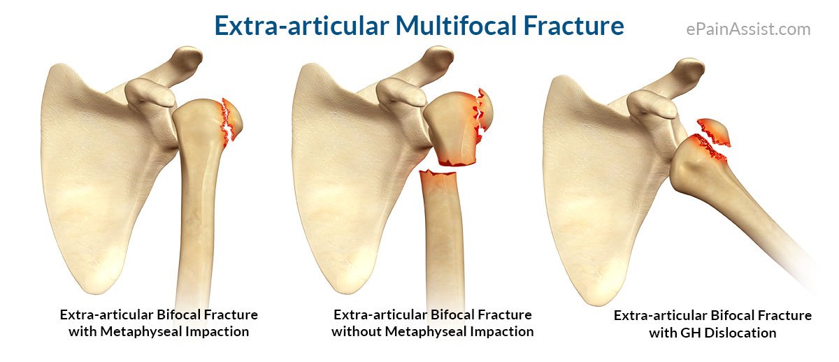
Classifications of Proximal Ulna Fractures

There are several classifications that describe different forms of olecranon fractures, yet none of them have gained widespread acceptance:[rx]

Mayo Classification

Based on the stability, the displacement and the comminution of the fracture. It is composed of three types, and each type is divided into two subtypes: subtype A (non-comminuted) and subtype B (comminuted).

  • Type I: Non-displaced fracture – It can be either non-comminuted ones (Type IA) or comminuted (Type IB).
  • Type II: Displaced, stable fractures – In this pattern, the proximal fracture fragment is displaced more than 3 mm, but the collateral ligaments are intact. That is why there is no elbow instability. It can be either non-comminuted ones (Type IIA) or comminuted (Type IIB).
  • Type III: Displaced unstable fracture – In this case, the fracture fragments are displaced and the forearm is unstable in relation to the humerus. It is a fracture -dislocation. It also may be either non-comminuted (Type IIIA) or comminuted (Type IIIB).

AO Classification

This classification incorporates all fractures of the proximal ulna and radius into one group, subdivided into three patterns:

  • Type A: Extra-articular fractures of the metadiaphysis of either the radius or the ulna
  • Type B: Intra-articular fractures of either the radius or ulna
  • Type C: Complex fractures of both the proximal radius and ulna

Colton Classification

  • Type I – Nondisplaced – Displacement does not increase with elbow flexion
  • Type II – Avulsion (displaced)
  • Type III – Oblique and Transverse (displaced)
  • Type IV – Comminuted (displaced)
  • Type V – Fracture dislocation

Schatzker Classification

  • Type A – Simple transverse fracture
  • Type B – Transverse impacted fracture
  • Type C – Oblique fracture
  • Type D – Comminuted fracture
  • Type E – More distal fracture, extra-articular
  • Type F – Fracture-dislocation

Mechanism of Proximal Ulna Fractures

Olecranon fractures are common. Typically they are caused by direct blows to the elbow (e.g. motor vehicle accidents), and due to falls when the triceps are contracted.[rx][rx] “Side-swipe” injury when driving a motor vehicle with an elbow projecting outside the vehicle resting on an open window’s edge is an example.[rx]

Direct trauma – This can happen in a fall with landing on the elbow or by being hit by a solid object. Trauma to the elbow often results in comminuted fractures of the olecranon.

Indirect trauma – by falling and landing with an outstretched arm.

Powerful pull of the triceps muscle can also cause avulsion fractures.

Causes of Proximal Ulna Fractures


  • Sudden forceful  fall down
  • Road traffic accident
  • Falls – Falling onto an outstretched hand is one of the most common causes of broken proximal ulna fractures.
  • Sports injuries – Many wrist fractures occur during contact sports or sports in which you might fall onto an outstretched hand — such as in-line skating or snowboarding.
  • Motor vehicle crashes – Motor vehicle crashes can cause wrist bones to break, sometimes into many pieces, and often require surgical repair.
  • Have osteoporosis – a disease that weakens your bones
  • Eave low muscle mass or poor muscle strength – or lack agility and have poor balance (these conditions make you more likely to fall)
  • Walk or do other activities in snow or on the ice –  or do activities that require a lot of forwarding momenta, such as in-line skating and skiing
  • Wave an inadequate intake of calcium or vitamin D
  • Football or soccer, especially on artificial turf
  • Rugby
  • Horseback riding
  • Hockey
  • Skiing
  • Snowboarding
  • In-line skating
  • Jumping on a trampoline

Symptoms of Proximal Ulna Fractures

People with olecranon fractures present with intense elbow pain after a direct blow or fall.[rx] Swelling over the bone site is seen and an inability to straighten the elbow is common. Due to the proximity of the olecranon to the ulnar nerve, the injury and swelling may cause numbness and tingle at the fourth and fifth fingers.[rx] The examination can bring out a palpable defect at the site of the fracture.[rx]

  • Severe pain that might worsen when gripping or squeezing or moving your hand or wrist
  • Swelling
  • Tenderness
  • Bruising
  • Obvious deformity, such as a bent wrist
  • Pain
  • Bruising
  • Tenderness
  • The wrist hanging in a deformed way
  • Pain, especially when flexing the ulnar styloid fracture

Diagnosis of Proximal Ulna Fractures

Diagnosis can be made upon the interpretation of anteroposterior and lateral views alone.[rx]

The classic proximal ulna fractures have the following characteristics:[rx]

  • Transverse fracture of the ulnar
  • dorsal displacement and dorsal angulation, together with radial tilt[rx]

Other characteristics[rx][rx]

  • Radial-ulnar styloid fracture shortening
  • Loss of ulnar inclination≤
  • Radial angulation of the wrist
  • Comminution at the fracture site
  • Associated fracture of the ulnar styloid process in more than 60% of cases.

Differential Diagnosis/ Associated Injuries

  • Scapholunate ligament tear
  • Median nerve injury
  • TFCC (triangular fibrocartilage complex) injury, up to 50% when ulnar styloid fx also present
  • Carpal ligament injury – Scapholunate Instability(most common), lunotriquetral ligament
  • Tendon injury, attritional EPL rupture, usually treated with EIP tendon transfer
  • Compartment syndrome
  • Ulnar styloid fracture
  • DRUJ (Distal Radial Ulnar Joint) Instability
  • Galeazzi Fracture: highly associated with distal 1/3 radial shaft fractures[rx]

Plain radiographs

  • Radiographic imaging is important in diagnosis, classification, treatment and follow-up assessment of these fractures. The routine minimal evaluation for proximal ulna fractures must include two views-a postero-anterior (PA) view and lateral view.[]
  • The PA view should be obtained with the humerus abducted 90 degrees from the chest wall, so that the elbow is at the same level as the shoulder and flexed 90 degrees.[] The palm is maintained flat against the cassette

Computed Tomography

  • CT may be useful and can give significant information in comparison with that obtained with conventional radiography in the evaluation of complex or occult fractures, distal radial articular surface, distal radio-ulnar joint, ventromedial fracture fragment,[] assessments of fracture healing as well as post-surgical evaluation.[]
  • CT may be indicated for the confirmation of occult fractures suspected on the basis of physical examination when plain films are normal.

Magnetic Resonance Imaging

  • Although this modality is not the first choice in evaluating acute distal radius fractures, it is a powerful diagnostic tool to assess bony, ligamentous and soft tissue abnormalities associated with these fractures.
  • MRI has proved to be a very important diagnostic tool for delineating perforation of triangular fibrocartilage complex (TFCC),[] perforation of interosseous ligaments of the proximal carpal row, evaluating occult fractures, post-traumatic or avascular necrosis of carpal bones.

Treatment of Ulnar Lower End Fracture

Ulnar Lower End Fracture

Treatment available can be broadly

  • Get medical help immediately – If you fall on an outstretched arm, get into a car accident or are hit while playing a sport and feel intense pain in your shoulder area, then get medical care immediately. Proximal ulna fractures cause significant pain in the front part of your shoulder, closer to the base of your neck. You’ll innately know that something is seriously wrong because you won’t be able to lift your arm up. Other symptoms include immediate swelling and/or bruising near the fracture, grinding sounds with arm movements and potential numbness and tingling in the arm/hand.
  • Apply ice to your fractured clavicle – After you get home from the hospital proximal ulna fractures (regardless if you had surgery or not), you should apply a bag of crushed ice (or something cold) to your injured in order to reduce the swelling and numb the pain. Ice therapy is effective for acute (recent) injuries that involve swelling because it reduces blood flow by constricting local blood vessels. Apply the crushed ice to your clavicle for 15 minutes three to five times daily until the soreness and inflammation eventually fades away
    Lightly exercise after the pain fades – After a couple of weeks when the swelling has subsided and the pain has faded away, remove your arm sling for short periods and carefully move your arm and shoulder in all different directions. Don’t aggravate the proximal ulna fractures so that it hurts, but gently reintroduce movements to the involved joints and muscles. Start cautiously, maybe starting with light calisthenics and then progress to holding light weights (five-pound weights to start). Your proximal ulna fractures need to move a little bit during the later phases of the injury to stimulate complete recovery.
  • Practice stretching and strengthening exercises – of the fingers, elbow, and shoulder if your doctor recommends them.
  • A splint – which you might use for a few days to a week while the swelling goes down; if a splint is used initially, a cast is usually put on about a week later.
  • A cast – which you might need for six to eight weeks or longer, depending on how bad the break is (you might need a second cast if the first one gets too loose after the swelling goes away.)
  • Get a supportive arm sling – Due to their anatomical position, proximal ulna fractures can’t be cast like a broken limb can. Instead, a supportive arm sling or “figure-eight” splint is typically used for support and comfort, either immediately after the injury if it’s just a hairline fracture or following surgery, if it’s a complicated fracture. A figure-eight splint wraps around both shoulders and the base of your neck in order to support the injured shoulder and keep it positioned up and back. Sometimes a larger swath of material is wrapped around the sling to keep it closer to your body. You’ll need to wear the sling constantly until there is no pain with arm movements, which takes between two to four weeks for children or four to eight weeks for adults.
  • Get a referral to physical therapy – Once you’ve recovered and able to remove your arm sling splint for good, you’ll likely notice that the muscles surrounding your shoulder and upper chest look smaller and feel weaker. That’s because muscle tissue atrophies without movement. If this occurs, then you’ll need to get a referral for some physical rehabilitation. Rehab can start once you are cleared by your orthopedist, are pain-free, and can perform all the basic arm and shoulder movements. A physiotherapist or athletic trainer can show you specific rehabilitation exercises and stretches to restore your muscle strength, joint movements and flexibility
  • Rigid fixation – osteosynthesis with locking plate, hook plate fixation, fixation with a distal radius locking plate, coracoclavicular screws, Knowles pin fixation.
  • Flexible fixation – simple k wire fixation, tension band wiring, suture anchors, vicryl tape, dacron arterial graft for coracoclavicular ligament reconstruction.

Rest your Hand

Once you’re discharged from the hospital in an arm sling, your top priority is to rest your shoulder and not further inflame the injury. Of course, the arm sling not only provides support, but it also restricts movement, which is why you should keep it on even during sleep. Avoiding the temptation to move your shoulder and arm will help the bone mend quicker and the pain fades away sooner.
  • Depending on what you do for a living and if the injury is to your dominant side, you may need to take a couple of weeks off work to recuperate.
  • Healing takes between four to six weeks in younger people and up to 12 weeks in the elderly, but it depends on the severity of the fractured clavicle.
  • Athletes in good health are typically able to resume their sporting activities within two months of breaking they’re ulnar styloid depending on the severity of the break and the specific sport.
  • Sleeping on your back (with the sling on) is necessary to keep the pressure off your shoulder and prevent stressing the clavicle injury.

Eat nutritiously during your recovery

All bones and tissues in the body need certain nutrients in order to heal properly and in a timely manner. Eating a nutritious and balanced diet that includes lots of minerals and vitamins is proven to help heal broken bones of all types, including ulnar styloid. Therefore, focus on eating lots of fresh produce (fruits and veggies), whole grains, lean meats, and fish to give your body the building blocks needed to properly repair your clavicle. In addition, drink plenty of purified water, milk, and other dairy-based beverages to augment what you eat.

  • Broken bones need ample minerals (calcium, phosphorus, magnesium, boron) and protein to become strong and healthy again.
  • Excellent sources of minerals/protein include dairy products, tofu, beans, broccoli, nuts and seeds, sardines and salmon.
  • Important vitamins that are needed for bone healing include vitamin C (needed to make collagen), vitamin D (crucial for mineral absorption), and vitamin K (binds calcium to bones and triggers collagen formation).
  • Conversely, don’t consume food or drink that is known to impair bone/tissue healing, such as alcoholic beverages, sodas, most fast food items and foods made with lots of refined sugars and preservatives.

Physical therapy

  • Although there will be some pain, it is important to maintain arm motion to prevent stiffness. Often, patients will begin doing exercises for elbow motion immediately after the injury.  After a proximal ulna fracture, it is common to lose some shoulder and arm strength. Once the bone begins to heal, your pain will decrease and your doctor may start gentle shoulder exercises. These exercises will help prevent stiffness and weakness. More strenuous exercises will be started gradually once the fracture is completely healed.

Follow-up care

  • You will need to see your doctor regularly until your fracture heals. During these visits, he or will take x-rays to make sure the bone is healing in a good position. After the bone has healed, you will be able to gradually return to your normal activities.

Breathing Exercise

  • To elevate breathing problem or remove the lung congestion.

Ulnar Lower End Fracture

Medications

Medication can be prescribed to ease the pain. 

Surgery

Fracture (left) and repair (right) with three pins, wires, and incision closure with staples

Nondisplaced Fractures

  • In fractures with little or no displacement, immobilization with a posterior splint may be sufficient.[rx] Elbows be immobilized at 45-90º of flexion for 3 weeks, followed by limited (90º) flexion exercises.

Displaced Fractures

  • Most olecranon fractures are displaced and are best treated surgically:

Tension Band Fixation

  • Tension band fixation is the most common form of internal fixation used for non-comminuted olecranon fractures.[rx] It is typically reserved for noncomminuted fractures that are proximal to the coronoid.[rx] This procedure is performed using Kirschner wire (K-wires) which converts tensile forces into compressive force.[rx]

Intramedullary Fixation and Plates

  • Single intramedullary screws can be used to treat simple transverse or oblique fractures.[rx] Plates can be used for all proximal ulna fracture types including Monteggia fractures, and comminuted fractures.[rx]

Excision and triceps advancement

  • This method is indicated for cases when open reduction and internal fixation is unlikely to be successful. For example: extensive comminutions, elderly patients with osteoporotic bone, and small or non-union fractures.[rx][rx]

Open reduction and internal fixation. This is the procedure most often used to treat ulnar styloid fracture fractures. During the procedure, the bone fragments are first repositioned (reduced) into their normal alignment. The pieces of bone are then held in place with special metal hardware.

Common methods of internal fixation include:

  • Plates and screws – After being repositioned into their normal alignment, the bone fragments are held in place with special screws and metal plates attached to the outer surface of the bone. After surgery, you may notice a small patch of numb skin below the incision. This numbness will become less noticeable with time. Because the clavicle lies directly under the skin, you may be able to feel the plate through your skin.
  • Pins or screws –  Pins or screws can also be used to hold the fracture in good position after the bone ends have been put back in place. The incisions for pin or screw placement are usually smaller than those used for plates.
    Pins or screws often irritate the skin where they have been inserted and are usually removed once the fracture has healed.
  • Precontoured locking plates
  • Hook plate
  • Distal radius plates
  • Ulnar styloid fracture screws
  • Flexible coracoclavicular fixation
  • Arthroscopic treatment
  • Intra-medullary fixation
  • Tension band fixation

Closed Reduction and Casting

  • All fractures characterized by minor comminution, without or with minimal displacements can be considered for closed reduction and cast immobilization. Mainly type I and type IIA Melone’s fracture can be managed conservatively. The fracture should be kept under closed observation to look for any re-displacement.
  • Despite the widespread acceptance of immobilization in a plaster cast, questions remain regarding the optimum position, the duration of immobilization and the need to extend the cast proximal to the ulna. No clear consensus exists as to the best position for immobilizing the wrist in plaster. Sarmiento et al.[] advocated immobilization in a position of supination to decrease the deforming force of the brachioradialis, which may cause loss of reduction.

Pins and Plaster Technique

  • Placement of pins in the metacarpals and forearm was initially advocated by Bohler in 1923, but it gained popularity after the report by Green, who showed good or excellent results in 86% of his patients.[]
  • However, he noted a high incidence of minor or major complications, one-third of which were related to pin site only. Other researchers also noted that one-third of the complications were related to pins and 16% of the patients needed reoperation for complications.[]

Percutaneous Pinning

  • Extra-articular fractures of the distal end of the ulna with extensive comminution or the fractures that have no more than two articular fragments, in which anatomical reduction is obtainable, are amenable to percutaneous pinning of the fracture fragments and application of a plaster cast. A single pin placed through the radial styloid as a means of stabilizing the displaced fracture fragment was first suggested by Lambotte in 1908.[,]

External Fixation

  • External fixation is generally accepted as superior to plaster immobilization in the young patients with an intra-articular comminuted fracture of the distal radius. Other indications for external fixation include some unstable extra-articular fractures with significant comminution and failure to maintain reduction after an initial attempt at closed management in a cast, certain situations of multiple trauma, the presence of dysfunctional contralateral limb, severe open fractures with significant soft tissue injury and neurovascular compromise, and bilateral injuries.[]

Limited Open Reduction

  • In intra-articular fractures that have more than 2 mm of displacement, the radio-carpal joint may be incongruent despite adequate attempts at reduction. The incongruency usually involves the lunate part of the distal end of the radius.
  • The ulna styloid process and scaphoid facet are more amenable to reduction through ligamentotaxis or by manipulation and reduction.
  • A new technique of combining external fixation with open reduction of the displaced lunate fossa through a small, longitudinal incision and elevation of the impacted fragment without direct visualization of the surface of the joint has been described.[]

Open Reduction and Internal Fixation

  • One of the recent advances in the treatment of distal ulna fractures is the more frequent application of open reduction and internal fixation, especially for intra-articular fractures. There are two groups of fractures for which open reduction and internal fixation is advisable.

Arthroscopic-Assisted Fracture Reduction

  • Intra-articular fractures of the radius can be arthroscopically assessed, and reduction of the particular components and assessment and repair of ligamentous injury can then be undertaken.[,,] The ideal timing for arthroscopically assisted distal radius surgery is 3 to 7 days after injury.

I. Non-operative methods of treating radial head fractures

A. Immediate mobilization vs. cast in flexion vs. cast in extension

  • One randomized controlled trial (CoE level II) was identified that compared patients treated with immediate mobilization with immobilization.6
  • The least pain and loss of elbow extension were seen in people treated with immobilization, but these differences were not statistically significant.
  • Comparisons between treatments may be limited by small sample sizes and study biases.

B. Aspiration of the elbow versus no aspiration

  • One randomized controlled trial (CoE level II) was identified that compared patients with radial head fractures treated with elbow aspiration versus those who were not treated with aspiration.7
  • Ninety-two percent of the patients who received aspiration reported immediate and lasting relief from pain following the treatment.

II. Non-operative versus operative methods

A. Closed reduction versus ORIF in children

  • One retrospective cohort study on children was identified that compared closed reduction versus open reduction for radial neck fractures.8, 9
  • Compared with children treated with open reduction and internal fixation, those treated with immobilization/closed reduction experienced significantly less pain (15% versus 65%; RR=0.2, 95% CI, 0.1–0.4), avascular necrosis (4% versus 24%; RR = 0.2, 95% CI, 0.05–0.6), and had more good or excellent overall clinical results (99% versus 55%); RR = 1.8, 95% CI, 1.3–2.5).

B. Closed reduction versus ORIF in adults

  • One retrospective cohort study in adults was identified that compared closed versus open reduction for radial head fractures.9
  • Patients who received closed reduction were more likely to report pain than those treated with open reduction, but this was not statistically significant (RR = 2.0, 95% CI, 0.9–4.5).
  • Patients treated with open reduction were more likely to have had a good or excellent clinical result (90%) than patients treated with closed reduction (46%), but, again, these were not statistically significantly different.

III. Operative methods of reducing olecranon fractures

A. Tension band wiring versus plate fixation

  • One randomized controlled trial was identified that compared tension band wiring with plate fixation as treatment of olecranon fractures.10
  • Plate fixation resulted in higher percentage of “good” clinical (no more than occasional pain; loss of movement in elbow less than 15°) and x-ray (no articular step-off or gap; no loss of reduction) results.
  • Many more tension band wiring patients reported complications (74%) compared with patients who received plate fixation (5%), a statistically significant difference (RR= 16.2, 95% CI, 2.3–112). To prevent one complication, one to 2 patients would need to be treated with plate fixation
  • Results may be limited by small sample size.

B. Netz pins versus Kirschner pins

  • One randomized controlled trial was identified that compared Netz pins with Kirschner pins as treatment of olecranon fractures.11
  • Median fracture healing time was similar for both groups (six weeks for Netz pins versus seven weeks for Kirschner pins).
  • More Kirschner (68%) than Netz (52%) pins had to be removed by the end of follow-up, but this difference was not statistically significant.

C. Tension band wiring versus figure of eight wiring

  • One study was found that compared tension band wiring with figure of eight wiring to treat olecranon fractures.2
  • No statistically or clinically significant differences were found between the two treatment groups in terms of pain, loss of strength, range of motion, or osteoarthritis.
  • Patients in the tension band wiring group (81%) experienced significantly more hardware removal than those in the figure of eight wiring group (43%); RR = 1.9, 95% CI, 1.3–1.9)).

IV. Operative versus operative methods for radial head fractures

A. Resection versus open reduction and fixation

  • One retrospective cohort study was identified that compared resection versus open reduction and internal fixation.12
  • Patients who received open reduction and internal fixation reported less pain (P = .02) and greater functional recovery (P = .003) than those who received resection.

B. Primary radial head excision versus delayed excision

  • One retrospective cohort study was identified that compared primary versus delayed excision as treatment of radial head fractures.13
  • No difference was found between immediate (10%) and delayed (11%) treatment in terms of daily pain (RR = 0.9, 95% CI, 0.2–4.6).

C. Biodegradable polylactide pins versus standard metal mini-fragment implants

  • One randomized controlled trial (CoE II) was identified that compared the use of polylactide pins with standard metal mini-fragment implants for treatment of displaced radial head fractures.14
  • No difference in postsurgical complications or in clinician-based outcomes at the two year follow-up was found between the use of biodegradable pins or standard metal lag screws.
  • At two years follow-up, a five times greater risk for osteoloysis was seen in polylactide patients (17%) compared with patients treated using standard metal devices (3%) (RR = 5.4, 95% CI, 1.3–23, NNT = 21). Of patients who developed osteolysis, the osteolysis was mild or moderate in eleven (92%) of those with polylactide pins and one of the two patients with metal devices.

V. Rehabilitation following radial head fractures

  • One study was identified that compared immediate to delayed mobilization for rehabilitation following radial head fractures.
  • No statistically or clinically significant differences were found between pain or range of motion scores at week twelve.
  • In olecranon fractures, osteoarthritis of the elbow joint, occurring after about 5% of these fractures, and the necessity of a second operation to remove hardware, required in 56-85% of olecranon fractures, are two common complications that result from operative treatments.
  • The most common complication of radial head fractures is a loss of joint motion as a result of avascular necrosis, which occurs in 10–20% of radial head fractures.1 No studies comparing prognosis were identified for radial head fractures.
  • Complications of treated Monteggia fractures include recurrent radial head dislocation, malunion, posterior interosseous nerve palsy, and Volkmann’s ischemic contracture. No comparative studies were found on prognosis following Monteggia fractures.

Overall, the quality of evidence on proximal forearm fractures is limited, and definitive statements regarding treatment are not possible. However, the following observations may be helpful:

  • Aspiration of radial head fractures consistently relieved pain.
  • Studies comparing operative to non-operative methods of treating proximal forearm fractures suggest non-operative methods may be better than operative methods in terms of pain, avascular necrosis, and clinical outcomes for radial neck fractures in children. However, in adults, operative methods had better results in terms of pain and clinical results.
  • Tension band wiring, compared with plate fixation, may result in more complications when used as a treatment for olecranon fractures.
  • Tension band wiring of forearm fractures also resulted in greater rates of hardware removal when compared with a figure of eight wiring.
  • Open reduction and internal fixation of radial head fractures seems to have better results than resection
  • Biodegradable pins or standard metal screws for ORIF may result in comparable outcomes but there may be a greater risk of developing a mild degree of osteolysis.

Rehabilitation Guideline for Non-Operative/Conservative rehabilitation [rx]

Acute Stage (0-8 weeks)

Goals

  • Protection with short-arm cast
  • Control pain and edema
  • Maintain range in digits, elbows, shoulder

Interventions

  • AROM and PROM of digits, elbow, shoulder
  • Elevation of hand and digits to control edema
  • Cast removal between 6-8 weeks

Sub Acute Stage

Goal

  • Control pain and edema (TENS, ice)
  • Increase ROM
  • Increase activities of daily living (ADLs)

Interventions

  • AROM and PROM of digits, elbow, shoulder
  • AROM wrist flexion/ extension, forearm supination/ pronation
  • PROM of low load and prolonged stretch

Settled Stage

Goals

  • Regain full ROM
  • Begin strengthening
  • Return to activity

Interventions

  • Continue all ROM exercises
  • Progress to the strengthening of all joints[rx]

Rehabilitation Guideline for External Fixation by Pho et al

Acute Stage (1-6 weeks)

Goals

  • Control pain and edema (TENS, ice)
  • Protect surgical site
  • Maintain ROM of digits, elbow, shoulder

Interventions

  • Elevation
  • AROM of digits, elbow, shoulder
  • AROM forearm supination/ pronation

Sub Acute (7-10 weeks)

Goal

  • Protect fracture site
  • Control pain and edema (TENS, ice)
  • ROM of involved and uninvolved joints

Interventions

  • AROM and PROM of wrist extension/ flexion, radial deviation, and supination/ pronation

Settled Stage (10-16 weeks)

Goal

  • Regain full ROM
  • Begin strengthening
  • Increase tolerance to ADLs

Interventions

  • ROM of wrist flexion/ extension, radial/ ulnar deviation, forearm supination/ pronation progressing to isometric exercises and resisted exercises using dumbbells or resistive bands
  • PROM of low load and prolonged stretching of wrist motions
  • Grip strengthening
  • ADL training within tolerance[rx]

Cryotherapy

  • Cryotherapy is an effective modality for controlling edema in the acute phase after trauma and during rehab due to its ability in helping to decrease blood flow through vasoconstriction limiting the amount of fluid escaping from capillaries to the interstitial fluid[rx]. Cryotherapy can also be combined with compression and elevation in the treatment of edema.[rx]
  • To control pain using cryotherapy, the modality should be applied to the area for 10-15 minutes which can result in pain control up to 2 hours post application.[rx]Precautions for the use of cryotherapy include: over a superficial branch of the nerve, over an open wound, poor sensation or mentation, and very young or very old patients.[rx] Contraindications for cryotherapy include; Acute febrile illness, Vasospasm e.g. Raynaud’s disease, Cryoglobulinemia, Cold urticaria.[rx]

Electrical Stimulation

  • The use of transcutaneous electrical nerve stimulation (TENS) may be used as an adjunct during any phase of rehab to address pain but can be particularly useful for patients that are increasing the level of activity of the wrist. Conventional (high-rate) TENS is useful for disrupting the pain cycle through a prolonged treatment session as great as 24 hours a day.[rx]
  • Low-rate TENS is another form of electrical stimulation that is successful in diminishing pain by targeting motor or nociceptive A-delta nerves. Low-rate TENS has been reported to be effective in pain control for up to 4-5 hours post-treatment.[rx]
  • The literature is still not conclusive on this topic and the results of one study may contradict or, on the contrary, reinforce the results of another study. Yet there is evidence supporting the beneficial effects of electrical stimulation, especially in combination with physiotherapy exercises.

Supervised Active rehabilitation program used in Study

ISOMETRIC EXERCISE

  • Wrist flexors and extensors

ACTIVE RANGE OF MOTION EXERCISE

  • Assisted stretch to forearm flexors and extensor musculature and radial/ulnar deviation
  • Weight-bearing wrist extension exercise(hand on the table with the patient leaning forward on them) to patient tolerance
  • Active stretch to shoulder girdle and rotator cuff musculature
  • Active stretch to elbow flexor and extensor musculature

INTRINSIC HAND MUSCLE EXERCISE

  • Thumb/digit opposition
  • Repetitive squeezing of therapy
  • repetitive towel wringing exercise

STRENGTHENING ROUTINE

  • Biceps curl with 1,5-2 pound weights bilaterally
  • Shoulder abduction, flexion and extension reps with 2-pound weights bilaterally
  • Repetitive squeezing of a rubber ball in affected wrist
  • Flexion and extension of wrist using 1,5-pound weights increasing as tolerated

FUNCTIONAL ACTIVITIES

  • The patient is encouraged to resume pre-accident activities that involve the affected extremity (eg. writing, typing, cooking, etc.)

Complications of 

There were no major complications such as neurovascular injury, infection, or impaired wound healing. Surgery-related complication at 2-year follow-up included nonunion in 3 patients (11%),

  • DRUJ subluxation in 3 patients (11%),
  • Implant migration in 4 patients (14%),
  • Radiographic resorption of the ulnar styloid in 4 patients (14%).
  • Radiographic nonunion was noted in 1 patient in group A (8%) and 2 in group B (13%). Residual DRUJ subluxation was noted in 3 patients; all were in group B (20%).
  • Partial or complete radiographic resorption of the ulna was found in 1 patient in group A (8%) and 3 in group B (20%).
  • Implant migration was noted in 1 patient in group A (8%), and 2 in group B (13%).
  • Subsequent removal surgery due to implant irritation occurred in 13 patients (46%), with 4 in group A (31%) and 8 in group B (53%).
  • A total of 11 patients (39%) with surgery-related complications included 5 (38%) in group A and 12 (80%) in group B, with a significant difference

There are risks associated with any type of surgery. These include

  • Nonunion (1-5%)
  • Infection (~4.8%)
  • 4% in the surgical group develop adhesive capsulitis requiring surgical intervention
  • Bleeding
  • Problems with wound healing
  • Blood clots
  • Damage to blood vessels or nerves
  • Reaction to anesthesia
  • Hardware prominence
  • Malunion with cosmetic deformity
  • Restriction of ROM
  • The difficulty with bone healing
  • Hardware irritation
  • Fracture comminution (Z deformity)
  • Fracture displacement
  • Increased fatigue with overhead activities
  • Dissatisfaction with appearance
  • The difficulty with shoulder straps, backpacks and the like
  • ~30% of patient request plate removal
  • Superior plates associated with increased irritation
  • Superior plates associated with increased risk of subclavian artery or vein penetration

References

Proximal ulna fractures

By

Ulnar Lower End Fracture; Causes, Diagnosis, Treatment

Ulnar Lower End Fracture/Ulnar styloid fracture is common and may be associated with a distal radial fracture (DRF) or occurs as an isolated injury []. In spite of acting as a unique strut on the ulnar end to stabilize the ulnar soft tissue and maintain the congruency of the distal radioulnar joint (DRUJ), the majority of these connected tissues including the triangular fibrocartilage complex (TFCC) are at the base of the ulnar styloid or fovea [].

An ulna fracture is a break of the ulna bone in the forearm. They are often associated with radius fractures.[rx] When the middle portion of the ulna is broken without other associated fractures, it may be called a nightstick fracture.

The ulnar styloid plays a crucial role in wrist biomechanics. The ulnar styloid base and the fovea are the insertion points for the primary stabilizers of the DRUJ, the superficial and deep portions of the palmar and dorsal radioulnar ligaments.,, In addition, the ulnar styloid functions as a strut, helping to stabilize the extensor carpi ulnaris (ECU), its sub-sheath, and the ulnocarpal ligaments. Because of its anatomic importance, the potential exists for ulnar styloid fractures to cause ulnar-sided wrist symptoms.

Ulnar Lower End Fracture

Anatomy of Ulnar Lower End Fracture

The management of acute ulnar styloid fractures is based on the long-term effect that they may have on the stability of the distal radioulnar (DRU) joint. The relationship of the ulnar styloid to the stabilizing ligaments determines whether a specific fracture type is likely to result in DRU joint instability.

The static stability of the DRU-joint is achieved by the bony congruity between the sigmoid notch of the radius and the ulnar head and the ligaments which hold the joint together []. The ulno-radial ligament represents the transverse, peripheral part of the Triangular Fibro-Cartilage Complex (TFCC) []. The ligaments run from the fovea of the ulnar head and the base of the ulnar styloid to the dorsal and palmar edges of the sigmoid notch on the distal radius [, ]. The ulno-radial ligament is the major stabilizer of DRU-joint in the dorsal/palmar direction [, ].

Ulnar Lower End Fracture

Types of Ulnar Lower End Fracture

  • Nightstick fracture – is a fracture of the middle portion of the ulna without other fractures.[rx]
  • Distal ulna fractures – typically occur along with distal radius fractures.[rx]
  • Monteggia fracture – a fracture of the proximal third of the ulna with the dislocation of the head of the radius
  • Hume fracture – a fracture of the olecranon with associated anterior dislocation of the radial head

Causes of Ulnar Lower End Fracture

Ulnar Lower End Fracture

  • Sudden forceful  fall down
  • Road traffic accident
  • Falls – Falling onto an outstretched hand is one of the most common causes of a broken ulnar styloid fracture.
  • Sports injuries – Many wrist fractures occur during contact sports or sports in which you might fall onto an outstretched hand — such as in-line skating or snowboarding.
  • Motor vehicle crashes – Motor vehicle crashes can cause wrist bones to break, sometimes into many pieces, and often require surgical repair.
  • Have osteoporosis – a disease that weakens your bones
  • Eave low muscle mass or poor muscle strength – or lack agility and have poor balance (these conditions make you more likely to fall)
  • Walk or do other activities in snow or on the ice –  or do activities that require a lot of forwarding momenta, such as in-line skating and skiing
  • Wave an inadequate intake of calcium or vitamin D
  • Football or soccer, especially on artificial turf
  • Rugby
  • Horseback riding
  • Hockey
  • Skiing
  • Snowboarding
  • In-line skating
  • Jumping on a trampoline

Symptoms of Ulnar Lower End Fracture

Common symptoms of a Colles fracture include:

  • Severe pain that might worsen when gripping or squeezing or moving your hand or wrist
  • Swelling
  • Tenderness
  • Bruising
  • Obvious deformity, such as a bent wrist
  • Pain
  • Bruising
  • Tenderness
  • The wrist hanging in a deformed way
  • Pain, especially when flexing the ulnar styloid fracture
  • Deformity of the wrist, causing it to look crooked and bent.
  • Your wrist is in great pain.
  • Your wrist, arm, or hand is numb.
  • Your fingers are pale.

Diagnosis of Ulnar Lower End Fracture

Diagnosis can be made upon the interpretation of anteroposterior and lateral views alone.[rx]

The classic ulnar styloid fracture has the following characteristics:[rx]

  • Transverse fracture of the ulnar styloid fracture
  • dorsal displacement and dorsal angulation, together with radial tilt[rx]

Other characteristics[rx][rx]

  • Radial-ulnar styloid fracture shortening
  • Loss of ulnar inclination≤
  • Radial angulation of the wrist
  • Comminution at the fracture site
  • Associated fracture of the ulnar styloid process in more than 60% of cases.

Differential Diagnosis/ Associated Injuries

  • Scapholunate ligament tear
  • Median nerve injury
  • TFCC (triangular fibrocartilage complex) injury, up to 50% when ulnar styloid fx also present
  • Carpal ligament injury – Scapholunate Instability(most common), lunotriquetral ligament
  • Tendon injury, attritional EPL rupture, usually treated with EIP tendon transfer
  • Compartment syndrome
  • Ulnar styloid fracture
  • DRUJ (Distal Radial Ulnar Joint) Instability
  • Galeazzi Fracture: highly associated with distal 1/3 radial shaft fractures[rx]

Plain radiographs

  • Radiographic imaging is important in diagnosis, classification, treatment and follow-up assessment of these fractures. The routine minimal evaluation for ulnar styloid fracture must include two views-a postero-anterior (PA) view and lateral view.[]
  • The PA view should be obtained with the humerus abducted 90 degrees from the chest wall, so that the elbow is at the same level as the shoulder and flexed 90 degrees.[] The palm is maintained flat against the cassette

Computed Tomography

  • CT may be useful and can give significant information in comparison with that obtained with conventional radiography in the evaluation of complex or occult fractures, distal radial articular surface, distal radio-ulnar joint, ventromedial fracture fragment (as described by Melone),[] assessments of fracture healing as well as post-surgical evaluation.[]
  • CT may be indicated for the confirmation of occult fractures suspected on the basis of physical examination when plain films are normal.

Magnetic Resonance Imaging

  • Although this modality is not the first choice in evaluating acute distal radius fractures, it is a powerful diagnostic tool to assess bony, ligamentous and soft tissue abnormalities associated with these fractures.
  • MRI has proved to be a very important diagnostic tool for delineating perforation of triangular fibrocartilage complex (TFCC),[] perforation of interosseous ligaments of the proximal carpal row, evaluating occult fractures, post-traumatic or avascular necrosis of carpal bones.

Treatment of Ulnar Lower End Fracture

Ulnar Lower End Fracture

Treatment available can be broadly

  • Get medical help immediately – If you fall on an outstretched arm, get into a car accident or are hit while playing a sport and feel intense pain in your shoulder area, then get medical care immediately. Ulnar styloid fracture causes significant pain in the front part of your shoulder, closer to the base of your neck. You’ll innately know that something is seriously wrong because you won’t be able to lift your arm up. Other symptoms include immediate swelling and/or bruising near the fracture, grinding sounds with arm movements and potential numbness and tingling in the arm/hand.
  • Apply ice to your fractured clavicle – After you get home from the hospital (regardless if you had surgery or not), you should apply a bag of crushed ice (or something cold) to your injured clavicle in order to reduce the swelling and numb the pain. Ice therapy is effective for acute (recent) injuries that involve swelling because it reduces blood flow by constricting local blood vessels. Apply the crushed ice to your clavicle for 15 minutes three to five times daily until the soreness and inflammation eventually fades away
    Lightly exercise after the pain fades – After a couple of weeks when the swelling has subsided and the pain has faded away, remove your arm sling for short periods and carefully move your arm and shoulder in all different directions. Don’t aggravate the ulnar styloid so that it hurts, but gently reintroduce movements to the involved joints and muscles. Start cautiously, maybe starting with light calisthenics and then progress to holding light weights (five-pound weights to start). Your ulnar styloid needs to move a little bit during the later phases of the injury to stimulate complete recovery.
  • Practice stretching and strengthening exercises – of the fingers, elbow, and shoulder if your doctor recommends them.
  • A splint – which you might use for a few days to a week while the swelling goes down; if a splint is used initially, a cast is usually put on about a week later.
  • A cast – which you might need for six to eight weeks or longer, depending on how bad the break is (you might need a second cast if the first one gets too loose after the swelling goes away.)
  • Get a supportive arm sling – Due to their anatomical position, an ulnar styloid fracture can’t be cast like a broken limb can. Instead, a supportive arm sling or “figure-eight” splint is typically used for support and comfort, either immediately after the injury if it’s just a hairline fracture or following surgery, if it’s a complicated fracture. A figure-eight splint wraps around both shoulders and the base of your neck in order to support the injured shoulder and keep it positioned up and back. Sometimes a larger swath of material is wrapped around the sling to keep it closer to your body. You’ll need to wear the sling constantly until there is no pain with arm movements, which takes between two to four weeks for children or four to eight weeks for adults.
  • Get a referral to physical therapy – Once you’ve recovered and able to remove your arm sling splint for good, you’ll likely notice that the muscles surrounding your shoulder and upper chest look smaller and feel weaker. That’s because muscle tissue atrophies without movement. If this occurs, then you’ll need to get a referral for some physical rehabilitation. Rehab can start once you are cleared by your orthopedist, are pain-free, and can perform all the basic arm and shoulder movements. A physiotherapist or athletic trainer can show you specific rehabilitation exercises and stretches to restore your muscle strength, joint movements and flexibility
  • Rigid fixation – osteosynthesis with locking plate, hook plate fixation, fixation with a distal radius locking plate, coracoclavicular screws, Knowles pin fixation.
  • Flexible fixation – simple k wire fixation, tension band wiring, suture anchors, vicryl tape, dacron arterial graft for coracoclavicular ligament reconstruction.

Rest your shoulder

Once you’re discharged from the hospital in an arm sling, your top priority is to rest your shoulder and not further inflame the injury. Of course, the arm sling not only provides support, but it also restricts movement, which is why you should keep it on even during sleep. Avoiding the temptation to move your shoulder and arm will help the bone mend quicker and the pain fades away sooner.
  • Depending on what you do for a living and if the injury is to your dominant side, you may need to take a couple of weeks off work to recuperate.
  • Healing takes between four to six weeks in younger people and up to 12 weeks in the elderly, but it depends on the severity of the fractured clavicle.
  • Athletes in good health are typically able to resume their sporting activities within two months of breaking they’re ulnar styloid depending on the severity of the break and the specific sport.
  • Sleeping on your back (with the sling on) is necessary to keep the pressure off your shoulder and prevent stressing the clavicle injury.

Eat nutritiously during your recovery

All bones and tissues in the body need certain nutrients in order to heal properly and in a timely manner. Eating a nutritious and balanced diet that includes lots of minerals and vitamins is proven to help heal broken bones of all types, including ulnar styloid. Therefore, focus on eating lots of fresh produce (fruits and veggies), whole grains, lean meats, and fish to give your body the building blocks needed to properly repair your clavicle. In addition, drink plenty of purified water, milk, and other dairy-based beverages to augment what you eat.

  • Broken bones need ample minerals (calcium, phosphorus, magnesium, boron) and protein to become strong and healthy again.
  • Excellent sources of minerals/protein include dairy products, tofu, beans, broccoli, nuts and seeds, sardines and salmon.
  • Important vitamins that are needed for bone healing include vitamin C (needed to make collagen), vitamin D (crucial for mineral absorption), and vitamin K (binds calcium to bones and triggers collagen formation).
  • Conversely, don’t consume food or drink that is known to impair bone/tissue healing, such as alcoholic beverages, sodas, most fast food items and foods made with lots of refined sugars and preservatives.

Physical therapy

  • Although there will be some pain, it is important to maintain arm motion to prevent stiffness. Often, patients will begin doing exercises for elbow motion immediately after the injury.  After an ulnar styloid, it is common to lose some shoulder and arm strength. Once the bone begins to heal, your pain will decrease and your doctor may start gentle shoulder exercises. These exercises will help prevent stiffness and weakness. More strenuous exercises will be started gradually once the fracture is completely healed.

Follow-up care

  • You will need to see your doctor regularly until your fracture heals. During these visits, he or will take x-rays to make sure the bone is healing in a good position. After the bone has healed, you will be able to gradually return to your normal activities.

Breathing Exercise

  • To elevate breathing problem or remove the lung congestion.

Ulnar Lower End Fracture

Medications

Medication can be prescribed to ease the pain. 

Surgery

The evidence for different types of surgery for breaks of the middle part of the clavicle is poor as of 2015.[10]

Surgery is considered when one or more of the following conditions present.

  • Comminution with separation (bone is broken into multiple pieces)
  • Significant foreshortening of the clavicle (indicated by shoulder forward)
  • Skin penetration (open fracture)
  • Associated nervous and vascular trauma (brachial plexus or supraclavicular nerves)
  • Nonunion after several months (3–6 months, typically)
  • Displaced distal third fractures (high risk of nonunion)

Open reduction and internal fixation. This is the procedure most often used to treat ulnar styloid fracture fractures. During the procedure, the bone fragments are first repositioned (reduced) into their normal alignment. The pieces of bone are then held in place with special metal hardware.

Common methods of internal fixation include:

  • Plates and screws – After being repositioned into their normal alignment, the bone fragments are held in place with special screws and metal plates attached to the outer surface of the bone. After surgery, you may notice a small patch of numb skin below the incision. This numbness will become less noticeable with time. Because the clavicle lies directly under the skin, you may be able to feel the plate through your skin.
  • Pins or screws –  Pins or screws can also be used to hold the fracture in good position after the bone ends have been put back in place. The incisions for pin or screw placement are usually smaller than those used for plates.
    Pins or screws often irritate the skin where they have been inserted and are usually removed once the fracture has healed.
  • Precontoured locking plates
  • Hook plate
  • Distal radius plates
  • Ulnar styloid fracture screws
  • Flexible coracoclavicular fixation
  • Arthroscopic treatment
  • Intra-medullary fixation
  • Tension band fixation

Closed Reduction and Casting

  • All fractures characterized by minor comminution, without or with minimal displacements can be considered for closed reduction and cast immobilization. Mainly type I and type IIA Melone’s fracture can be managed conservatively. The fracture should be kept under closed observation to look for any re-displacement.
  • Despite the widespread acceptance of immobilization in a plaster cast, questions remain regarding the optimum position, the duration of immobilization and the need to extend the cast proximal to the ulna. No clear consensus exists as to the best position for immobilizing the wrist in plaster. Sarmiento et al.[] advocated immobilization in a position of supination to decrease the deforming force of the brachioradialis, which may cause loss of reduction.

Pins and Plaster Technique

  • Placement of pins in the metacarpals and forearm was initially advocated by Bohler in 1923, but it gained popularity after the report by Green, who showed good or excellent results in 86% of his patients.[]
  • However, he noted a high incidence of minor or major complications, one-third of which were related to pin site only. Other researchers also noted that one-third of the complications were related to pins and 16% of the patients needed reoperation for complications.[]

Percutaneous Pinning

  • Extra-articular fractures of the distal end of the ulna with extensive comminution or the fractures that have no more than two articular fragments, in which anatomical reduction is obtainable, are amenable to percutaneous pinning of the fracture fragments and application of a plaster cast. A single pin placed through the radial styloid as a means of stabilizing the displaced fracture fragment was first suggested by Lambotte in 1908.[,]

External Fixation

  • External fixation is generally accepted as superior to plaster immobilization in the young patients with an intra-articular comminuted fracture of the distal radius. Other indications for external fixation include some unstable extra-articular fractures with significant comminution and failure to maintain reduction after an initial attempt at closed management in a cast, certain situations of multiple trauma, the presence of dysfunctional contralateral limb, severe open fractures with significant soft tissue injury and neurovascular compromise, and bilateral injuries.[]

Limited Open Reduction

  • In intra-articular fractures that have more than 2 mm of displacement, the radio-carpal joint may be incongruent despite adequate attempts at reduction. The incongruency usually involves the lunate part of the distal end of the radius.
  • The ulna styloid process and scaphoid facet are more amenable to reduction through ligamentotaxis or by manipulation and reduction.
  • A new technique of combining external fixation with open reduction of the displaced lunate fossa through a small, longitudinal incision and elevation of the impacted fragment without direct visualization of the surface of the joint has been described.[]

Open Reduction and Internal Fixation

  • One of the recent advances in the treatment of distal ulna fractures is the more frequent application of open reduction and internal fixation, especially for intra-articular fractures. There are two groups of fractures for which open reduction and internal fixation is advisable.

Arthroscopic-Assisted Fracture Reduction

  • Intra-articular fractures of the radius can be arthroscopically assessed, and reduction of the particular components and assessment and repair of ligamentous injury can then be undertaken.[,,] The ideal timing for arthroscopically assisted distal radius surgery is 3 to 7 days after injury.

Rehabilitation Guideline for Non-Operative/Conservative rehabilitation [rx]

Acute Stage (0-8 weeks)

Goals

  • Protection with short-arm cast
  • Control pain and edema
  • Maintain range in digits, elbows, shoulder

Interventions

  • AROM and PROM of digits, elbow, shoulder
  • Elevation of hand and digits to control edema
  • Cast removal between 6-8 weeks

Sub Acute Stage

Goal

  • Control pain and edema (TENS, ice)
  • Increase ROM
  • Increase activities of daily living (ADLs)

Interventions

  • AROM and PROM of digits, elbow, shoulder
  • AROM wrist flexion/ extension, forearm supination/ pronation
  • PROM of low load and prolonged stretch

Settled Stage

Goals

  • Regain full ROM
  • Begin strengthening
  • Return to activity

Interventions

  • Continue all ROM exercises
  • Progress to the strengthening of all joints[rx]

Rehabilitation Guideline for External Fixation by Pho et al

Acute Stage (1-6 weeks)

Goals

  • Control pain and edema (TENS, ice)
  • Protect surgical site
  • Maintain ROM of digits, elbow, shoulder

Interventions

  • Elevation
  • AROM of digits, elbow, shoulder
  • AROM forearm supination/ pronation

Sub Acute (7-10 weeks)

Goal

  • Protect fracture site
  • Control pain and edema (TENS, ice)
  • ROM of involved and uninvolved joints

Interventions

  • AROM and PROM of wrist extension/ flexion, radial deviation, and supination/ pronation

Settled Stage (10-16 weeks)

Goal

  • Regain full ROM
  • Begin strengthening
  • Increase tolerance to ADLs

Interventions

  • ROM of wrist flexion/ extension, radial/ ulnar deviation, forearm supination/ pronation progressing to isometric exercises and resisted exercises using dumbbells or resistive bands
  • PROM of low load and prolonged stretching of wrist motions
  • Grip strengthening
  • ADL training within tolerance[rx]

Cryotherapy

  • Cryotherapy is an effective modality for controlling edema in the acute phase after trauma and during rehab due to its ability in helping to decrease blood flow through vasoconstriction limiting the amount of fluid escaping from capillaries to the interstitial fluid[rx]. Cryotherapy can also be combined with compression and elevation in the treatment of edema.[rx]
  • To control pain using cryotherapy, the modality should be applied to the area for 10-15 minutes which can result in pain control up to 2 hours post application.[rx]Precautions for the use of cryotherapy include: over a superficial branch of the nerve, over an open wound, poor sensation or mentation, and very young or very old patients.[rx] Contraindications for cryotherapy include; Acute febrile illness, Vasospasm e.g. Raynaud’s disease, Cryoglobulinemia, Cold urticaria.[rx]

Electrical Stimulation

  • The use of transcutaneous electrical nerve stimulation (TENS) may be used as an adjunct during any phase of rehab to address pain but can be particularly useful for patients that are increasing the level of activity of the wrist. Conventional (high-rate) TENS is useful for disrupting the pain cycle through a prolonged treatment session as great as 24 hours a day.[rx]
  • Low-rate TENS is another form of electrical stimulation that is successful in diminishing pain by targeting motor or nociceptive A-delta nerves. Low-rate TENS has been reported to be effective in pain control for up to 4-5 hours post-treatment.[rx]
  • The literature is still not conclusive on this topic and the results of one study may contradict or, on the contrary, reinforce the results of another study. Yet there is evidence supporting the beneficial effects of electrical stimulation, especially in combination with physiotherapy exercises.

Supervised Active rehabilitation program used in Study

ISOMETRIC EXERCISE

  • Wrist flexors and extensors

ACTIVE RANGE OF MOTION EXERCISE

  • Assisted stretch to forearm flexors and extensor musculature and radial/ulnar deviation
  • Weight-bearing wrist extension exercise(hand on the table with the patient leaning forward on them) to patient tolerance
  • Active stretch to shoulder girdle and rotator cuff musculature
  • Active stretch to elbow flexor and extensor musculature

INTRINSIC HAND MUSCLE EXERCISE

  • Thumb/digit opposition
  • Repetitive squeezing of therapy
  • repetitive towel wringing exercise

STRENGTHENING ROUTINE

  • Biceps curl with 1,5-2 pound weights bilaterally
  • Shoulder abduction, flexion and extension reps with 2-pound weights bilaterally
  • Repetitive squeezing of a rubber ball in affected wrist
  • Flexion and extension of wrist using 1,5-pound weights increasing as tolerated

FUNCTIONAL ACTIVITIES

  • Patient is encouraged to resume pre-accident activities that involve the affected extremity (eg. writing, typing, cooking, etc.)

Complications of Ulnar Lower End Fracture

There were no major complications such as neurovascular injury, infection, or impaired wound healing. Surgery-related complication at 2-year follow-up included nonunion in 3 patients (11%),

  • DRUJ subluxation in 3 patients (11%),
  • implant migration in 4 patients (14%),
  • Radiographic resorption of the ulnar styloid in 4 patients (14%).
  • Radiographic nonunion was noted in 1 patient in group A (8%) and 2 in group B (13%). Residual DRUJ subluxation was noted in 3 patients; all were in group B (20%).
  • Partial or complete radiographic resorption of the ulnar styloid was found in 1 patient in group A (8%) and 3 in group B (20%).
  • Implant migration was noted in 1 patient in group A (8%), and 2 in group B (13%).
  • Subsequent removal surgery due to implant irritation occurred in 13 patients (46%), with 4 in group A (31%) and 8 in group B (53%).
  • A total of 11 patients (39%) with surgery-related complications included 5 (38%) in group A and 12 (80%) in group B, with a significant difference

There are risks associated with any type of surgery. These include

  • Nonunion (1-5%)
  • Infection (~4.8%)
  • 4% in the surgical group develop adhesive capsulitis requiring surgical intervention
  • Bleeding
  • Problems with wound healing
  • Blood clots
  • Damage to blood vessels or nerves
  • Reaction to anesthesia
  • Hardware prominence
  • Malunion with cosmetic deformity
  • Restriction of ROM
  • Difficulty with bone healing
  • Hardware irritation
  • Fracture comminution (Z deformity)
  • Fracture displacement
  • Increased fatigue with overhead activities
  • Dissatisfaction with appearance
  • The difficulty with shoulder straps, backpacks and the like
  • ~30% of patient request plate removal
  • Superior plates associated with increased irritation
  • Superior plates associated with increased risk of subclavian artery or vein penetration

References

Hume fracture

By

Hume Fracture; Causes, Symptoms, Diagnosis, Treatment 

Hume fracture /Ulnar styloid fracture is common and may be associated with a distal radial fracture (DRF) or occurs as an isolated injury []. In spite of acting as a unique strut on the ulnar end to stabilize the ulnar soft tissue and maintain the congruency of the distal radioulnar joint (DRUJ), the majority of these connected tissues including the triangular fibrocartilage complex (TFCC) are at the base of the ulnar styloid or fovea [].

An ulna fracture is a break of the ulna bone in the forearm. They are often associated with radius fractures.[rx] When the middle portion of the ulna is broken without other associated fractures, it may be called a nightstick fracture.

The ulnar styloid plays a crucial role in wrist biomechanics. The ulnar styloid base and the fovea are the insertion points for the primary stabilizers of the DRUJ, the superficial and deep portions of the palmar and dorsal radioulnar ligaments.,, In addition, the ulnar styloid functions as a strut, helping to stabilize the extensor carpi ulnaris (ECU), its sub-sheath, and the ulnocarpal ligaments. Because of its anatomic importance, the potential exists for ulnar styloid fractures to cause ulnar-sided wrist symptoms.

Hume fracture

Anatomy of Hume fracture

The management of acute ulnar styloid fractures is based on the long-term effect that they may have on the stability of the distal radioulnar (DRU) joint. The relationship of the ulnar styloid to the stabilizing ligaments determines whether a specific fracture type is likely to result in DRU joint instability.

The static stability of the DRU-joint is achieved by the bony congruity between the sigmoid notch of the radius and the ulnar head and the ligaments which hold the joint together []. The ulno-radial ligament represents the transverse, peripheral part of the Triangular Fibro-Cartilage Complex (TFCC) []. The ligaments run from the fovea of the ulnar head and the base of the ulnar styloid to the dorsal and palmar edges of the sigmoid notch on the distal radius [, ]. The ulno-radial ligament is the major stabilizer of DRU-joint in the dorsal/palmar direction [, ].

Hume fracture

Types of Hume fracture

  • Nightstick fracture – is a fracture of the middle portion of the ulna without other fractures.[rx]
  • Distal ulna fractures – typically occur along with distal radius fractures.[rx]
  • Monteggia fracture – a fracture of the proximal third of the ulna with the dislocation of the head of the radius
  • Hume fracture – a fracture of the olecranon with associated anterior dislocation of the radial head

Causes of Hume fracture

Hume fracture

  • Sudden forceful  fall down
  • Road traffic accident
  • Falls – Falling onto an outstretched hand is one of the most common causes of a broken ulnar styloid fracture.
  • Sports injuries – Many wrist fractures occur during contact sports or sports in which you might fall onto an outstretched hand — such as in-line skating or snowboarding.
  • Motor vehicle crashes – Motor vehicle crashes can cause wrist bones to break, sometimes into many pieces, and often require surgical repair.
  • Have osteoporosis – a disease that weakens your bones
  • Eave low muscle mass or poor muscle strength – or lack agility and have poor balance (these conditions make you more likely to fall)
  • Walk or do other activities in snow or on the ice –  or do activities that require a lot of forwarding momenta, such as in-line skating and skiing
  • Wave an inadequate intake of calcium or vitamin D
  • Football or soccer, especially on artificial turf
  • Rugby
  • Horseback riding
  • Hockey
  • Skiing
  • Snowboarding
  • In-line skating
  • Jumping on a trampoline

Symptoms of Hume fracture

Common symptoms of a Colles fracture include:

  • Severe pain that might worsen when gripping or squeezing or moving your hand or wrist
  • Swelling
  • Tenderness
  • Bruising
  • Obvious deformity, such as a bent wrist
  • Pain
  • Bruising
  • Tenderness
  • The wrist hanging in a deformed way
  • Pain, especially when flexing the ulnar styloid fracture
  • Deformity of the wrist, causing it to look crooked and bent.
  • Your wrist is in great pain.
  • Your wrist, arm, or hand is numb.
  • Your fingers are pale.

Diagnosis of Hume fracture

Diagnosis can be made upon the interpretation of anteroposterior and lateral views alone.[rx]

The classic ulnar styloid fracture has the following characteristics:[rx]

  • Transverse fracture of the ulnar styloid fracture
  • dorsal displacement and dorsal angulation, together with radial tilt[rx]

Other characteristics[rx][rx]

  • Radial-ulnar styloid fracture shortening
  • Loss of ulnar inclination≤
  • Radial angulation of the wrist
  • Comminution at the fracture site
  • Associated fracture of the ulnar styloid process in more than 60% of cases.

Differential Diagnosis/ Associated Injuries

  • Scapholunate ligament tear
  • Median nerve injury
  • TFCC (triangular fibrocartilage complex) injury, up to 50% when ulnar styloid fx also present
  • Carpal ligament injury – Scapholunate Instability(most common), lunotriquetral ligament
  • Tendon injury, attritional EPL rupture, usually treated with EIP tendon transfer
  • Compartment syndrome
  • Ulnar styloid fracture
  • DRUJ (Distal Radial Ulnar Joint) Instability
  • Galeazzi Fracture: highly associated with distal 1/3 radial shaft fractures[rx]

Plain radiographs

  • Radiographic imaging is important in diagnosis, classification, treatment and follow-up assessment of these fractures. The routine minimal evaluation for ulnar styloid fracture must include two views-a postero-anterior (PA) view and lateral view.[]
  • The PA view should be obtained with the humerus abducted 90 degrees from the chest wall, so that the elbow is at the same level as the shoulder and flexed 90 degrees.[] The palm is maintained flat against the cassette

Computed Tomography

  • CT may be useful and can give significant information in comparison with that obtained with conventional radiography in the evaluation of complex or occult fractures, distal radial articular surface, distal radio-ulnar joint, ventromedial fracture fragment (as described by Melone),[] assessments of fracture healing as well as post-surgical evaluation.[]
  • CT may be indicated for the confirmation of occult fractures suspected on the basis of physical examination when plain films are normal.

Magnetic Resonance Imaging

  • Although this modality is not the first choice in evaluating acute distal radius fractures, it is a powerful diagnostic tool to assess bony, ligamentous and soft tissue abnormalities associated with these fractures.
  • MRI has proved to be a very important diagnostic tool for delineating perforation of triangular fibrocartilage complex (TFCC),[] perforation of interosseous ligaments of the proximal carpal row, evaluating occult fractures, post-traumatic or avascular necrosis of carpal bones.

Treatment of Hume fracture

Hume fracture

Treatment available can be broadly

  • Get medical help immediately – If you fall on an outstretched arm, get into a car accident or are hit while playing a sport and feel intense pain in your shoulder area, then get medical care immediately. Ulnar styloid fracture causes significant pain in the front part of your shoulder, closer to the base of your neck. You’ll innately know that something is seriously wrong because you won’t be able to lift your arm up. Other symptoms include immediate swelling and/or bruising near the fracture, grinding sounds with arm movements and potential numbness and tingling in the arm/hand.
  • Apply ice to your fractured clavicle – After you get home from the hospital (regardless if you had surgery or not), you should apply a bag of crushed ice (or something cold) to your injured clavicle in order to reduce the swelling and numb the pain. Ice therapy is effective for acute (recent) injuries that involve swelling because it reduces blood flow by constricting local blood vessels. Apply the crushed ice to your clavicle for 15 minutes three to five times daily until the soreness and inflammation eventually fades away
    Lightly exercise after the pain fades – After a couple of weeks when the swelling has subsided and the pain has faded away, remove your arm sling for short periods and carefully move your arm and shoulder in all different directions. Don’t aggravate the ulnar styloid so that it hurts, but gently reintroduce movements to the involved joints and muscles. Start cautiously, maybe starting with light calisthenics and then progress to holding light weights (five-pound weights to start). Your ulnar styloid needs to move a little bit during the later phases of the injury to stimulate complete recovery.
  • Practice stretching and strengthening exercises – of the fingers, elbow, and shoulder if your doctor recommends them.
  • A splint – which you might use for a few days to a week while the swelling goes down; if a splint is used initially, a cast is usually put on about a week later.
  • A cast – which you might need for six to eight weeks or longer, depending on how bad the break is (you might need a second cast if the first one gets too loose after the swelling goes away.)
  • Get a supportive arm sling – Due to their anatomical position, an ulnar styloid fracture can’t be cast like a broken limb can. Instead, a supportive arm sling or “figure-eight” splint is typically used for support and comfort, either immediately after the injury if it’s just a hairline fracture or following surgery, if it’s a complicated fracture. A figure-eight splint wraps around both shoulders and the base of your neck in order to support the injured shoulder and keep it positioned up and back. Sometimes a larger swath of material is wrapped around the sling to keep it closer to your body. You’ll need to wear the sling constantly until there is no pain with arm movements, which takes between two to four weeks for children or four to eight weeks for adults.
  • Get a referral to physical therapy – Once you’ve recovered and able to remove your arm sling splint for good, you’ll likely notice that the muscles surrounding your shoulder and upper chest look smaller and feel weaker. That’s because muscle tissue atrophies without movement. If this occurs, then you’ll need to get a referral for some physical rehabilitation. Rehab can start once you are cleared by your orthopedist, are pain-free, and can perform all the basic arm and shoulder movements. A physiotherapist or athletic trainer can show you specific rehabilitation exercises and stretches to restore your muscle strength, joint movements and flexibility
  • Rigid fixation – osteosynthesis with locking plate, hook plate fixation, fixation with a distal radius locking plate, coracoclavicular screws, Knowles pin fixation.
  • Flexible fixation – simple k wire fixation, tension band wiring, suture anchors, vicryl tape, dacron arterial graft for coracoclavicular ligament reconstruction.

Rest your shoulder

Once you’re discharged from the hospital in an arm sling, your top priority is to rest your shoulder and not further inflame the injury. Of course, the arm sling not only provides support, but it also restricts movement, which is why you should keep it on even during sleep. Avoiding the temptation to move your shoulder and arm will help the bone mend quicker and the pain fades away sooner.
  • Depending on what you do for a living and if the injury is to your dominant side, you may need to take a couple of weeks off work to recuperate.
  • Healing takes between four to six weeks in younger people and up to 12 weeks in the elderly, but it depends on the severity of the fractured clavicle.
  • Athletes in good health are typically able to resume their sporting activities within two months of breaking they’re ulnar styloid depending on the severity of the break and the specific sport.
  • Sleeping on your back (with the sling on) is necessary to keep the pressure off your shoulder and prevent stressing the clavicle injury.

Eat nutritiously during your recovery

All bones and tissues in the body need certain nutrients in order to heal properly and in a timely manner. Eating a nutritious and balanced diet that includes lots of minerals and vitamins is proven to help heal broken bones of all types, including ulnar styloid. Therefore, focus on eating lots of fresh produce (fruits and veggies), whole grains, lean meats, and fish to give your body the building blocks needed to properly repair your clavicle. In addition, drink plenty of purified water, milk, and other dairy-based beverages to augment what you eat.

  • Broken bones need ample minerals (calcium, phosphorus, magnesium, boron) and protein to become strong and healthy again.
  • Excellent sources of minerals/protein include dairy products, tofu, beans, broccoli, nuts and seeds, sardines and salmon.
  • Important vitamins that are needed for bone healing include vitamin C (needed to make collagen), vitamin D (crucial for mineral absorption), and vitamin K (binds calcium to bones and triggers collagen formation).
  • Conversely, don’t consume food or drink that is known to impair bone/tissue healing, such as alcoholic beverages, sodas, most fast food items and foods made with lots of refined sugars and preservatives.

Physical therapy

  • Although there will be some pain, it is important to maintain arm motion to prevent stiffness. Often, patients will begin doing exercises for elbow motion immediately after the injury.  After an ulnar styloid, it is common to lose some shoulder and arm strength. Once the bone begins to heal, your pain will decrease and your doctor may start gentle shoulder exercises. These exercises will help prevent stiffness and weakness. More strenuous exercises will be started gradually once the fracture is completely healed.

Follow-up care

  • You will need to see your doctor regularly until your fracture heals. During these visits, he or will take x-rays to make sure the bone is healing in a good position. After the bone has healed, you will be able to gradually return to your normal activities.

Breathing Exercise

  • To elevate breathing problem or remove the lung congestion.

Hume fracture

Medications

Medication can be prescribed to ease the pain. 

Surgery

The evidence for different types of surgery for breaks of the middle part of the clavicle is poor as of 2015.[10]

Surgery is considered when one or more of the following conditions present.

  • Comminution with separation (bone is broken into multiple pieces)
  • Significant foreshortening of the clavicle (indicated by shoulder forward)
  • Skin penetration (open fracture)
  • Associated nervous and vascular trauma (brachial plexus or supraclavicular nerves)
  • Nonunion after several months (3–6 months, typically)
  • Displaced distal third fractures (high risk of nonunion)

Open reduction and internal fixation. This is the procedure most often used to treat ulnar styloid fracture fractures. During the procedure, the bone fragments are first repositioned (reduced) into their normal alignment. The pieces of bone are then held in place with special metal hardware.

Common methods of internal fixation include:

  • Plates and screws – After being repositioned into their normal alignment, the bone fragments are held in place with special screws and metal plates attached to the outer surface of the bone. After surgery, you may notice a small patch of numb skin below the incision. This numbness will become less noticeable with time. Because the clavicle lies directly under the skin, you may be able to feel the plate through your skin.
  • Pins or screws –  Pins or screws can also be used to hold the fracture in good position after the bone ends have been put back in place. The incisions for pin or screw placement are usually smaller than those used for plates.
    Pins or screws often irritate the skin where they have been inserted and are usually removed once the fracture has healed.
  • Precontoured locking plates
  • Hook plate
  • Distal radius plates
  • Ulnar styloid fracture screws
  • Flexible coracoclavicular fixation
  • Arthroscopic treatment
  • Intra-medullary fixation
  • Tension band fixation

Closed Reduction and Casting

  • All fractures characterized by minor comminution, without or with minimal displacements can be considered for closed reduction and cast immobilization. Mainly type I and type IIA Melone’s fracture can be managed conservatively. The fracture should be kept under closed observation to look for any re-displacement.
  • Despite the widespread acceptance of immobilization in a plaster cast, questions remain regarding the optimum position, the duration of immobilization and the need to extend the cast proximal to the ulna. No clear consensus exists as to the best position for immobilizing the wrist in plaster. Sarmiento et al.[] advocated immobilization in a position of supination to decrease the deforming force of the brachioradialis, which may cause loss of reduction.

Pins and Plaster Technique

  • Placement of pins in the metacarpals and forearm was initially advocated by Bohler in 1923, but it gained popularity after the report by Green, who showed good or excellent results in 86% of his patients.[]
  • However, he noted a high incidence of minor or major complications, one-third of which were related to pin site only. Other researchers also noted that one-third of the complications were related to pins and 16% of the patients needed reoperation for complications.[]

Percutaneous Pinning

  • Extra-articular fractures of the distal end of the ulna with extensive comminution or the fractures that have no more than two articular fragments, in which anatomical reduction is obtainable, are amenable to percutaneous pinning of the fracture fragments and application of a plaster cast. A single pin placed through the radial styloid as a means of stabilizing the displaced fracture fragment was first suggested by Lambotte in 1908.[,]

External Fixation

  • External fixation is generally accepted as superior to plaster immobilization in the young patients with an intra-articular comminuted fracture of the distal radius. Other indications for external fixation include some unstable extra-articular fractures with significant comminution and failure to maintain reduction after an initial attempt at closed management in a cast, certain situations of multiple trauma, the presence of dysfunctional contralateral limb, severe open fractures with significant soft tissue injury and neurovascular compromise, and bilateral injuries.[]

Limited Open Reduction

  • In intra-articular fractures that have more than 2 mm of displacement, the radio-carpal joint may be incongruent despite adequate attempts at reduction. The incongruency usually involves the lunate part of the distal end of the radius.
  • The ulna styloid process and scaphoid facet are more amenable to reduction through ligamentotaxis or by manipulation and reduction.
  • A new technique of combining external fixation with open reduction of the displaced lunate fossa through a small, longitudinal incision and elevation of the impacted fragment without direct visualization of the surface of the joint has been described.[]

Open Reduction and Internal Fixation

  • One of the recent advances in the treatment of distal ulna fractures is the more frequent application of open reduction and internal fixation, especially for intra-articular fractures. There are two groups of fractures for which open reduction and internal fixation is advisable.

Arthroscopic-Assisted Fracture Reduction

  • Intra-articular fractures of the radius can be arthroscopically assessed, and reduction of the particular components and assessment and repair of ligamentous injury can then be undertaken.[,,] The ideal timing for arthroscopically assisted distal radius surgery is 3 to 7 days after injury.

Rehabilitation Guideline for Non-Operative/Conservative rehabilitation [rx]

Acute Stage (0-8 weeks)

Goals

  • Protection with short-arm cast
  • Control pain and edema
  • Maintain range in digits, elbows, shoulder

Interventions

  • AROM and PROM of digits, elbow, shoulder
  • Elevation of hand and digits to control edema
  • Cast removal between 6-8 weeks

Sub Acute Stage

Goal

  • Control pain and edema (TENS, ice)
  • Increase ROM
  • Increase activities of daily living (ADLs)

Interventions

  • AROM and PROM of digits, elbow, shoulder
  • AROM wrist flexion/ extension, forearm supination/ pronation
  • PROM of low load and prolonged stretch

Settled Stage

Goals

  • Regain full ROM
  • Begin strengthening
  • Return to activity

Interventions

  • Continue all ROM exercises
  • Progress to the strengthening of all joints[rx]

Rehabilitation Guideline for External Fixation by Pho et al

Acute Stage (1-6 weeks)

Goals

  • Control pain and edema (TENS, ice)
  • Protect surgical site
  • Maintain ROM of digits, elbow, shoulder

Interventions

  • Elevation
  • AROM of digits, elbow, shoulder
  • AROM forearm supination/ pronation

Sub Acute (7-10 weeks)

Goal

  • Protect fracture site
  • Control pain and edema (TENS, ice)
  • ROM of involved and uninvolved joints

Interventions

  • AROM and PROM of wrist extension/ flexion, radial deviation, and supination/ pronation

Settled Stage (10-16 weeks)

Goal

  • Regain full ROM
  • Begin strengthening
  • Increase tolerance to ADLs

Interventions

  • ROM of wrist flexion/ extension, radial/ ulnar deviation, forearm supination/ pronation progressing to isometric exercises and resisted exercises using dumbbells or resistive bands
  • PROM of low load and prolonged stretching of wrist motions
  • Grip strengthening
  • ADL training within tolerance[rx]

Cryotherapy

  • Cryotherapy is an effective modality for controlling edema in the acute phase after trauma and during rehab due to its ability in helping to decrease blood flow through vasoconstriction limiting the amount of fluid escaping from capillaries to the interstitial fluid[rx]. Cryotherapy can also be combined with compression and elevation in the treatment of edema.[rx]
  • To control pain using cryotherapy, the modality should be applied to the area for 10-15 minutes which can result in pain control up to 2 hours post application.[rx]Precautions for the use of cryotherapy include: over a superficial branch of the nerve, over an open wound, poor sensation or mentation, and very young or very old patients.[rx] Contraindications for cryotherapy include; Acute febrile illness, Vasospasm e.g. Raynaud’s disease, Cryoglobulinemia, Cold urticaria.[rx]

Electrical Stimulation

  • The use of transcutaneous electrical nerve stimulation (TENS) may be used as an adjunct during any phase of rehab to address pain but can be particularly useful for patients that are increasing the level of activity of the wrist. Conventional (high-rate) TENS is useful for disrupting the pain cycle through a prolonged treatment session as great as 24 hours a day.[rx]
  • Low-rate TENS is another form of electrical stimulation that is successful in diminishing pain by targeting motor or nociceptive A-delta nerves. Low-rate TENS has been reported to be effective in pain control for up to 4-5 hours post-treatment.[rx]
  • The literature is still not conclusive on this topic and the results of one study may contradict or, on the contrary, reinforce the results of another study. Yet there is evidence supporting the beneficial effects of electrical stimulation, especially in combination with physiotherapy exercises.

Supervised Active rehabilitation program used in Study

ISOMETRIC EXERCISE

  • Wrist flexors and extensors

ACTIVE RANGE OF MOTION EXERCISE

  • Assisted stretch to forearm flexors and extensor musculature and radial/ulnar deviation
  • Weight-bearing wrist extension exercise(hand on the table with the patient leaning forward on them) to patient tolerance
  • Active stretch to shoulder girdle and rotator cuff musculature
  • Active stretch to elbow flexor and extensor musculature

INTRINSIC HAND MUSCLE EXERCISE

  • Thumb/digit opposition
  • Repetitive squeezing of therapy
  • repetitive towel wringing exercise

STRENGTHENING ROUTINE

  • Biceps curl with 1,5-2 pound weights bilaterally
  • Shoulder abduction, flexion and extension reps with 2-pound weights bilaterally
  • Repetitive squeezing of a rubber ball in affected wrist
  • Flexion and extension of wrist using 1,5-pound weights increasing as tolerated

FUNCTIONAL ACTIVITIES

  • Patient is encouraged to resume pre-accident activities that involve the affected extremity (eg. writing, typing, cooking, etc.)

Complications of Hume fracture

There were no major complications such as neurovascular injury, infection, or impaired wound healing. Surgery-related complication at 2-year follow-up included nonunion in 3 patients (11%),

  • DRUJ subluxation in 3 patients (11%),
  • implant migration in 4 patients (14%),
  • Radiographic resorption of the ulnar styloid in 4 patients (14%).
  • Radiographic nonunion was noted in 1 patient in group A (8%) and 2 in group B (13%). Residual DRUJ subluxation was noted in 3 patients; all were in group B (20%).
  • Partial or complete radiographic resorption of the ulnar styloid was found in 1 patient in group A (8%) and 3 in group B (20%).
  • Implant migration was noted in 1 patient in group A (8%), and 2 in group B (13%).
  • Subsequent removal surgery due to implant irritation occurred in 13 patients (46%), with 4 in group A (31%) and 8 in group B (53%).
  • A total of 11 patients (39%) with surgery-related complications included 5 (38%) in group A and 12 (80%) in group B, with a significant difference

There are risks associated with any type of surgery. These include

  • Nonunion (1-5%)
  • Infection (~4.8%)
  • 4% in the surgical group develop adhesive capsulitis requiring surgical intervention
  • Bleeding
  • Problems with wound healing
  • Blood clots
  • Damage to blood vessels or nerves
  • Reaction to anesthesia
  • Hardware prominence
  • Malunion with cosmetic deformity
  • Restriction of ROM
  • Difficulty with bone healing
  • Hardware irritation
  • Fracture comminution (Z deformity)
  • Fracture displacement
  • Increased fatigue with overhead activities
  • Dissatisfaction with appearance
  • The difficulty with shoulder straps, backpacks and the like
  • ~30% of patient request plate removal
  • Superior plates associated with increased irritation
  • Superior plates associated with increased risk of subclavian artery or vein penetration

References

Hume fracture

By

Monteggia Fracture; Causes, Diagnosis, Treatment

Monteggia Fracture/Ulnar styloid fracture is common and may be associated with a distal radial fracture (DRF) or occurs as an isolated injury []. In spite of acting as a unique strut on the ulnar end to stabilize the ulnar soft tissue and maintain the congruency of the distal radioulnar joint (DRUJ), the majority of these connected tissues including the triangular fibrocartilage complex (TFCC) are at the base of the ulnar styloid or fovea [].

An ulna fracture is a break of the ulna bone in the forearm. They are often associated with radius fractures.[rx] When the middle portion of the ulna is broken without other associated fractures, it may be called a nightstick fracture.

The ulnar styloid plays a crucial role in wrist biomechanics. The ulnar styloid base and the fovea are the insertion points for the primary stabilizers of the DRUJ, the superficial and deep portions of the palmar and dorsal radioulnar ligaments.,, In addition, the ulnar styloid functions as a strut, helping to stabilize the extensor carpi ulnaris (ECU), its sub-sheath, and the ulnocarpal ligaments. Because of its anatomic importance, the potential exists for ulnar styloid fractures to cause ulnar-sided wrist symptoms.

Monteggia Fracture

Anatomy of Monteggia Fracture

The management of acute ulnar styloid fractures is based on the long-term effect that they may have on the stability of the distal radioulnar (DRU) joint. The relationship of the ulnar styloid to the stabilizing ligaments determines whether a specific fracture type is likely to result in DRU joint instability.

The static stability of the DRU-joint is achieved by the bony congruity between the sigmoid notch of the radius and the ulnar head and the ligaments which hold the joint together []. The ulno-radial ligament represents the transverse, peripheral part of the Triangular Fibro-Cartilage Complex (TFCC) []. The ligaments run from the fovea of the ulnar head and the base of the ulnar styloid to the dorsal and palmar edges of the sigmoid notch on the distal radius [, ]. The ulno-radial ligament is the major stabilizer of DRU-joint in the dorsal/palmar direction [, ].

Monteggia Fracture

Types of Monteggia Fracture

  • Nightstick fracture – is a fracture of the middle portion of the ulna without other fractures.[rx]
  • Distal ulna fractures – typically occur along with distal radius fractures.[rx]
  • Monteggia fracture – a fracture of the proximal third of the ulna with the dislocation of the head of the radius
  • Hume fracture – a fracture of the olecranon with associated anterior dislocation of the radial head

Causes of Monteggia Fracture

Monteggia Fracture

  • Sudden forceful  fall down
  • Road traffic accident
  • Falls – Falling onto an outstretched hand is one of the most common causes of a broken ulnar styloid fracture.
  • Sports injuries – Many wrist fractures occur during contact sports or sports in which you might fall onto an outstretched hand — such as in-line skating or snowboarding.
  • Motor vehicle crashes – Motor vehicle crashes can cause wrist bones to break, sometimes into many pieces, and often require surgical repair.
  • Have osteoporosis – a disease that weakens your bones
  • Eave low muscle mass or poor muscle strength – or lack agility and have poor balance (these conditions make you more likely to fall)
  • Walk or do other activities in snow or on the ice –  or do activities that require a lot of forwarding momenta, such as in-line skating and skiing
  • Wave an inadequate intake of calcium or vitamin D
  • Football or soccer, especially on artificial turf
  • Rugby
  • Horseback riding
  • Hockey
  • Skiing
  • Snowboarding
  • In-line skating
  • Jumping on a trampoline

Symptoms of Monteggia Fracture

Common symptoms of a Colles fracture include:

  • Severe pain that might worsen when gripping or squeezing or moving your hand or wrist
  • Swelling
  • Tenderness
  • Bruising
  • Obvious deformity, such as a bent wrist
  • Pain
  • Bruising
  • Tenderness
  • The wrist hanging in a deformed way
  • Pain, especially when flexing the ulnar styloid fracture
  • Deformity of the wrist, causing it to look crooked and bent.
  • Your wrist is in great pain.
  • Your wrist, arm, or hand is numb.
  • Your fingers are pale.

Diagnosis of Monteggia Fracture

Diagnosis can be made upon the interpretation of anteroposterior and lateral views alone.[rx]

The classic ulnar styloid fracture has the following characteristics:[rx]

  • Transverse fracture of the ulnar styloid fracture
  • dorsal displacement and dorsal angulation, together with radial tilt[rx]

Other characteristics[rx][rx]

  • Radial-ulnar styloid fracture shortening
  • Loss of ulnar inclination≤
  • Radial angulation of the wrist
  • Comminution at the fracture site
  • Associated fracture of the ulnar styloid process in more than 60% of cases.

Differential Diagnosis/ Associated Injuries

  • Scapholunate ligament tear
  • Median nerve injury
  • TFCC (triangular fibrocartilage complex) injury, up to 50% when ulnar styloid fx also present
  • Carpal ligament injury – Scapholunate Instability(most common), lunotriquetral ligament
  • Tendon injury, attritional EPL rupture, usually treated with EIP tendon transfer
  • Compartment syndrome
  • Ulnar styloid fracture
  • DRUJ (Distal Radial Ulnar Joint) Instability
  • Galeazzi Fracture: highly associated with distal 1/3 radial shaft fractures[rx]

Plain radiographs

  • Radiographic imaging is important in diagnosis, classification, treatment and follow-up assessment of these fractures. The routine minimal evaluation for ulnar styloid fracture must include two views-a postero-anterior (PA) view and lateral view.[]
  • The PA view should be obtained with the humerus abducted 90 degrees from the chest wall, so that the elbow is at the same level as the shoulder and flexed 90 degrees.[] The palm is maintained flat against the cassette

Computed Tomography

  • CT may be useful and can give significant information in comparison with that obtained with conventional radiography in the evaluation of complex or occult fractures, distal radial articular surface, distal radio-ulnar joint, ventromedial fracture fragment (as described by Melone),[] assessments of fracture healing as well as post-surgical evaluation.[]
  • CT may be indicated for the confirmation of occult fractures suspected on the basis of physical examination when plain films are normal.

Magnetic Resonance Imaging

  • Although this modality is not the first choice in evaluating acute distal radius fractures, it is a powerful diagnostic tool to assess bony, ligamentous and soft tissue abnormalities associated with these fractures.
  • MRI has proved to be a very important diagnostic tool for delineating perforation of triangular fibrocartilage complex (TFCC),[] perforation of interosseous ligaments of the proximal carpal row, evaluating occult fractures, post-traumatic or avascular necrosis of carpal bones.

Treatment of Monteggia Fracture

Monteggia Fracture

Treatment available can be broadly

  • Get medical help immediately – If you fall on an outstretched arm, get into a car accident or are hit while playing a sport and feel intense pain in your shoulder area, then get medical care immediately. Ulnar styloid fracture causes significant pain in the front part of your shoulder, closer to the base of your neck. You’ll innately know that something is seriously wrong because you won’t be able to lift your arm up. Other symptoms include immediate swelling and/or bruising near the fracture, grinding sounds with arm movements and potential numbness and tingling in the arm/hand.
  • Apply ice to your fractured clavicle – After you get home from the hospital (regardless if you had surgery or not), you should apply a bag of crushed ice (or something cold) to your injured clavicle in order to reduce the swelling and numb the pain. Ice therapy is effective for acute (recent) injuries that involve swelling because it reduces blood flow by constricting local blood vessels. Apply the crushed ice to your clavicle for 15 minutes three to five times daily until the soreness and inflammation eventually fades away
    Lightly exercise after the pain fades – After a couple of weeks when the swelling has subsided and the pain has faded away, remove your arm sling for short periods and carefully move your arm and shoulder in all different directions. Don’t aggravate the ulnar styloid so that it hurts, but gently reintroduce movements to the involved joints and muscles. Start cautiously, maybe starting with light calisthenics and then progress to holding light weights (five-pound weights to start). Your ulnar styloid needs to move a little bit during the later phases of the injury to stimulate complete recovery.
  • Practice stretching and strengthening exercises – of the fingers, elbow, and shoulder if your doctor recommends them.
  • A splint – which you might use for a few days to a week while the swelling goes down; if a splint is used initially, a cast is usually put on about a week later.
  • A cast – which you might need for six to eight weeks or longer, depending on how bad the break is (you might need a second cast if the first one gets too loose after the swelling goes away.)
  • Get a supportive arm sling – Due to their anatomical position, an ulnar styloid fracture can’t be cast like a broken limb can. Instead, a supportive arm sling or “figure-eight” splint is typically used for support and comfort, either immediately after the injury if it’s just a hairline fracture or following surgery, if it’s a complicated fracture. A figure-eight splint wraps around both shoulders and the base of your neck in order to support the injured shoulder and keep it positioned up and back. Sometimes a larger swath of material is wrapped around the sling to keep it closer to your body. You’ll need to wear the sling constantly until there is no pain with arm movements, which takes between two to four weeks for children or four to eight weeks for adults.
  • Get a referral to physical therapy – Once you’ve recovered and able to remove your arm sling splint for good, you’ll likely notice that the muscles surrounding your shoulder and upper chest look smaller and feel weaker. That’s because muscle tissue atrophies without movement. If this occurs, then you’ll need to get a referral for some physical rehabilitation. Rehab can start once you are cleared by your orthopedist, are pain-free, and can perform all the basic arm and shoulder movements. A physiotherapist or athletic trainer can show you specific rehabilitation exercises and stretches to restore your muscle strength, joint movements and flexibility
  • Rigid fixation – osteosynthesis with locking plate, hook plate fixation, fixation with a distal radius locking plate, coracoclavicular screws, Knowles pin fixation.
  • Flexible fixation – simple k wire fixation, tension band wiring, suture anchors, vicryl tape, dacron arterial graft for coracoclavicular ligament reconstruction.

Rest your shoulder

Once you’re discharged from the hospital in an arm sling, your top priority is to rest your shoulder and not further inflame the injury. Of course, the arm sling not only provides support, but it also restricts movement, which is why you should keep it on even during sleep. Avoiding the temptation to move your shoulder and arm will help the bone mend quicker and the pain fades away sooner.
  • Depending on what you do for a living and if the injury is to your dominant side, you may need to take a couple of weeks off work to recuperate.
  • Healing takes between four to six weeks in younger people and up to 12 weeks in the elderly, but it depends on the severity of the fractured clavicle.
  • Athletes in good health are typically able to resume their sporting activities within two months of breaking they’re ulnar styloid depending on the severity of the break and the specific sport.
  • Sleeping on your back (with the sling on) is necessary to keep the pressure off your shoulder and prevent stressing the clavicle injury.

Eat nutritiously during your recovery

All bones and tissues in the body need certain nutrients in order to heal properly and in a timely manner. Eating a nutritious and balanced diet that includes lots of minerals and vitamins is proven to help heal broken bones of all types, including ulnar styloid. Therefore, focus on eating lots of fresh produce (fruits and veggies), whole grains, lean meats, and fish to give your body the building blocks needed to properly repair your clavicle. In addition, drink plenty of purified water, milk, and other dairy-based beverages to augment what you eat.

  • Broken bones need ample minerals (calcium, phosphorus, magnesium, boron) and protein to become strong and healthy again.
  • Excellent sources of minerals/protein include dairy products, tofu, beans, broccoli, nuts and seeds, sardines and salmon.
  • Important vitamins that are needed for bone healing include vitamin C (needed to make collagen), vitamin D (crucial for mineral absorption), and vitamin K (binds calcium to bones and triggers collagen formation).
  • Conversely, don’t consume food or drink that is known to impair bone/tissue healing, such as alcoholic beverages, sodas, most fast food items and foods made with lots of refined sugars and preservatives.

Physical therapy

  • Although there will be some pain, it is important to maintain arm motion to prevent stiffness. Often, patients will begin doing exercises for elbow motion immediately after the injury.  After an ulnar styloid, it is common to lose some shoulder and arm strength. Once the bone begins to heal, your pain will decrease and your doctor may start gentle shoulder exercises. These exercises will help prevent stiffness and weakness. More strenuous exercises will be started gradually once the fracture is completely healed.

Follow-up care

  • You will need to see your doctor regularly until your fracture heals. During these visits, he or will take x-rays to make sure the bone is healing in a good position. After the bone has healed, you will be able to gradually return to your normal activities.

Breathing Exercise

  • To elevate breathing problem or remove the lung congestion.

Monteggia Fracture

Medications

Medication can be prescribed to ease the pain. 

Surgery

The evidence for different types of surgery for breaks of the middle part of the clavicle is poor as of 2015.[10]

Surgery is considered when one or more of the following conditions present.

  • Comminution with separation (bone is broken into multiple pieces)
  • Significant foreshortening of the clavicle (indicated by shoulder forward)
  • Skin penetration (open fracture)
  • Associated nervous and vascular trauma (brachial plexus or supraclavicular nerves)
  • Nonunion after several months (3–6 months, typically)
  • Displaced distal third fractures (high risk of nonunion)

Open reduction and internal fixation. This is the procedure most often used to treat ulnar styloid fracture fractures. During the procedure, the bone fragments are first repositioned (reduced) into their normal alignment. The pieces of bone are then held in place with special metal hardware.

Common methods of internal fixation include:

  • Plates and screws – After being repositioned into their normal alignment, the bone fragments are held in place with special screws and metal plates attached to the outer surface of the bone. After surgery, you may notice a small patch of numb skin below the incision. This numbness will become less noticeable with time. Because the clavicle lies directly under the skin, you may be able to feel the plate through your skin.
  • Pins or screws –  Pins or screws can also be used to hold the fracture in good position after the bone ends have been put back in place. The incisions for pin or screw placement are usually smaller than those used for plates.
    Pins or screws often irritate the skin where they have been inserted and are usually removed once the fracture has healed.
  • Precontoured locking plates
  • Hook plate
  • Distal radius plates
  • Ulnar styloid fracture screws
  • Flexible coracoclavicular fixation
  • Arthroscopic treatment
  • Intra-medullary fixation
  • Tension band fixation

Closed Reduction and Casting

  • All fractures characterized by minor comminution, without or with minimal displacements can be considered for closed reduction and cast immobilization. Mainly type I and type IIA Melone’s fracture can be managed conservatively. The fracture should be kept under closed observation to look for any re-displacement.
  • Despite the widespread acceptance of immobilization in a plaster cast, questions remain regarding the optimum position, the duration of immobilization and the need to extend the cast proximal to the ulna. No clear consensus exists as to the best position for immobilizing the wrist in plaster. Sarmiento et al.[] advocated immobilization in a position of supination to decrease the deforming force of the brachioradialis, which may cause loss of reduction.

Pins and Plaster Technique

  • Placement of pins in the metacarpals and forearm was initially advocated by Bohler in 1923, but it gained popularity after the report by Green, who showed good or excellent results in 86% of his patients.[]
  • However, he noted a high incidence of minor or major complications, one-third of which were related to pin site only. Other researchers also noted that one-third of the complications were related to pins and 16% of the patients needed reoperation for complications.[]

Percutaneous Pinning

  • Extra-articular fractures of the distal end of the ulna with extensive comminution or the fractures that have no more than two articular fragments, in which anatomical reduction is obtainable, are amenable to percutaneous pinning of the fracture fragments and application of a plaster cast. A single pin placed through the radial styloid as a means of stabilizing the displaced fracture fragment was first suggested by Lambotte in 1908.[,]

External Fixation

  • External fixation is generally accepted as superior to plaster immobilization in the young patients with an intra-articular comminuted fracture of the distal radius. Other indications for external fixation include some unstable extra-articular fractures with significant comminution and failure to maintain reduction after an initial attempt at closed management in a cast, certain situations of multiple trauma, the presence of dysfunctional contralateral limb, severe open fractures with significant soft tissue injury and neurovascular compromise, and bilateral injuries.[]

Limited Open Reduction

  • In intra-articular fractures that have more than 2 mm of displacement, the radio-carpal joint may be incongruent despite adequate attempts at reduction. The incongruency usually involves the lunate part of the distal end of the radius.
  • The ulna styloid process and scaphoid facet are more amenable to reduction through ligamentotaxis or by manipulation and reduction.
  • A new technique of combining external fixation with open reduction of the displaced lunate fossa through a small, longitudinal incision and elevation of the impacted fragment without direct visualization of the surface of the joint has been described.[]

Open Reduction and Internal Fixation

  • One of the recent advances in the treatment of distal ulna fractures is the more frequent application of open reduction and internal fixation, especially for intra-articular fractures. There are two groups of fractures for which open reduction and internal fixation is advisable.

Arthroscopic-Assisted Fracture Reduction

  • Intra-articular fractures of the radius can be arthroscopically assessed, and reduction of the particular components and assessment and repair of ligamentous injury can then be undertaken.[,,] The ideal timing for arthroscopically assisted distal radius surgery is 3 to 7 days after injury.

Rehabilitation Guideline for Non-Operative/Conservative rehabilitation [rx]

Acute Stage (0-8 weeks)

Goals

  • Protection with short-arm cast
  • Control pain and edema
  • Maintain range in digits, elbows, shoulder

Interventions

  • AROM and PROM of digits, elbow, shoulder
  • Elevation of hand and digits to control edema
  • Cast removal between 6-8 weeks

Sub Acute Stage

Goal

  • Control pain and edema (TENS, ice)
  • Increase ROM
  • Increase activities of daily living (ADLs)

Interventions

  • AROM and PROM of digits, elbow, shoulder
  • AROM wrist flexion/ extension, forearm supination/ pronation
  • PROM of low load and prolonged stretch

Settled Stage

Goals

  • Regain full ROM
  • Begin strengthening
  • Return to activity

Interventions

  • Continue all ROM exercises
  • Progress to the strengthening of all joints[rx]

Rehabilitation Guideline for External Fixation by Pho et al

Acute Stage (1-6 weeks)

Goals

  • Control pain and edema (TENS, ice)
  • Protect surgical site
  • Maintain ROM of digits, elbow, shoulder

Interventions

  • Elevation
  • AROM of digits, elbow, shoulder
  • AROM forearm supination/ pronation

Sub Acute (7-10 weeks)

Goal

  • Protect fracture site
  • Control pain and edema (TENS, ice)
  • ROM of involved and uninvolved joints

Interventions

  • AROM and PROM of wrist extension/ flexion, radial deviation, and supination/ pronation

Settled Stage (10-16 weeks)

Goal

  • Regain full ROM
  • Begin strengthening
  • Increase tolerance to ADLs

Interventions

  • ROM of wrist flexion/ extension, radial/ ulnar deviation, forearm supination/ pronation progressing to isometric exercises and resisted exercises using dumbbells or resistive bands
  • PROM of low load and prolonged stretching of wrist motions
  • Grip strengthening
  • ADL training within tolerance[rx]

Cryotherapy

  • Cryotherapy is an effective modality for controlling edema in the acute phase after trauma and during rehab due to its ability in helping to decrease blood flow through vasoconstriction limiting the amount of fluid escaping from capillaries to the interstitial fluid[rx]. Cryotherapy can also be combined with compression and elevation in the treatment of edema.[rx]
  • To control pain using cryotherapy, the modality should be applied to the area for 10-15 minutes which can result in pain control up to 2 hours post application.[rx]Precautions for the use of cryotherapy include: over a superficial branch of the nerve, over an open wound, poor sensation or mentation, and very young or very old patients.[rx] Contraindications for cryotherapy include; Acute febrile illness, Vasospasm e.g. Raynaud’s disease, Cryoglobulinemia, Cold urticaria.[rx]

Electrical Stimulation

  • The use of transcutaneous electrical nerve stimulation (TENS) may be used as an adjunct during any phase of rehab to address pain but can be particularly useful for patients that are increasing the level of activity of the wrist. Conventional (high-rate) TENS is useful for disrupting the pain cycle through a prolonged treatment session as great as 24 hours a day.[rx]
  • Low-rate TENS is another form of electrical stimulation that is successful in diminishing pain by targeting motor or nociceptive A-delta nerves. Low-rate TENS has been reported to be effective in pain control for up to 4-5 hours post-treatment.[rx]
  • The literature is still not conclusive on this topic and the results of one study may contradict or, on the contrary, reinforce the results of another study. Yet there is evidence supporting the beneficial effects of electrical stimulation, especially in combination with physiotherapy exercises.

Supervised Active rehabilitation program used in Study

ISOMETRIC EXERCISE

  • Wrist flexors and extensors

ACTIVE RANGE OF MOTION EXERCISE

  • Assisted stretch to forearm flexors and extensor musculature and radial/ulnar deviation
  • Weight-bearing wrist extension exercise(hand on the table with the patient leaning forward on them) to patient tolerance
  • Active stretch to shoulder girdle and rotator cuff musculature
  • Active stretch to elbow flexor and extensor musculature

INTRINSIC HAND MUSCLE EXERCISE

  • Thumb/digit opposition
  • Repetitive squeezing of therapy
  • repetitive towel wringing exercise

STRENGTHENING ROUTINE

  • Biceps curl with 1,5-2 pound weights bilaterally
  • Shoulder abduction, flexion and extension reps with 2-pound weights bilaterally
  • Repetitive squeezing of a rubber ball in affected wrist
  • Flexion and extension of wrist using 1,5-pound weights increasing as tolerated

FUNCTIONAL ACTIVITIES

  • Patient is encouraged to resume pre-accident activities that involve the affected extremity (eg. writing, typing, cooking, etc.)

Complications of Monteggia Fracture

There were no major complications such as neurovascular injury, infection, or impaired wound healing. Surgery-related complication at 2-year follow-up included nonunion in 3 patients (11%),

  • DRUJ subluxation in 3 patients (11%),
  • implant migration in 4 patients (14%),
  • Radiographic resorption of the ulnar styloid in 4 patients (14%).
  • Radiographic nonunion was noted in 1 patient in group A (8%) and 2 in group B (13%). Residual DRUJ subluxation was noted in 3 patients; all were in group B (20%).
  • Partial or complete radiographic resorption of the ulnar styloid was found in 1 patient in group A (8%) and 3 in group B (20%).
  • Implant migration was noted in 1 patient in group A (8%), and 2 in group B (13%).
  • Subsequent removal surgery due to implant irritation occurred in 13 patients (46%), with 4 in group A (31%) and 8 in group B (53%).
  • A total of 11 patients (39%) with surgery-related complications included 5 (38%) in group A and 12 (80%) in group B, with a significant difference

There are risks associated with any type of surgery. These include

  • Nonunion (1-5%)
  • Infection (~4.8%)
  • 4% in the surgical group develop adhesive capsulitis requiring surgical intervention
  • Bleeding
  • Problems with wound healing
  • Blood clots
  • Damage to blood vessels or nerves
  • Reaction to anesthesia
  • Hardware prominence
  • Malunion with cosmetic deformity
  • Restriction of ROM
  • Difficulty with bone healing
  • Hardware irritation
  • Fracture comminution (Z deformity)
  • Fracture displacement
  • Increased fatigue with overhead activities
  • Dissatisfaction with appearance
  • The difficulty with shoulder straps, backpacks and the like
  • ~30% of patient request plate removal
  • Superior plates associated with increased irritation
  • Superior plates associated with increased risk of subclavian artery or vein penetration

References

Monteggia Fracture

By

Nightstick Fracture; Causes, Symptoms, Diagnosis, Treatment

Nightstick Fracture/Ulnar styloid fracture is common and may be associated with a distal radial fracture (DRF) or occurs as an isolated injury []. In spite of acting as a unique strut on the ulnar end to stabilize the ulnar soft tissue and maintain the congruency of the distal radioulnar joint (DRUJ), the majority of these connected tissues including the triangular fibrocartilage complex (TFCC) are at the base of the ulnar styloid or fovea [].

An ulna fracture is a break of the ulna bone in the forearm. They are often associated with radius fractures.[rx] When the middle portion of the ulna is broken without other associated fractures, it may be called a nightstick fracture.

The ulnar styloid plays a crucial role in wrist biomechanics. The ulnar styloid base and the fovea are the insertion points for the primary stabilizers of the DRUJ, the superficial and deep portions of the palmar and dorsal radioulnar ligaments.,, In addition, the ulnar styloid functions as a strut, helping to stabilize the extensor carpi ulnaris (ECU), its sub-sheath, and the ulnocarpal ligaments. Because of its anatomic importance, the potential exists for ulnar styloid fractures to cause ulnar-sided wrist symptoms.

Nightstick Fracture

Anatomy of Distal Ulna Fracture

The management of acute ulnar styloid fractures is based on the long-term effect that they may have on the stability of the distal radioulnar (DRU) joint. The relationship of the ulnar styloid to the stabilizing ligaments determines whether a specific fracture type is likely to result in DRU joint instability.

The static stability of the DRU-joint is achieved by the bony congruity between the sigmoid notch of the radius and the ulnar head and the ligaments which hold the joint together []. The ulno-radial ligament represents the transverse, peripheral part of the Triangular Fibro-Cartilage Complex (TFCC) []. The ligaments run from the fovea of the ulnar head and the base of the ulnar styloid to the dorsal and palmar edges of the sigmoid notch on the distal radius [, ]. The ulno-radial ligament is the major stabilizer of DRU-joint in the dorsal/palmar direction [, ].

Types of Ulnar Styloid Fracture

  • Nightstick fracture – is a fracture of the middle portion of the ulna without other fractures.[rx]
  • Distal ulna fractures – typically occur along with distal radius fractures.[rx]
  • Monteggia fracture – a fracture of the proximal third of the ulna with the dislocation of the head of the radius
  • Hume fracture – a fracture of the olecranon with associated anterior dislocation of the radial head

Causes of Distal Ulna Fracture

Nightstick Fracture

  • Sudden forceful  fall down
  • Road traffic accident
  • Falls – Falling onto an outstretched hand is one of the most common causes of a broken ulnar styloid fracture.
  • Sports injuries – Many wrist fractures occur during contact sports or sports in which you might fall onto an outstretched hand — such as in-line skating or snowboarding.
  • Motor vehicle crashes – Motor vehicle crashes can cause wrist bones to break, sometimes into many pieces, and often require surgical repair.
  • Have osteoporosis – a disease that weakens your bones
  • Eave low muscle mass or poor muscle strength – or lack agility and have poor balance (these conditions make you more likely to fall)
  • Walk or do other activities in snow or on the ice –  or do activities that require a lot of forwarding momenta, such as in-line skating and skiing
  • Wave an inadequate intake of calcium or vitamin D
  • Football or soccer, especially on artificial turf
  • Rugby
  • Horseback riding
  • Hockey
  • Skiing
  • Snowboarding
  • In-line skating
  • Jumping on a trampoline

Symptoms of Distal Ulna Fracture

Common symptoms of a Colles fracture include:

  • Severe pain that might worsen when gripping or squeezing or moving your hand or wrist
  • Swelling
  • Tenderness
  • Bruising
  • Obvious deformity, such as a bent wrist
  • Pain
  • Bruising
  • Tenderness
  • The wrist hanging in a deformed way
  • Pain, especially when flexing the ulnar styloid fracture
  • Deformity of the wrist, causing it to look crooked and bent.
  • Your wrist is in great pain.
  • Your wrist, arm, or hand is numb.
  • Your fingers are pale.

Diagnosis of Distal Ulna Fracture

Diagnosis can be made upon the interpretation of anteroposterior and lateral views alone.[rx]

The classic ulnar styloid fracture has the following characteristics:[rx]

  • Transverse fracture of the ulnar styloid fracture
  • dorsal displacement and dorsal angulation, together with radial tilt[rx]

Other characteristics[rx][rx]

  • Radial-ulnar styloid fracture shortening
  • Loss of ulnar inclination≤
  • Radial angulation of the wrist
  • Comminution at the fracture site
  • Associated fracture of the ulnar styloid process in more than 60% of cases.

Differential Diagnosis/ Associated Injuries

  • Scapholunate ligament tear
  • Median nerve injury
  • TFCC (triangular fibrocartilage complex) injury, up to 50% when ulnar styloid fx also present
  • Carpal ligament injury – Scapholunate Instability(most common), lunotriquetral ligament
  • Tendon injury, attritional EPL rupture, usually treated with EIP tendon transfer
  • Compartment syndrome
  • Ulnar styloid fracture
  • DRUJ (Distal Radial Ulnar Joint) Instability
  • Galeazzi Fracture: highly associated with distal 1/3 radial shaft fractures[rx]

Plain radiographs

  • Radiographic imaging is important in diagnosis, classification, treatment and follow-up assessment of these fractures. The routine minimal evaluation for ulnar styloid fracture must include two views-a postero-anterior (PA) view and lateral view.[]
  • The PA view should be obtained with the humerus abducted 90 degrees from the chest wall, so that the elbow is at the same level as the shoulder and flexed 90 degrees.[] The palm is maintained flat against the cassette

Computed Tomography

  • CT may be useful and can give significant information in comparison with that obtained with conventional radiography in the evaluation of complex or occult fractures, distal radial articular surface, distal radio-ulnar joint, ventromedial fracture fragment (as described by Melone),[] assessments of fracture healing as well as post-surgical evaluation.[]
  • CT may be indicated for the confirmation of occult fractures suspected on the basis of physical examination when plain films are normal.

Magnetic Resonance Imaging

  • Although this modality is not the first choice in evaluating acute distal radius fractures, it is a powerful diagnostic tool to assess bony, ligamentous and soft tissue abnormalities associated with these fractures.
  • MRI has proved to be a very important diagnostic tool for delineating perforation of triangular fibrocartilage complex (TFCC),[] perforation of interosseous ligaments of the proximal carpal row, evaluating occult fractures, post-traumatic or avascular necrosis of carpal bones.

Treatment of Distal Ulna Fracture

Nightstick Fracture

Treatment available can be broadly

  • Get medical help immediately – If you fall on an outstretched arm, get into a car accident or are hit while playing a sport and feel intense pain in your shoulder area, then get medical care immediately. Ulnar styloid fracture causes significant pain in the front part of your shoulder, closer to the base of your neck. You’ll innately know that something is seriously wrong because you won’t be able to lift your arm up. Other symptoms include immediate swelling and/or bruising near the fracture, grinding sounds with arm movements and potential numbness and tingling in the arm/hand.
  • Apply ice to your fractured clavicle – After you get home from the hospital (regardless if you had surgery or not), you should apply a bag of crushed ice (or something cold) to your injured clavicle in order to reduce the swelling and numb the pain. Ice therapy is effective for acute (recent) injuries that involve swelling because it reduces blood flow by constricting local blood vessels. Apply the crushed ice to your clavicle for 15 minutes three to five times daily until the soreness and inflammation eventually fades away
    Lightly exercise after the pain fades – After a couple of weeks when the swelling has subsided and the pain has faded away, remove your arm sling for short periods and carefully move your arm and shoulder in all different directions. Don’t aggravate the ulnar styloid so that it hurts, but gently reintroduce movements to the involved joints and muscles. Start cautiously, maybe starting with light calisthenics and then progress to holding light weights (five-pound weights to start). Your ulnar styloid needs to move a little bit during the later phases of the injury to stimulate complete recovery.
  • Practice stretching and strengthening exercises – of the fingers, elbow, and shoulder if your doctor recommends them.
  • A splint – which you might use for a few days to a week while the swelling goes down; if a splint is used initially, a cast is usually put on about a week later.
  • A cast – which you might need for six to eight weeks or longer, depending on how bad the break is (you might need a second cast if the first one gets too loose after the swelling goes away.)
  • Get a supportive arm sling – Due to their anatomical position, an ulnar styloid fracture can’t be casted like a broken limb can. Instead, a supportive arm sling or “figure-eight” splint is typically used for support and comfort, either immediately after the injury if it’s just a hairline fracture or following surgery, if it’s a complicated fracture. A figure-eight splint wraps around both shoulders and the base of your neck in order to support the injured shoulder and keep it positioned up and back. Sometimes a larger swath of material is wrapped around the sling to keep it closer to your body. You’ll need to wear the sling constantly until there is no pain with arm movements, which takes between two to four weeks for children or four to eight weeks for adults.
  • Get a referral to physical therapy – Once you’ve recovered and able to remove your arm sling splint for good, you’ll likely notice that the muscles surrounding your shoulder and upper chest look smaller and feel weaker. That’s because muscle tissue atrophies without movement. If this occurs, then you’ll need to get a referral for some physical rehabilitation. Rehab can start once you are cleared by your orthopedist, are pain-free, and can perform all the basic arm and shoulder movements. A physiotherapist or athletic trainer can show you specific rehabilitation exercises and stretches to restore your muscle strength, joint movements and flexibility
  • Rigid fixation – osteosynthesis with locking plate, hook plate fixation, fixation with a distal radius locking plate, coracoclavicular screws, Knowles pin fixation.
  • Flexible fixation – simple k wire fixation, tension band wiring, suture anchors, vicryl tape, dacron arterial graft for coracoclavicular ligament reconstruction.

Rest your shoulder

Once you’re discharged from the hospital in an arm sling, your top priority is to rest your shoulder and not further inflame the injury. Of course, the arm sling not only provides support, but it also restricts movement, which is why you should keep it on even during sleep. Avoiding the temptation to move your shoulder and arm will help the bone mend quicker and the pain fades away sooner.
  • Depending on what you do for a living and if the injury is to your dominant side, you may need to take a couple of weeks off work to recuperate.
  • Healing takes between four to six weeks in younger people and up to 12 weeks in the elderly, but it depends on the severity of the fractured clavicle.
  • Athletes in good health are typically able to resume their sporting activities within two months of breaking they’re ulnar styloid depending on the severity of the break and the specific sport.
  • Sleeping on your back (with the sling on) is necessary to keep the pressure off your shoulder and prevent stressing the clavicle injury.

Eat nutritiously during your recovery

All bones and tissues in the body need certain nutrients in order to heal properly and in a timely manner. Eating a nutritious and balanced diet that includes lots of minerals and vitamins is proven to help heal broken bones of all types, including ulnar styloid. Therefore, focus on eating lots of fresh produce (fruits and veggies), whole grains, lean meats, and fish to give your body the building blocks needed to properly repair your clavicle. In addition, drink plenty of purified water, milk, and other dairy-based beverages to augment what you eat.

  • Broken bones need ample minerals (calcium, phosphorus, magnesium, boron) and protein to become strong and healthy again.
  • Excellent sources of minerals/protein include dairy products, tofu, beans, broccoli, nuts and seeds, sardines and salmon.
  • Important vitamins that are needed for bone healing include vitamin C (needed to make collagen), vitamin D (crucial for mineral absorption), and vitamin K (binds calcium to bones and triggers collagen formation).
  • Conversely, don’t consume food or drink that is known to impair bone/tissue healing, such as alcoholic beverages, sodas, most fast food items and foods made with lots of refined sugars and preservatives.

Physical therapy

  • Although there will be some pain, it is important to maintain arm motion to prevent stiffness. Often, patients will begin doing exercises for elbow motion immediately after the injury.  After an ulnar styloid, it is common to lose some shoulder and arm strength. Once the bone begins to heal, your pain will decrease and your doctor may start gentle shoulder exercises. These exercises will help prevent stiffness and weakness. More strenuous exercises will be started gradually once the fracture is completely healed.

Follow-up care

  • You will need to see your doctor regularly until your fracture heals. During these visits, he or will take x-rays to make sure the bone is healing in a good position. After the bone has healed, you will be able to gradually return to your normal activities.

Breathing Exercise

  • To elevate breathing problem or remove the lung congestion.

Nightstick Fracture

Medications

Medication can be prescribed to ease the pain. 

Surgery

The evidence for different types of surgery for breaks of the middle part of the clavicle is poor as of 2015.[10]

Surgery is considered when one or more of the following conditions present.

  • Comminution with separation (bone is broken into multiple pieces)
  • Significant foreshortening of the clavicle (indicated by shoulder forward)
  • Skin penetration (open fracture)
  • Associated nervous and vascular trauma (brachial plexus or supraclavicular nerves)
  • Nonunion after several months (3–6 months, typically)
  • Displaced distal third fractures (high risk of nonunion)

Open reduction and internal fixation. This is the procedure most often used to treat ulnar styloid fracture fractures. During the procedure, the bone fragments are first repositioned (reduced) into their normal alignment. The pieces of bone are then held in place with special metal hardware.

Common methods of internal fixation include:

  • Plates and screws – After being repositioned into their normal alignment, the bone fragments are held in place with special screws and metal plates attached to the outer surface of the bone. After surgery, you may notice a small patch of numb skin below the incision. This numbness will become less noticeable with time. Because the clavicle lies directly under the skin, you may be able to feel the plate through your skin.
  • Pins or screws –  Pins or screws can also be used to hold the fracture in good position after the bone ends have been put back in place. The incisions for pin or screw placement are usually smaller than those used for plates.
    Pins or screws often irritate the skin where they have been inserted and are usually removed once the fracture has healed.
  • Precontoured locking plates
  • Hook plate
  • Distal radius plates
  • Ulnar styloid fracture screws
  • Flexible coracoclavicular fixation
  • Arthroscopic treatment
  • Intra-medullary fixation
  • Tension band fixation

Closed Reduction and Casting

  • All fractures characterized by minor comminution, without or with minimal displacements can be considered for closed reduction and cast immobilization. Mainly type I and type IIA Melone’s fracture can be managed conservatively. The fracture should be kept under closed observation to look for any re-displacement.
  • Despite the widespread acceptance of immobilization in a plaster cast, questions remain regarding the optimum position, the duration of immobilization and the need to extend the cast proximal to the ulna. No clear consensus exists as to the best position for immobilizing the wrist in plaster. Sarmiento et al.[] advocated immobilization in a position of supination to decrease the deforming force of the brachioradialis, which may cause loss of reduction.

Pins and Plaster Technique

  • Placement of pins in the metacarpals and forearm was initially advocated by Bohler in 1923, but it gained popularity after the report by Green, who showed good or excellent results in 86% of his patients.[]
  • However, he noted a high incidence of minor or major complications, one-third of which were related to pin site only. Other researchers also noted that one-third of the complications were related to pins and 16% of the patients needed reoperation for complications.[]

Percutaneous Pinning

  • Extra-articular fractures of the distal end of the ulna with extensive comminution or the fractures that have no more than two articular fragments, in which anatomical reduction is obtainable, are amenable to percutaneous pinning of the fracture fragments and application of a plaster cast. A single pin placed through the radial styloid as a means of stabilizing the displaced fracture fragment was first suggested by Lambotte in 1908.[,]

External Fixation

  • External fixation is generally accepted as superior to plaster immobilization in the young patients with an intra-articular comminuted fracture of the distal radius. Other indications for external fixation include some unstable extra-articular fractures with significant comminution and failure to maintain reduction after an initial attempt at closed management in a cast, certain situations of multiple trauma, the presence of dysfunctional contralateral limb, severe open fractures with significant soft tissue injury and neurovascular compromise, and bilateral injuries.[]

Limited Open Reduction

  • In intra-articular fractures that have more than 2 mm of displacement, the radio-carpal joint may be incongruent despite adequate attempts at reduction. The incongruency usually involves the lunate part of the distal end of the radius.
  • The ulna styloid process and scaphoid facet are more amenable to reduction through ligamentotaxis or by manipulation and reduction.
  • A new technique of combining external fixation with open reduction of the displaced lunate fossa through a small, longitudinal incision and elevation of the impacted fragment without direct visualization of the surface of the joint has been described.[]

Open Reduction and Internal Fixation

  • One of the recent advances in the treatment of distal ulna fractures is the more frequent application of open reduction and internal fixation, especially for intra-articular fractures. There are two groups of fractures for which open reduction and internal fixation is advisable.

Arthroscopic-Assisted Fracture Reduction

  • Intra-articular fractures of the radius can be arthroscopically assessed, and reduction of the particular components and assessment and repair of ligamentous injury can then be undertaken.[,,] The ideal timing for arthroscopically assisted distal radius surgery is 3 to 7 days after injury.

Rehabilitation Guideline for Non-Operative/Conservative rehabilitation [rx]

Acute Stage (0-8 weeks)

Goals

  • Protection with short-arm cast
  • Control pain and edema
  • Maintain range in digits, elbows, shoulder

Interventions

  • AROM and PROM of digits, elbow, shoulder
  • Elevation of hand and digits to control edema
  • Cast removal between 6-8 weeks

Sub Acute Stage

Goal

  • Control pain and edema (TENS, ice)
  • Increase ROM
  • Increase activities of daily living (ADLs)

Interventions

  • AROM and PROM of digits, elbow, shoulder
  • AROM wrist flexion/ extension, forearm supination/ pronation
  • PROM of low load and prolonged stretch

Settled Stage

Goals

  • Regain full ROM
  • Begin strengthening
  • Return to activity

Interventions

  • Continue all ROM exercises
  • Progress to the strengthening of all joints[rx]

Rehabilitation Guideline for External Fixation by Pho et al

Acute Stage (1-6 weeks)

Goals

  • Control pain and edema (TENS, ice)
  • Protect surgical site
  • Maintain ROM of digits, elbow, shoulder

Interventions

  • Elevation
  • AROM of digits, elbow, shoulder
  • AROM forearm supination/ pronation

Sub Acute (7-10 weeks)

Goal

  • Protect fracture site
  • Control pain and edema (TENS, ice)
  • ROM of involved and uninvolved joints

Interventions

  • AROM and PROM of wrist extension/ flexion, radial deviation, and supination/ pronation

Settled Stage (10-16 weeks)

Goal

  • Regain full ROM
  • Begin strengthening
  • Increase tolerance to ADLs

Interventions

  • ROM of wrist flexion/ extension, radial/ ulnar deviation, forearm supination/ pronation progressing to isometric exercises and resisted exercises using dumbbells or resistive bands
  • PROM of low load and prolonged stretching of wrist motions
  • Grip strengthening
  • ADL training within tolerance[rx]

Cryotherapy

  • Cryotherapy is an effective modality for controlling edema in the acute phase after trauma and during rehab due to its ability in helping to decrease blood flow through vasoconstriction limiting the amount of fluid escaping from capillaries to the interstitial fluid[rx]. Cryotherapy can also be combined with compression and elevation in the treatment of edema.[rx]
  • To control pain using cryotherapy, the modality should be applied to the area for 10-15 minutes which can result in pain control up to 2 hours post application.[rx]Precautions for the use of cryotherapy include: over a superficial branch of the nerve, over an open wound, poor sensation or mentation, and very young or very old patients.[rx] Contraindications for cryotherapy include; Acute febrile illness, Vasospasm e.g. Raynaud’s disease, Cryoglobulinemia, Cold urticaria.[rx]

Electrical Stimulation

  • The use of transcutaneous electrical nerve stimulation (TENS) may be used as an adjunct during any phase of rehab to address pain but can be particularly useful for patients that are increasing the level of activity of the wrist. Conventional (high-rate) TENS is useful for disrupting the pain cycle through a prolonged treatment session as great as 24 hours a day.[rx]
  • Low-rate TENS is another form of electrical stimulation that is successful in diminishing pain by targeting motor or nociceptive A-delta nerves. Low-rate TENS has been reported to be effective in pain control for up to 4-5 hours post-treatment.[rx]
  • The literature is still not conclusive on this topic and the results of one study may contradict or, on the contrary, reinforce the results of another study. Yet there is evidence supporting the beneficial effects of electrical stimulation, especially in combination with physiotherapy exercises.

Supervised Active rehabilitation program used in Study

ISOMETRIC EXERCISE

  • Wrist flexors and extensors

ACTIVE RANGE OF MOTION EXERCISE

  • Assisted stretch to forearm flexors and extensor musculature and radial/ulnar deviation
  • Weight-bearing wrist extension exercise(hand on the table with the patient leaning forward on them) to patient tolerance
  • Active stretch to shoulder girdle and rotator cuff musculature
  • Active stretch to elbow flexor and extensor musculature

INTRINSIC HAND MUSCLE EXERCISE

  • Thumb/digit opposition
  • Repetitive squeezing of theraputy
  • repetitive towel wringing exercise

STRENGTHENING ROUTINE

  • Biceps curl with 1,5-2 pound weights bilaterally
  • Shoulder abduction, flexion and extension reps with 2-pound weights bilaterally
  • Repetitive squeezing of a rubber ball in affected wrist
  • Flexion and extension of wrist using 1,5-pound weights increasing as tolerated

FUNCTIONAL ACTIVITIES

  • Patient is encouraged to resume pre-accident activities that involve the affected extremity (eg. writing, typing, cooking, etc.)

Complications of Distal Ulna Fracture

There were no major complications such as neurovascular injury, infection, or impaired wound healing. Surgery-related complication at 2-year follow-up included nonunion in 3 patients (11%),

  • DRUJ subluxation in 3 patients (11%),
  • implant migration in 4 patients (14%),
  • Radiographic resorption of the ulnar styloid in 4 patients (14%).
  • Radiographic nonunion was noted in 1 patient in group A (8%) and 2 in group B (13%). Residual DRUJ subluxation was noted in 3 patients; all were in group B (20%).
  • Partial or complete radiographic resorption of the ulnar styloid was found in 1 patient in group A (8%) and 3 in group B (20%).
  • Implant migration was noted in 1 patient in group A (8%), and 2 in group B (13%).
  • Subsequent removal surgery due to implant irritation occurred in 13 patients (46%), with 4 in group A (31%) and 8 in group B (53%).
  • A total of 11 patients (39%) with surgery-related complications included 5 (38%) in group A and 12 (80%) in group B, with a significant difference

There are risks associated with any type of surgery. These include

  • Nonunion (1-5%)
  • Infection (~4.8%)
  • 4% in the surgical group develop adhesive capsulitis requiring surgical intervention
  • Bleeding
  • Problems with wound healing
  • Blood clots
  • Damage to blood vessels or nerves
  • Reaction to anesthesia
  • Hardware prominence
  • Malunion with cosmetic deformity
  • Restriction of ROM
  • Difficulty with bone healing
  • Hardware irritation
  • Fracture comminution (Z deformity)
  • Fracture displacement
  • Increased fatigue with overhead activities
  • Dissatisfaction with appearance
  • The difficulty with shoulder straps, backpacks and the like
  • ~30% of patient request plate removal
  • Superior plates associated with increased irritation
  • Superior plates associated with increased risk of subclavian artery or vein penetration

References

Nightstick Fracture

By

Distal Ulna Fracture; Causes, Diagnosis, Treatment

Distal Ulna Fracture/Ulnar styloid fracture is common and may be associated with a distal radial fracture (DRF) or occurs as an isolated injury []. In spite of acting as a unique strut on the ulnar end to stabilize the ulnar soft tissue and maintain the congruency of the distal radioulnar joint (DRUJ), the majority of these connected tissues including the triangular fibrocartilage complex (TFCC) are at the base of the ulnar styloid or fovea [].

An ulna fracture is a break of the ulna bone in the forearm. They are often associated with radius fractures.[rx] When the middle portion of the ulna is broken without other associated fractures, it may be called a nightstick fracture.

The ulnar styloid plays a crucial role in wrist biomechanics. The ulnar styloid base and the fovea are the insertion points for the primary stabilizers of the DRUJ, the superficial and deep portions of the palmar and dorsal radioulnar ligaments.,, In addition, the ulnar styloid functions as a strut, helping to stabilize the extensor carpi ulnaris (ECU), its sub-sheath, and the ulnocarpal ligaments. Because of its anatomic importance, the potential exists for ulnar styloid fractures to cause ulnar-sided wrist symptoms.

Ulnar styloid fracture

Anatomy of Distal Ulna Fracture

The management of acute ulnar styloid fractures is based on the long-term effect that they may have on the stability of the distal radioulnar (DRU) joint. The relationship of the ulnar styloid to the stabilizing ligaments determines whether a specific fracture type is likely to result in DRU joint instability.

The static stability of the DRU-joint is achieved by the bony congruity between the sigmoid notch of the radius and the ulnar head and the ligaments which hold the joint together []. The ulno-radial ligament represents the transverse, peripheral part of the Triangular Fibro-Cartilage Complex (TFCC) []. The ligaments run from the fovea of the ulnar head and the base of the ulnar styloid to the dorsal and palmar edges of the sigmoid notch on the distal radius [, ]. The ulno-radial ligament is the major stabilizer of DRU-joint in the dorsal/palmar direction [, ].

Types of Ulnar Styloid Fracture

  • Nightstick fracture – is a fracture of the middle portion of the ulna without other fractures.[rx]
  • Distal ulna fractures – typically occur along with distal radius fractures.[rx]
  • Monteggia fracture – a fracture of the proximal third of the ulna with the dislocation of the head of the radius
  • Hume fracture – a fracture of the olecranon with associated anterior dislocation of the radial head

Causes of Distal Ulna Fracture

Ulnar styloid fracture

  • Sudden forceful  fall down
  • Road traffic accident
  • Falls – Falling onto an outstretched hand is one of the most common causes of a broken ulnar styloid fracture.
  • Sports injuries – Many wrist fractures occur during contact sports or sports in which you might fall onto an outstretched hand — such as in-line skating or snowboarding.
  • Motor vehicle crashes – Motor vehicle crashes can cause wrist bones to break, sometimes into many pieces, and often require surgical repair.
  • Have osteoporosis – a disease that weakens your bones
  • Eave low muscle mass or poor muscle strength – or lack agility and have poor balance (these conditions make you more likely to fall)
  • Walk or do other activities in snow or on the ice –  or do activities that require a lot of forwarding momenta, such as in-line skating and skiing
  • Wave an inadequate intake of calcium or vitamin D
  • Football or soccer, especially on artificial turf
  • Rugby
  • Horseback riding
  • Hockey
  • Skiing
  • Snowboarding
  • In-line skating
  • Jumping on a trampoline

Symptoms of Distal Ulna Fracture

Common symptoms of a Colles fracture include:

  • Severe pain that might worsen when gripping or squeezing or moving your hand or wrist
  • Swelling
  • Tenderness
  • Bruising
  • Obvious deformity, such as a bent wrist
  • Pain
  • Bruising
  • Tenderness
  • The wrist hanging in a deformed way
  • Pain, especially when flexing the ulnar styloid fracture
  • Deformity of the wrist, causing it to look crooked and bent.
  • Your wrist is in great pain.
  • Your wrist, arm, or hand is numb.
  • Your fingers are pale.

Diagnosis of Distal Ulna Fracture

Diagnosis can be made upon the interpretation of anteroposterior and lateral views alone.[rx]

The classic ulnar styloid fracture has the following characteristics:[rx]

  • Transverse fracture of the ulnar styloid fracture
  • dorsal displacement and dorsal angulation, together with radial tilt[rx]

Other characteristics[rx][rx]

  • Radial-ulnar styloid fracture shortening
  • Loss of ulnar inclination≤
  • Radial angulation of the wrist
  • Comminution at the fracture site
  • Associated fracture of the ulnar styloid process in more than 60% of cases.

Differential Diagnosis/ Associated Injuries

  • Scapholunate ligament tear
  • Median nerve injury
  • TFCC (triangular fibrocartilage complex) injury, up to 50% when ulnar styloid fx also present
  • Carpal ligament injury – Scapholunate Instability(most common), lunotriquetral ligament
  • Tendon injury, attritional EPL rupture, usually treated with EIP tendon transfer
  • Compartment syndrome
  • Ulnar styloid fracture
  • DRUJ (Distal Radial Ulnar Joint) Instability
  • Galeazzi Fracture: highly associated with distal 1/3 radial shaft fractures[rx]

Plain radiographs

  • Radiographic imaging is important in diagnosis, classification, treatment and follow-up assessment of these fractures. The routine minimal evaluation for ulnar styloid fracture must include two views-a postero-anterior (PA) view and lateral view.[]
  • The PA view should be obtained with the humerus abducted 90 degrees from the chest wall, so that the elbow is at the same level as the shoulder and flexed 90 degrees.[] The palm is maintained flat against the cassette

Computed Tomography

  • CT may be useful and can give significant information in comparison with that obtained with conventional radiography in the evaluation of complex or occult fractures, distal radial articular surface, distal radio-ulnar joint, ventromedial fracture fragment (as described by Melone),[] assessments of fracture healing as well as post-surgical evaluation.[]
  • CT may be indicated for the confirmation of occult fractures suspected on the basis of physical examination when plain films are normal.

Magnetic Resonance Imaging

  • Although this modality is not the first choice in evaluating acute distal radius fractures, it is a powerful diagnostic tool to assess bony, ligamentous and soft tissue abnormalities associated with these fractures.
  • MRI has proved to be a very important diagnostic tool for delineating perforation of triangular fibrocartilage complex (TFCC),[] perforation of interosseous ligaments of the proximal carpal row, evaluating occult fractures, post-traumatic or avascular necrosis of carpal bones.

Treatment of Distal Ulna Fracture

Ulnar styloid fracture

Treatment available can be broadly

  • Get medical help immediately – If you fall on an outstretched arm, get into a car accident or are hit while playing a sport and feel intense pain in your shoulder area, then get medical care immediately. Ulnar styloid fracture causes significant pain in the front part of your shoulder, closer to the base of your neck. You’ll innately know that something is seriously wrong because you won’t be able to lift your arm up. Other symptoms include immediate swelling and/or bruising near the fracture, grinding sounds with arm movements and potential numbness and tingling in the arm/hand.
  • Apply ice to your fractured clavicle – After you get home from the hospital (regardless if you had surgery or not), you should apply a bag of crushed ice (or something cold) to your injured clavicle in order to reduce the swelling and numb the pain. Ice therapy is effective for acute (recent) injuries that involve swelling because it reduces blood flow by constricting local blood vessels. Apply the crushed ice to your clavicle for 15 minutes three to five times daily until the soreness and inflammation eventually fades away
    Lightly exercise after the pain fades – After a couple of weeks when the swelling has subsided and the pain has faded away, remove your arm sling for short periods and carefully move your arm and shoulder in all different directions. Don’t aggravate the ulnar styloid so that it hurts, but gently reintroduce movements to the involved joints and muscles. Start cautiously, maybe starting with light calisthenics and then progress to holding light weights (five-pound weights to start). Your ulnar styloid needs to move a little bit during the later phases of the injury to stimulate complete recovery.
  • Practice stretching and strengthening exercises – of the fingers, elbow, and shoulder if your doctor recommends them.
  • A splint – which you might use for a few days to a week while the swelling goes down; if a splint is used initially, a cast is usually put on about a week later.
  • A cast – which you might need for six to eight weeks or longer, depending on how bad the break is (you might need a second cast if the first one gets too loose after the swelling goes away.)
  • Get a supportive arm sling – Due to their anatomical position, an ulnar styloid fracture can’t be casted like a broken limb can. Instead, a supportive arm sling or “figure-eight” splint is typically used for support and comfort, either immediately after the injury if it’s just a hairline fracture or following surgery, if it’s a complicated fracture. A figure-eight splint wraps around both shoulders and the base of your neck in order to support the injured shoulder and keep it positioned up and back. Sometimes a larger swath of material is wrapped around the sling to keep it closer to your body. You’ll need to wear the sling constantly until there is no pain with arm movements, which takes between two to four weeks for children or four to eight weeks for adults.
  • Get a referral to physical therapy – Once you’ve recovered and able to remove your arm sling splint for good, you’ll likely notice that the muscles surrounding your shoulder and upper chest look smaller and feel weaker. That’s because muscle tissue atrophies without movement. If this occurs, then you’ll need to get a referral for some physical rehabilitation. Rehab can start once you are cleared by your orthopedist, are pain-free, and can perform all the basic arm and shoulder movements. A physiotherapist or athletic trainer can show you specific rehabilitation exercises and stretches to restore your muscle strength, joint movements and flexibility
  • Rigid fixation – osteosynthesis with locking plate, hook plate fixation, fixation with a distal radius locking plate, coracoclavicular screws, Knowles pin fixation.
  • Flexible fixation – simple k wire fixation, tension band wiring, suture anchors, vicryl tape, dacron arterial graft for coracoclavicular ligament reconstruction.

Rest your shoulder

Once you’re discharged from the hospital in an arm sling, your top priority is to rest your shoulder and not further inflame the injury. Of course, the arm sling not only provides support, but it also restricts movement, which is why you should keep it on even during sleep. Avoiding the temptation to move your shoulder and arm will help the bone mend quicker and the pain fades away sooner.
  • Depending on what you do for a living and if the injury is to your dominant side, you may need to take a couple of weeks off work to recuperate.
  • Healing takes between four to six weeks in younger people and up to 12 weeks in the elderly, but it depends on the severity of the fractured clavicle.
  • Athletes in good health are typically able to resume their sporting activities within two months of breaking they’re ulnar styloid depending on the severity of the break and the specific sport.
  • Sleeping on your back (with the sling on) is necessary to keep the pressure off your shoulder and prevent stressing the clavicle injury.

Eat nutritiously during your recovery

All bones and tissues in the body need certain nutrients in order to heal properly and in a timely manner. Eating a nutritious and balanced diet that includes lots of minerals and vitamins is proven to help heal broken bones of all types, including ulnar styloid. Therefore, focus on eating lots of fresh produce (fruits and veggies), whole grains, lean meats, and fish to give your body the building blocks needed to properly repair your clavicle. In addition, drink plenty of purified water, milk, and other dairy-based beverages to augment what you eat.

  • Broken bones need ample minerals (calcium, phosphorus, magnesium, boron) and protein to become strong and healthy again.
  • Excellent sources of minerals/protein include dairy products, tofu, beans, broccoli, nuts and seeds, sardines and salmon.
  • Important vitamins that are needed for bone healing include vitamin C (needed to make collagen), vitamin D (crucial for mineral absorption), and vitamin K (binds calcium to bones and triggers collagen formation).
  • Conversely, don’t consume food or drink that is known to impair bone/tissue healing, such as alcoholic beverages, sodas, most fast food items and foods made with lots of refined sugars and preservatives.

Physical therapy

  • Although there will be some pain, it is important to maintain arm motion to prevent stiffness. Often, patients will begin doing exercises for elbow motion immediately after the injury.  After an ulnar styloid, it is common to lose some shoulder and arm strength. Once the bone begins to heal, your pain will decrease and your doctor may start gentle shoulder exercises. These exercises will help prevent stiffness and weakness. More strenuous exercises will be started gradually once the fracture is completely healed.

Follow-up care

  • You will need to see your doctor regularly until your fracture heals. During these visits, he or will take x-rays to make sure the bone is healing in a good position. After the bone has healed, you will be able to gradually return to your normal activities.

Breathing Exercise

  • To elevate breathing problem or remove the lung congestion.

Distal Ulna Fracture

Medications

Medication can be prescribed to ease the pain. 

Surgery

The evidence for different types of surgery for breaks of the middle part of the clavicle is poor as of 2015.[10]

Surgery is considered when one or more of the following conditions present.

  • Comminution with separation (bone is broken into multiple pieces)
  • Significant foreshortening of the clavicle (indicated by shoulder forward)
  • Skin penetration (open fracture)
  • Associated nervous and vascular trauma (brachial plexus or supraclavicular nerves)
  • Nonunion after several months (3–6 months, typically)
  • Displaced distal third fractures (high risk of nonunion)

Open reduction and internal fixation. This is the procedure most often used to treat ulnar styloid fracture fractures. During the procedure, the bone fragments are first repositioned (reduced) into their normal alignment. The pieces of bone are then held in place with special metal hardware.

Common methods of internal fixation include:

  • Plates and screws – After being repositioned into their normal alignment, the bone fragments are held in place with special screws and metal plates attached to the outer surface of the bone. After surgery, you may notice a small patch of numb skin below the incision. This numbness will become less noticeable with time. Because the clavicle lies directly under the skin, you may be able to feel the plate through your skin.
  • Pins or screws –  Pins or screws can also be used to hold the fracture in good position after the bone ends have been put back in place. The incisions for pin or screw placement are usually smaller than those used for plates.
    Pins or screws often irritate the skin where they have been inserted and are usually removed once the fracture has healed.
  • Precontoured locking plates
  • Hook plate
  • Distal radius plates
  • Ulnar styloid fracture screws
  • Flexible coracoclavicular fixation
  • Arthroscopic treatment
  • Intra-medullary fixation
  • Tension band fixation

Closed Reduction and Casting

  • All fractures characterized by minor comminution, without or with minimal displacements can be considered for closed reduction and cast immobilization. Mainly type I and type IIA Melone’s fracture can be managed conservatively. The fracture should be kept under closed observation to look for any re-displacement.
  • Despite the widespread acceptance of immobilization in a plaster cast, questions remain regarding the optimum position, the duration of immobilization and the need to extend the cast proximal to the ulna. No clear consensus exists as to the best position for immobilizing the wrist in plaster. Sarmiento et al.[] advocated immobilization in a position of supination to decrease the deforming force of the brachioradialis, which may cause loss of reduction.

Pins and Plaster Technique

  • Placement of pins in the metacarpals and forearm was initially advocated by Bohler in 1923, but it gained popularity after the report by Green, who showed good or excellent results in 86% of his patients.[]
  • However, he noted a high incidence of minor or major complications, one-third of which were related to pin site only. Other researchers also noted that one-third of the complications were related to pins and 16% of the patients needed reoperation for complications.[]

Percutaneous Pinning

  • Extra-articular fractures of the distal end of the ulna with extensive comminution or the fractures that have no more than two articular fragments, in which anatomical reduction is obtainable, are amenable to percutaneous pinning of the fracture fragments and application of a plaster cast. A single pin placed through the radial styloid as a means of stabilizing the displaced fracture fragment was first suggested by Lambotte in 1908.[,]

External Fixation

  • External fixation is generally accepted as superior to plaster immobilization in the young patients with an intra-articular comminuted fracture of the distal radius. Other indications for external fixation include some unstable extra-articular fractures with significant comminution and failure to maintain reduction after an initial attempt at closed management in a cast, certain situations of multiple trauma, the presence of dysfunctional contralateral limb, severe open fractures with significant soft tissue injury and neurovascular compromise, and bilateral injuries.[]

Limited Open Reduction

  • In intra-articular fractures that have more than 2 mm of displacement, the radio-carpal joint may be incongruent despite adequate attempts at reduction. The incongruency usually involves the lunate part of the distal end of the radius.
  • The ulna styloid process and scaphoid facet are more amenable to reduction through ligamentotaxis or by manipulation and reduction.
  • A new technique of combining external fixation with open reduction of the displaced lunate fossa through a small, longitudinal incision and elevation of the impacted fragment without direct visualization of the surface of the joint has been described.[]

Open Reduction and Internal Fixation

  • One of the recent advances in the treatment of distal ulna fractures is the more frequent application of open reduction and internal fixation, especially for intra-articular fractures. There are two groups of fractures for which open reduction and internal fixation is advisable.

Arthroscopic-Assisted Fracture Reduction

  • Intra-articular fractures of the radius can be arthroscopically assessed, and reduction of the particular components and assessment and repair of ligamentous injury can then be undertaken.[,,] The ideal timing for arthroscopically assisted distal radius surgery is 3 to 7 days after injury.

Rehabilitation Guideline for Non-Operative/Conservative rehabilitation [rx]

Acute Stage (0-8 weeks)

Goals

  • Protection with short-arm cast
  • Control pain and edema
  • Maintain range in digits, elbows, shoulder

Interventions

  • AROM and PROM of digits, elbow, shoulder
  • Elevation of hand and digits to control edema
  • Cast removal between 6-8 weeks

Sub Acute Stage

Goal

  • Control pain and edema (TENS, ice)
  • Increase ROM
  • Increase activities of daily living (ADLs)

Interventions

  • AROM and PROM of digits, elbow, shoulder
  • AROM wrist flexion/ extension, forearm supination/ pronation
  • PROM of low load and prolonged stretch

Settled Stage

Goals

  • Regain full ROM
  • Begin strengthening
  • Return to activity

Interventions

  • Continue all ROM exercises
  • Progress to the strengthening of all joints[rx]

Rehabilitation Guideline for External Fixation by Pho et al

Acute Stage (1-6 weeks)

Goals

  • Control pain and edema (TENS, ice)
  • Protect surgical site
  • Maintain ROM of digits, elbow, shoulder

Interventions

  • Elevation
  • AROM of digits, elbow, shoulder
  • AROM forearm supination/ pronation

Sub Acute (7-10 weeks)

Goal

  • Protect fracture site
  • Control pain and edema (TENS, ice)
  • ROM of involved and uninvolved joints

Interventions

  • AROM and PROM of wrist extension/ flexion, radial deviation, and supination/ pronation

Settled Stage (10-16 weeks)

Goal

  • Regain full ROM
  • Begin strengthening
  • Increase tolerance to ADLs

Interventions

  • ROM of wrist flexion/ extension, radial/ ulnar deviation, forearm supination/ pronation progressing to isometric exercises and resisted exercises using dumbbells or resistive bands
  • PROM of low load and prolonged stretching of wrist motions
  • Grip strengthening
  • ADL training within tolerance[rx]

Cryotherapy

  • Cryotherapy is an effective modality for controlling edema in the acute phase after trauma and during rehab due to its ability in helping to decrease blood flow through vasoconstriction limiting the amount of fluid escaping from capillaries to the interstitial fluid[rx]. Cryotherapy can also be combined with compression and elevation in the treatment of edema.[rx]
  • To control pain using cryotherapy, the modality should be applied to the area for 10-15 minutes which can result in pain control up to 2 hours post application.[rx]Precautions for the use of cryotherapy include: over a superficial branch of the nerve, over an open wound, poor sensation or mentation, and very young or very old patients.[rx] Contraindications for cryotherapy include; Acute febrile illness, Vasospasm e.g. Raynaud’s disease, Cryoglobulinemia, Cold urticaria.[rx]

Electrical Stimulation

  • The use of transcutaneous electrical nerve stimulation (TENS) may be used as an adjunct during any phase of rehab to address pain but can be particularly useful for patients that are increasing the level of activity of the wrist. Conventional (high-rate) TENS is useful for disrupting the pain cycle through a prolonged treatment session as great as 24 hours a day.[rx]
  • Low-rate TENS is another form of electrical stimulation that is successful in diminishing pain by targeting motor or nociceptive A-delta nerves. Low-rate TENS has been reported to be effective in pain control for up to 4-5 hours post-treatment.[rx]
  • The literature is still not conclusive on this topic and the results of one study may contradict or, on the contrary, reinforce the results of another study. Yet there is evidence supporting the beneficial effects of electrical stimulation, especially in combination with physiotherapy exercises.

Supervised Active rehabilitation program used in Study

ISOMETRIC EXERCISE

  • Wrist flexors and extensors

ACTIVE RANGE OF MOTION EXERCISE

  • Assisted stretch to forearm flexors and extensor musculature and radial/ulnar deviation
  • Weight-bearing wrist extension exercise(hand on the table with the patient leaning forward on them) to patient tolerance
  • Active stretch to shoulder girdle and rotator cuff musculature
  • Active stretch to elbow flexor and extensor musculature

INTRINSIC HAND MUSCLE EXERCISE

  • Thumb/digit opposition
  • Repetitive squeezing of theraputy
  • repetitive towel wringing exercise

STRENGTHENING ROUTINE

  • Biceps curl with 1,5-2 pound weights bilaterally
  • Shoulder abduction, flexion and extension reps with 2-pound weights bilaterally
  • Repetitive squeezing of a rubber ball in affected wrist
  • Flexion and extension of wrist using 1,5-pound weights increasing as tolerated

FUNCTIONAL ACTIVITIES

  • Patient is encouraged to resume pre-accident activities that involve the affected extremity (eg. writing, typing, cooking, etc.)

Complications of Distal Ulna Fracture

There were no major complications such as neurovascular injury, infection, or impaired wound healing. Surgery-related complication at 2-year follow-up included nonunion in 3 patients (11%),

  • DRUJ subluxation in 3 patients (11%),
  • implant migration in 4 patients (14%),
  • Radiographic resorption of the ulnar styloid in 4 patients (14%).
  • Radiographic nonunion was noted in 1 patient in group A (8%) and 2 in group B (13%). Residual DRUJ subluxation was noted in 3 patients; all were in group B (20%).
  • Partial or complete radiographic resorption of the ulnar styloid was found in 1 patient in group A (8%) and 3 in group B (20%).
  • Implant migration was noted in 1 patient in group A (8%), and 2 in group B (13%).
  • Subsequent removal surgery due to implant irritation occurred in 13 patients (46%), with 4 in group A (31%) and 8 in group B (53%).
  • A total of 11 patients (39%) with surgery-related complications included 5 (38%) in group A and 12 (80%) in group B, with a significant difference

There are risks associated with any type of surgery. These include

  • Nonunion (1-5%)
  • Infection (~4.8%)
  • 4% in the surgical group develop adhesive capsulitis requiring surgical intervention
  • Bleeding
  • Problems with wound healing
  • Blood clots
  • Damage to blood vessels or nerves
  • Reaction to anesthesia
  • Hardware prominence
  • Malunion with cosmetic deformity
  • Restriction of ROM
  • Difficulty with bone healing
  • Hardware irritation
  • Fracture comminution (Z deformity)
  • Fracture displacement
  • Increased fatigue with overhead activities
  • Dissatisfaction with appearance
  • The difficulty with shoulder straps, backpacks and the like
  • ~30% of patient request plate removal
  • Superior plates associated with increased irritation
  • Superior plates associated with increased risk of subclavian artery or vein penetration

References

Distal Ulna Fracture

By

Ulnar Styloid Fracture; Causes, Diagnosis, Treatment

Ulnar styloid fracture is common and may be associated with a distal radial fracture (DRF) or occurs as an isolated injury []. In spite of acting as a unique strut on the ulnar end to stabilize the ulnar soft tissue and maintain the congruency of the distal radioulnar joint (DRUJ), the majority of these connected tissues including the triangular fibrocartilage complex (TFCC) are at the base of the ulnar styloid or fovea [].

An ulna fracture is a break of the ulna bone in the forearm. They are often associated with radius fractures.[rx] When the middle portion of the ulna is broken without other associated fractures, it may be called a nightstick fracture.

The ulnar styloid plays a crucial role in wrist biomechanics. The ulnar styloid base and the fovea are the insertion points for the primary stabilizers of the DRUJ, the superficial and deep portions of the palmar and dorsal radioulnar ligaments.,, In addition, the ulnar styloid functions as a strut, helping to stabilize the extensor carpi ulnaris (ECU), its sub-sheath, and the ulnocarpal ligaments. Because of its anatomic importance, the potential exists for ulnar styloid fractures to cause ulnar-sided wrist symptoms.

Ulnar styloid fracture

Anatomy of Ulnar Styloid Fracture

The management of acute ulnar styloid fractures is based on the long-term effect that they may have on the stability of the distal radioulnar (DRU) joint. The relationship of the ulnar styloid to the stabilizing ligaments determines whether a specific fracture type is likely to result in DRU joint instability.

The static stability of the DRU-joint is achieved by the bony congruity between the sigmoid notch of the radius and the ulnar head and the ligaments which hold the joint together []. The ulno-radial ligament represents the transverse, peripheral part of the Triangular Fibro-Cartilage Complex (TFCC) []. The ligaments run from the fovea of the ulnar head and the base of the ulnar styloid to the dorsal and palmar edges of the sigmoid notch on the distal radius [, ]. The ulno-radial ligament is the major stabilizer of DRU-joint in the dorsal/palmar direction [, ].

Types of Ulnar Styloid Fracture

  • Nightstick fracture – is a fracture of the middle portion of the ulna without other fractures.[rx]
  • Distal ulna fractures – typically occur along with distal radius fractures.[rx]
  • Monteggia fracture – a fracture of the proximal third of the ulna with the dislocation of the head of the radius
  • Hume fracture – a fracture of the olecranon with associated anterior dislocation of the radial head

Causes of Ulnar Styloid Fracture

Ulnar styloid fracture

  • Sudden forceful  fall down
  • Road traffic accident
  • Falls – Falling onto an outstretched hand is one of the most common causes of a broken ulnar styloid fracture.
  • Sports injuries – Many wrist fractures occur during contact sports or sports in which you might fall onto an outstretched hand — such as in-line skating or snowboarding.
  • Motor vehicle crashes – Motor vehicle crashes can cause wrist bones to break, sometimes into many pieces, and often require surgical repair.
  • Have osteoporosis – a disease that weakens your bones
  • Eave low muscle mass or poor muscle strength – or lack agility and have poor balance (these conditions make you more likely to fall)
  • Walk or do other activities in snow or on the ice –  or do activities that require a lot of forwarding momenta, such as in-line skating and skiing
  • Wave an inadequate intake of calcium or vitamin D
  • Football or soccer, especially on artificial turf
  • Rugby
  • Horseback riding
  • Hockey
  • Skiing
  • Snowboarding
  • In-line skating
  • Jumping on a trampoline

Symptoms of Ulnar Styloid Fracture

Common symptoms of a Colles fracture include:

  • Severe pain that might worsen when gripping or squeezing or moving your hand or wrist
  • Swelling
  • Tenderness
  • Bruising
  • Obvious deformity, such as a bent wrist
  • Pain
  • Bruising
  • Tenderness
  • The wrist hanging in a deformed way
  • Pain, especially when flexing the ulnar styloid fracture
  • Deformity of the wrist, causing it to look crooked and bent.
  • Your wrist is in great pain.
  • Your wrist, arm, or hand is numb.
  • Your fingers are pale.

Diagnosis of Ulnar Styloid Fracture

Diagnosis can be made upon the interpretation of anteroposterior and lateral views alone.[rx]

The classic ulnar styloid fracture has the following characteristics:[rx]

  • Transverse fracture of the ulnar styloid fracture
  • dorsal displacement and dorsal angulation, together with radial tilt[rx]

Other characteristics[rx][rx]

  • Radial-ulnar styloid fracture shortening
  • Loss of ulnar inclination≤
  • Radial angulation of the wrist
  • Comminution at the fracture site
  • Associated fracture of the ulnar styloid process in more than 60% of cases.

Differential Diagnosis/ Associated Injuries

  • Scapholunate ligament tear
  • Median nerve injury
  • TFCC (triangular fibrocartilage complex) injury, up to 50% when ulnar styloid fx also present
  • Carpal ligament injury – Scapholunate Instability(most common), lunotriquetral ligament
  • Tendon injury, attritional EPL rupture, usually treated with EIP tendon transfer
  • Compartment syndrome
  • Ulnar styloid fracture
  • DRUJ (Distal Radial Ulnar Joint) Instability
  • Galeazzi Fracture: highly associated with distal 1/3 radial shaft fractures[rx]

Plain radiographs

  • Radiographic imaging is important in diagnosis, classification, treatment and follow-up assessment of these fractures. The routine minimal evaluation for ulnar styloid fracture must include two views-a postero-anterior (PA) view and lateral view.[]
  • The PA view should be obtained with the humerus abducted 90 degrees from the chest wall, so that the elbow is at the same level as the shoulder and flexed 90 degrees.[] The palm is maintained flat against the cassette

Computed Tomography

  • CT may be useful and can give significant information in comparison with that obtained with conventional radiography in the evaluation of complex or occult fractures, distal radial articular surface, distal radio-ulnar joint, ventromedial fracture fragment (as described by Melone),[] assessments of fracture healing as well as post-surgical evaluation.[]
  • CT may be indicated for the confirmation of occult fractures suspected on the basis of physical examination when plain films are normal.

Magnetic Resonance Imaging

  • Although this modality is not the first choice in evaluating acute distal radius fractures, it is a powerful diagnostic tool to assess bony, ligamentous and soft tissue abnormalities associated with these fractures.
  • MRI has proved to be a very important diagnostic tool for delineating perforation of triangular fibrocartilage complex (TFCC),[] perforation of interosseous ligaments of the proximal carpal row, evaluating occult fractures, post-traumatic or avascular necrosis of carpal bones.

Treatment of Ulnar Styloid Fracture

Ulnar styloid fracture

Treatment available can be broadly

  • Get medical help immediately – If you fall on an outstretched arm, get into a car accident or are hit while playing a sport and feel intense pain in your shoulder area, then get medical care immediately. Ulnar styloid fracture causes significant pain in the front part of your shoulder, closer to the base of your neck. You’ll innately know that something is seriously wrong because you won’t be able to lift your arm up. Other symptoms include immediate swelling and/or bruising near the fracture, grinding sounds with arm movements and potential numbness and tingling in the arm/hand.
  • Apply ice to your fractured clavicle – After you get home from the hospital (regardless if you had surgery or not), you should apply a bag of crushed ice (or something cold) to your injured clavicle in order to reduce the swelling and numb the pain. Ice therapy is effective for acute (recent) injuries that involve swelling because it reduces blood flow by constricting local blood vessels. Apply the crushed ice to your clavicle for 15 minutes three to five times daily until the soreness and inflammation eventually fades away
    Lightly exercise after the pain fades – After a couple of weeks when the swelling has subsided and the pain has faded away, remove your arm sling for short periods and carefully move your arm and shoulder in all different directions. Don’t aggravate the ulnar styloid so that it hurts, but gently reintroduce movements to the involved joints and muscles. Start cautiously, maybe starting with light calisthenics and then progress to holding light weights (five-pound weights to start). Your ulnar styloid needs to move a little bit during the later phases of the injury to stimulate complete recovery.
  • Practice stretching and strengthening exercises – of the fingers, elbow, and shoulder if your doctor recommends them.
  • A splint – which you might use for a few days to a week while the swelling goes down; if a splint is used initially, a cast is usually put on about a week later.
  • A cast – which you might need for six to eight weeks or longer, depending on how bad the break is (you might need a second cast if the first one gets too loose after the swelling goes away.)
  • Get a supportive arm sling – Due to their anatomical position, an ulnar styloid fracture can’t be casted like a broken limb can. Instead, a supportive arm sling or “figure-eight” splint is typically used for support and comfort, either immediately after the injury if it’s just a hairline fracture or following surgery, if it’s a complicated fracture. A figure-eight splint wraps around both shoulders and the base of your neck in order to support the injured shoulder and keep it positioned up and back. Sometimes a larger swath of material is wrapped around the sling to keep it closer to your body. You’ll need to wear the sling constantly until there is no pain with arm movements, which takes between two to four weeks for children or four to eight weeks for adults.
  • Get a referral to physical therapy – Once you’ve recovered and able to remove your arm sling splint for good, you’ll likely notice that the muscles surrounding your shoulder and upper chest look smaller and feel weaker. That’s because muscle tissue atrophies without movement. If this occurs, then you’ll need to get a referral for some physical rehabilitation. Rehab can start once you are cleared by your orthopedist, are pain-free, and can perform all the basic arm and shoulder movements. A physiotherapist or athletic trainer can show you specific rehabilitation exercises and stretches to restore your muscle strength, joint movements and flexibility
  • Rigid fixation – osteosynthesis with locking plate, hook plate fixation, fixation with a distal radius locking plate, coracoclavicular screws, Knowles pin fixation.
  • Flexible fixation – simple k wire fixation, tension band wiring, suture anchors, vicryl tape, dacron arterial graft for coracoclavicular ligament reconstruction.

Rest your shoulder

Once you’re discharged from the hospital in an arm sling, your top priority is to rest your shoulder and not further inflame the injury. Of course, the arm sling not only provides support, but it also restricts movement, which is why you should keep it on even during sleep. Avoiding the temptation to move your shoulder and arm will help the bone mend quicker and the pain fades away sooner.
  • Depending on what you do for a living and if the injury is to your dominant side, you may need to take a couple of weeks off work to recuperate.
  • Healing takes between four to six weeks in younger people and up to 12 weeks in the elderly, but it depends on the severity of the fractured clavicle.
  • Athletes in good health are typically able to resume their sporting activities within two months of breaking they’re ulnar styloid depending on the severity of the break and the specific sport.
  • Sleeping on your back (with the sling on) is necessary to keep the pressure off your shoulder and prevent stressing the clavicle injury.

Eat nutritiously during your recovery

All bones and tissues in the body need certain nutrients in order to heal properly and in a timely manner. Eating a nutritious and balanced diet that includes lots of minerals and vitamins is proven to help heal broken bones of all types, including ulnar styloid. Therefore, focus on eating lots of fresh produce (fruits and veggies), whole grains, lean meats, and fish to give your body the building blocks needed to properly repair your clavicle. In addition, drink plenty of purified water, milk, and other dairy-based beverages to augment what you eat.

  • Broken bones need ample minerals (calcium, phosphorus, magnesium, boron) and protein to become strong and healthy again.
  • Excellent sources of minerals/protein include dairy products, tofu, beans, broccoli, nuts and seeds, sardines and salmon.
  • Important vitamins that are needed for bone healing include vitamin C (needed to make collagen), vitamin D (crucial for mineral absorption), and vitamin K (binds calcium to bones and triggers collagen formation).
  • Conversely, don’t consume food or drink that is known to impair bone/tissue healing, such as alcoholic beverages, sodas, most fast food items and foods made with lots of refined sugars and preservatives.

Physical therapy

  • Although there will be some pain, it is important to maintain arm motion to prevent stiffness. Often, patients will begin doing exercises for elbow motion immediately after the injury.  After an ulnar styloid, it is common to lose some shoulder and arm strength. Once the bone begins to heal, your pain will decrease and your doctor may start gentle shoulder exercises. These exercises will help prevent stiffness and weakness. More strenuous exercises will be started gradually once the fracture is completely healed.

Follow-up care

  • You will need to see your doctor regularly until your fracture heals. During these visits, he or will take x-rays to make sure the bone is healing in a good position. After the bone has healed, you will be able to gradually return to your normal activities.

Breathing Exercise

  • To elevate breathing problem or remove the lung congestion.

Ulnar styloid fracture

Medications

Medication can be prescribed to ease the pain. 

Surgery

The evidence for different types of surgery for breaks of the middle part of the clavicle is poor as of 2015.[10]

Surgery is considered when one or more of the following conditions present.

  • Comminution with separation (bone is broken into multiple pieces)
  • Significant foreshortening of the clavicle (indicated by shoulder forward)
  • Skin penetration (open fracture)
  • Associated nervous and vascular trauma (brachial plexus or supraclavicular nerves)
  • Nonunion after several months (3–6 months, typically)
  • Displaced distal third fractures (high risk of nonunion)

Open reduction and internal fixation. This is the procedure most often used to treat ulnar styloid fracture fractures. During the procedure, the bone fragments are first repositioned (reduced) into their normal alignment. The pieces of bone are then held in place with special metal hardware.

Common methods of internal fixation include:

  • Plates and screws – After being repositioned into their normal alignment, the bone fragments are held in place with special screws and metal plates attached to the outer surface of the bone. After surgery, you may notice a small patch of numb skin below the incision. This numbness will become less noticeable with time. Because the clavicle lies directly under the skin, you may be able to feel the plate through your skin.
  • Pins or screws –  Pins or screws can also be used to hold the fracture in good position after the bone ends have been put back in place. The incisions for pin or screw placement are usually smaller than those used for plates.
    Pins or screws often irritate the skin where they have been inserted and are usually removed once the fracture has healed.
  • Precontoured locking plates
  • Hook plate
  • Distal radius plates
  • Ulnar styloid fracture screws
  • Flexible coracoclavicular fixation
  • Arthroscopic treatment
  • Intra-medullary fixation
  • Tension band fixation

Closed Reduction and Casting

  • All fractures characterized by minor comminution, without or with minimal displacements can be considered for closed reduction and cast immobilization. Mainly type I and type IIA Melone’s fracture can be managed conservatively. The fracture should be kept under closed observation to look for any re-displacement.
  • Despite the widespread acceptance of immobilization in a plaster cast, questions remain regarding the optimum position, the duration of immobilization and the need to extend the cast proximal to the ulna. No clear consensus exists as to the best position for immobilizing the wrist in plaster. Sarmiento et al.[] advocated immobilization in a position of supination to decrease the deforming force of the brachioradialis, which may cause loss of reduction.

Pins and Plaster Technique

  • Placement of pins in the metacarpals and forearm was initially advocated by Bohler in 1923, but it gained popularity after the report by Green, who showed good or excellent results in 86% of his patients.[]
  • However, he noted a high incidence of minor or major complications, one-third of which were related to pin site only. Other researchers also noted that one-third of the complications were related to pins and 16% of the patients needed reoperation for complications.[]

Percutaneous Pinning

  • Extra-articular fractures of the distal end of the ulna with extensive comminution or the fractures that have no more than two articular fragments, in which anatomical reduction is obtainable, are amenable to percutaneous pinning of the fracture fragments and application of a plaster cast. A single pin placed through the radial styloid as a means of stabilizing the displaced fracture fragment was first suggested by Lambotte in 1908.[,]

External Fixation

  • External fixation is generally accepted as superior to plaster immobilization in the young patients with an intra-articular comminuted fracture of the distal radius. Other indications for external fixation include some unstable extra-articular fractures with significant comminution and failure to maintain reduction after an initial attempt at closed management in a cast, certain situations of multiple trauma, the presence of dysfunctional contralateral limb, severe open fractures with significant soft tissue injury and neurovascular compromise, and bilateral injuries.[]

Limited Open Reduction

  • In intra-articular fractures that have more than 2 mm of displacement, the radio-carpal joint may be incongruent despite adequate attempts at reduction. The incongruency usually involves the lunate part of the distal end of the radius.
  • The ulna styloid process and scaphoid facet are more amenable to reduction through ligamentotaxis or by manipulation and reduction.
  • A new technique of combining external fixation with open reduction of the displaced lunate fossa through a small, longitudinal incision and elevation of the impacted fragment without direct visualization of the surface of the joint has been described.[]

Open Reduction and Internal Fixation

  • One of the recent advances in the treatment of distal ulna fractures is the more frequent application of open reduction and internal fixation, especially for intra-articular fractures. There are two groups of fractures for which open reduction and internal fixation is advisable.

Arthroscopic-Assisted Fracture Reduction

  • Intra-articular fractures of the radius can be arthroscopically assessed, and reduction of the particular components and assessment and repair of ligamentous injury can then be undertaken.[,,] The ideal timing for arthroscopically assisted distal radius surgery is 3 to 7 days after injury.

Rehabilitation Guideline for Non-Operative/Conservative rehabilitation [rx]

Acute Stage (0-8 weeks)

Goals

  • Protection with short-arm cast
  • Control pain and edema
  • Maintain range in digits, elbows, shoulder

Interventions

  • AROM and PROM of digits, elbow, shoulder
  • Elevation of hand and digits to control edema
  • Cast removal between 6-8 weeks

Sub Acute Stage

Goal

  • Control pain and edema (TENS, ice)
  • Increase ROM
  • Increase activities of daily living (ADLs)

Interventions

  • AROM and PROM of digits, elbow, shoulder
  • AROM wrist flexion/ extension, forearm supination/ pronation
  • PROM of low load and prolonged stretch

Settled Stage

Goals

  • Regain full ROM
  • Begin strengthening
  • Return to activity

Interventions

  • Continue all ROM exercises
  • Progress to the strengthening of all joints[rx]

Rehabilitation Guideline for External Fixation by Pho et al

Acute Stage (1-6 weeks)

Goals

  • Control pain and edema (TENS, ice)
  • Protect surgical site
  • Maintain ROM of digits, elbow, shoulder

Interventions

  • Elevation
  • AROM of digits, elbow, shoulder
  • AROM forearm supination/ pronation

Sub Acute (7-10 weeks)

Goal

  • Protect fracture site
  • Control pain and edema (TENS, ice)
  • ROM of involved and uninvolved joints

Interventions

  • AROM and PROM of wrist extension/ flexion, radial deviation, and supination/ pronation

Settled Stage (10-16 weeks)

Goal

  • Regain full ROM
  • Begin strengthening
  • Increase tolerance to ADLs

Interventions

  • ROM of wrist flexion/ extension, radial/ ulnar deviation, forearm supination/ pronation progressing to isometric exercises and resisted exercises using dumbbells or resistive bands
  • PROM of low load and prolonged stretching of wrist motions
  • Grip strengthening
  • ADL training within tolerance[rx]

Cryotherapy

  • Cryotherapy is an effective modality for controlling edema in the acute phase after trauma and during rehab due to its ability in helping to decrease blood flow through vasoconstriction limiting the amount of fluid escaping from capillaries to the interstitial fluid[rx]. Cryotherapy can also be combined with compression and elevation in the treatment of edema.[rx]
  • To control pain using cryotherapy, the modality should be applied to the area for 10-15 minutes which can result in pain control up to 2 hours post application.[rx]Precautions for the use of cryotherapy include: over a superficial branch of the nerve, over an open wound, poor sensation or mentation, and very young or very old patients.[rx] Contraindications for cryotherapy include; Acute febrile illness, Vasospasm e.g. Raynaud’s disease, Cryoglobulinemia, Cold urticaria.[rx]

Electrical Stimulation

  • The use of transcutaneous electrical nerve stimulation (TENS) may be used as an adjunct during any phase of rehab to address pain but can be particularly useful for patients that are increasing the level of activity of the wrist. Conventional (high-rate) TENS is useful for disrupting the pain cycle through a prolonged treatment session as great as 24 hours a day.[rx]
  • Low-rate TENS is another form of electrical stimulation that is successful in diminishing pain by targeting motor or nociceptive A-delta nerves. Low-rate TENS has been reported to be effective in pain control for up to 4-5 hours post-treatment.[rx]
  • The literature is still not conclusive on this topic and the results of one study may contradict or, on the contrary, reinforce the results of another study. Yet there is evidence supporting the beneficial effects of electrical stimulation, especially in combination with physiotherapy exercises.

Supervised Active rehabilitation program used in Study

ISOMETRIC EXERCISE

  • Wrist flexors and extensors

ACTIVE RANGE OF MOTION EXERCISE

  • Assisted stretch to forearm flexors and extensor musculature and radial/ulnar deviation
  • Weight-bearing wrist extension exercise(hand on the table with the patient leaning forward on them) to patient tolerance
  • Active stretch to shoulder girdle and rotator cuff musculature
  • Active stretch to elbow flexor and extensor musculature

INTRINSIC HAND MUSCLE EXERCISE

  • Thumb/digit opposition
  • Repetitive squeezing of theraputy
  • repetitive towel wringing exercise

STRENGTHENING ROUTINE

  • Biceps curl with 1,5-2 pound weights bilaterally
  • Shoulder abduction, flexion and extension reps with 2-pound weights bilaterally
  • Repetitive squeezing of a rubber ball in affected wrist
  • Flexion and extension of wrist using 1,5-pound weights increasing as tolerated

FUNCTIONAL ACTIVITIES

  • Patient is encouraged to resume pre-accident activities that involve the affected extremity (eg. writing, typing, cooking, etc.)

Complications of Ulnar Styloid Fracture

There were no major complications such as neurovascular injury, infection, or impaired wound healing. Surgery-related complication at 2-year follow-up included nonunion in 3 patients (11%),

  • DRUJ subluxation in 3 patients (11%),
  • implant migration in 4 patients (14%),
  • Radiographic resorption of the ulnar styloid in 4 patients (14%).
  • Radiographic nonunion was noted in 1 patient in group A (8%) and 2 in group B (13%). Residual DRUJ subluxation was noted in 3 patients; all were in group B (20%).
  • Partial or complete radiographic resorption of the ulnar styloid was found in 1 patient in group A (8%) and 3 in group B (20%).
  • Implant migration was noted in 1 patient in group A (8%), and 2 in group B (13%).
  • Subsequent removal surgery due to implant irritation occurred in 13 patients (46%), with 4 in group A (31%) and 8 in group B (53%).
  • A total of 11 patients (39%) with surgery-related complications included 5 (38%) in group A and 12 (80%) in group B, with a significant difference

There are risks associated with any type of surgery. These include

  • Nonunion (1-5%)
  • Infection (~4.8%)
  • 4% in the surgical group develop adhesive capsulitis requiring surgical intervention
  • Bleeding
  • Problems with wound healing
  • Blood clots
  • Damage to blood vessels or nerves
  • Reaction to anesthesia
  • Hardware prominence
  • Malunion with cosmetic deformity
  • Restriction of ROM
  • Difficulty with bone healing
  • Hardware irritation
  • Fracture comminution (Z deformity)
  • Fracture displacement
  • Increased fatigue with overhead activities
  • Dissatisfaction with appearance
  • The difficulty with shoulder straps, backpacks and the like
  • ~30% of patient request plate removal
  • Superior plates associated with increased irritation
  • Superior plates associated with increased risk of subclavian artery or vein penetration

References

Ulnar Styloid Fracture

By

Humerus Fractures Types, Causes, Diagnosis, Treatment

Humerus Fractures Types/Proximal humeral fractures are common, particularly in the elderly. Along with proximal femoral, distal radial, and vertebral-body fractures, they are a common type of osteoporotic fracture. Women are affected two to three times as often as men [. Proximal humerus and humerus fractures account for 4% to 6% and 1% to 3% of all fractures respectively in both young and elderly patients.

The proximal humerus has two necks. The anatomic neck is the old epiphyseal plate, and the surgical neck is the metaphyseal area below the humeral head. The blood supply is the anterior and posterior humeral circumflex artery with the axillary nerve as the major nerve of this region. The humeral shaft is a cylindrical bone that gradually becomes triangular distally. This bone serves as an insertion site for the pectoralis major, deltoid, and coracobrachialis and is the site of origin for the brachialis, triceps, and brachioradialis. The radial nerve is the major nerve of the humerus shaft which is seen in the spiral groove and is approximately 14 cm from the lateral epicondyle and 20 cm from the medial epicondyle. The major nerves of the distal humerus are the ulnar nerve and radial nerve. Distal humerus fractures comprise the supracondylar fractures, single condyle fractures, bi-column fractures, and coronal shear fractures. Treatment options include open reduction and internal fixation, closed reduction and percutaneous pinning, and intramedullary nailing or bracing.

Humeral Neck Fracture

Anatomy of Humerus Fractures

The proximal humerus includes the humeral head, greater tuberosity, lesser tuberosity, and the humeral shaft. In the sagittal plane, the humeral head has retroverted an average of 30 degrees relative to the humeral shaft []. In the coronal plane, it is angled 130 to 150 degrees cephalad relative to the diaphysis. Fractures through the anatomic neck can result in significant vascular compromise to humeral head leading to avascular necrosis [].

In the neutral rotation, the greater tuberosity forms the lateral border of the proximal humerus. The lesser tuberosity, which sits directly anterior in this position, becomes profiled medially when the humerus is internally rotated—this creates a rounded silhouette “lightbulb sign” on the radiograph. The long head of the biceps passes between the two tuberosities in the intertubercular groove, approximately 1 cm lateral to the midline of the humerus, and its relationship is an important landmark during fracture reduction [].

The supraspinatus muscle, innervated by the suprascapular nerve, attaches to the superior facet of the greater tuberosity with a force vector that pulls predominantly in a medial direction. The infraspinatus muscle, also innervated by the suprascapular nerve, inserts on the middle facet of the greater tuberosity. The teres minor muscle, innervated by the axillary nerve, attaches to the inferior facet. Together, these three externally rotate and yield a posteromedially directed deforming force. Therefore, if the greater tuberosity is fractured, it is displaced posteromedially. If it remains intact, and there is a surgical neck fracture, the resulting deformity is typically varus and external rotation. Anteriorly, the subscapularis, innervated by the upper and lower subscapular nerves, attaches to the lesser tuberosity, resulting in the anteromedial displacement of this osseous fragment if fractured [, ]. The pectoralis major tendon insertion is an important landmark, especially during hemiarthroplasty.

The ulnar nerve arises from the medial cord of the brachial plexus and begins in the anterior compartment. It travels anterior to the medial intermuscular septum and posterior to the brachial artery. At the arcade of Struthers, approximately 8 cm from the medial epicondyle, it crosses to the posterior compartment to enter the cubital tunnel. It also provides no innervation to muscles proximal to the elbow.

The radial nerve is a terminal branch of the posterior cord of the brachial plexus. It begins in the posterior compartment and then passes through to the anterior compartment. It begins anteromedially and travels along the subscapularis proximally to join with the profound brachii at the triangular interval. About 10–14 cm from the lateral acromion, the nerve and artery travel along the posterior humerus in the spiral groove, separating the medial and lateral heads of the triceps at about the level of the deltoid tuberosity. It enters the anterior compartment through the lateral intermuscular septum approximately 10 cm from the distal articular surface, here it is tightly bound by the septum and therefore susceptible to traction injuries and radial nerve palsies [, ].

Proximal humeral fractures

Humerus Fractures Types

AO/OTA Classification of Distal Humerus Fractures

Type A Extraarticular (supracondylar fracture),80% are extension type; epicondyle
Type B Intraarticular-Single column (partial articular-isolated condylar, coronal shear, epicondyle with particular extension)
Type C Intraarticular-Both columns fractured and no portion of the joint contiguous with the shaft (complete articular)
Each type further divided by  degree and location of fracture comminution

The AO classification divides proximal humeral fractures into three groups, A, B and C, each with subgroups, and places more emphasis on the blood supply to the articular surface. The assumption is that if either the lesser or greater tuberosity remains attached to the particular segment, then blood supply is probably adequate to avoid avascular necrosis.

Type A: extra-articular unifocal (either tuberosity +/- surgical neck of the humerus)

  • A1: extra-articular unifocal fracture
  • A2: extra-articular unifocal fracture with an impacted metaphyseal fracture
  • A3: extra-articular unifocal fracture with a non-impacted metaphyseal fracture

Type B: extra-articular bifocal (both tuberosities +/- surgical neck of the humerus or glenohumeral dislocation)

  • B1: extra-articular bifocal fractures with impacted metaphyseal fracture
  • B2: extra-articular bifocal fractures with non-impacted metaphyseal fracture
  • B3: extra-articular bifocal fractures with glenohumeral joint dislocation

Type C: extra-articular (anatomical neck) but compromise the vascular supply of the particular segment

  • C1: anatomical neck fracture, minimally displaced
  • C2: anatomical neck fracture, displaced and impacted
  • C3: anatomical neck fracture with glenohumeral joint dislocation

The risk of avascular necrosis increases from type A (very low) to type C (high risk) and thus determines treatment.

Milch Classification of Single Column Condyle Fractures

Milch Type I Lateral trochlear ridge intact
Milch Type II Fracture through the lateral trochlear ridge

Jupiter Classification of Two-Column Distal Humerus Fractures

High-T Transverse fx proximal to or at upper olecranon fossa
Low-T Transverse fx just proximal to trochlea (common)
Y Oblique fx line through both columns with distal vertical fx line
H Trochlea is a free fragment (risk of AVN)
Medial lambda Proximal fx line exists medially
Lateral lambda Proximal fx line exists laterally
Multiplane T T type with an additional fracture in the coronal plane

The Gartland classification is a system of categorizing supracondylar humerus fractures, clinically useful as it predicts the likelihood of associated neurovascular injuries, such as anterior interosseous nerve neurapraxia or brachial artery disruption.

Supracondylar fractures: Gartland classification
Type Description[rx]
I Non-displaced
II Angulated with intact posterior cortex
IIA Angulation
IIB Angulation with rotation
III Complete displacement but have periosteal (medial/lateral) contact
IIIA The medial periosteal hinge is intact. Distal fragment goes posteromedially
IIIB Lateral periosteal hinge intact. Distal fragment goes posterolaterally
IV Periostial disruption with instability in both flexion and extension

The classification has been variably adapted by multiple authors. What is presented below is what will probably be understood by most surgeons and radiologists and consists of four major groupings, based on the number of displaced parts?

No fragments meet the criteria for displacement; a fracture with no fragments considered displaced is defined as a one-part fracture regardless of the actual number of fracture lines or their location.

  • fracture lines involve 1-4 parts
  • none of the parts are displaced (i.e <1cm and <45 degrees)

These undisplaced/minimally displaced fractures account for ~70-80% of all proximal humeral fractures and are almost always treated conservatively.

One segment is displaced, which may be the greater tuberosity, lesser tuberosity, or articular segment at the level of the anatomic neck or surgical neck.

  • fracture lines involve 2-4 parts
  • one part is displaced (i.e >1cm or >45 degrees)

Four possible types of two-part fractures exist (one for each part):

  • surgical neck: most common
  • greater tuberosity
    • frequently seen in the setting of anterior shoulder dislocation
    • a lower threshold of displacement (> 5mm) has been proposed
  • anatomical neck
  • lesser tuberosity: uncommon

These fractures account for approximately 20% of proximal humeral fractures.

With a three-part fracture, one tuberosity is displaced and the surgical neck fracture is displaced. The remaining tuberosity is attached, which produces a rotational deformity.

  • fracture lines involve 3-4 parts
  • two parts are displaced (i.e >1cm or >45 degrees)

Two three-part fracture patterns are encountered:

  • greater tuberosity and shaft are displaced with respect to the lesser tuberosity and articular surface which remain together
    • most common three-part pattern
  • lesser tuberosity and shaft are displaced with respect to the greater tuberosity and articular surface which remain together

These fractures account for approximately 5% of proximal humeral fractures.

All four segments (both tuberosities, the articular surface, and the shaft) meet criteria for displacement. The articular segment typically is laterally displaced and out of contact with the glenoid [rx]. This is a severe injury and carries a high risk of avascular necrosis.

  • fracture lines involve more than 4 parts
  • three parts are displaced (i.e., >1 cm or >45 degrees) with respect to the 4th

These fractures are uncommon (<1% of proximal humeral fractures).

This pattern has poor non-operative results, and as the articular surface is no longer attached to any parts of the humerus which are attached to soft tissues, it has a high incidence of osteonecrosis.

Valgus-Impacted Four-Part Fractures

Neer added this pattern [rx] as a separate category in 2002 []. In this situation, the head is rotated into a valgus posture and driven down between the tuberosities, which splay out to accommodate the head. Unlike in the classic four-part fracture, the articular surface maintains contact with the glenoid, and is not laterally displaced. This four-part fracture warranted its own category because the prognosis and treatment for this injury are different than those for the classic four-part fracture [, ].

Humerus Upper End Fracture

Causes of Humerus Fractures

Humerus fractures usually occur after physical trauma, falls, excess physical stress, or pathological conditions. Falls that produce humerus fractures among the elderly are usually accompanied by a preexisting risk factor for bone fractures, such as osteoporosis, a low bone density, or vitamin B deficiency.[rx]

  • Proximal humerus fractures –  most often occur among elderly people with osteoporosis who fall on an outstretched arm.[rx] Less frequently, proximal fractures occur from motor vehicle accidents, gunshots, and violent muscle contractions from an electric shock or seizure.[rx]
  • A stress fracture of the proximal – and shaft regions can occur after an excessive amount of throwing, such as pitching in baseball.[rx]
  • Sudden forceful  fall down
  • Road traffic accident
  • Falls – Falling onto an outstretched hand is one of the most common causes of a broken clavicle.
  • Sports injuries – Many Scapular fractures occur during contact sports or sports in which you might fall onto an outstretched hand — such as in-line skating or snowboarding.
  • Motor vehicle crashes – Motor vehicle crashes can cause wrist bones to break, sometimes into many pieces, and often require surgical repair.
  • Have osteoporosis – a disease that weakens your bones
  • Eave low muscle mass or poor muscle strength – or lack agility and have poor balance (these conditions make you more likely to fall)
  • Walk or do other activities in snow or on the ice – or do activities that require a lot of forwarding momenta, such as in-line skating and skiing
  • Wave an inadequate – intake of calcium or vitamin D
  • Football or soccer – especially on artificial turf

In younger patients, proximal humeral fractures are usually caused by high-energy trauma, such as traffic accidents or sporting accidents. In older patients, the most common cause is a fall onto the outstretched arm from a standing position, which is a type of low-energy trauma [,

Symptoms of Humerus Fractures

  • Typical signs and symptoms include pain, swelling, bruising, and limited range of motion at the shoulder. Deformity may be present in severe fractures, however, a musculature may cause absence of deformity on inspection.[rx]
  • Numbness over the outside part of the upper arm and deltoid muscle weakness may indicate axillary nerve injury.[rx]
  • Symptoms from poor blood circulation in the arm are uncommon due to collateral circulation in the arm.[rx]

Diagnosis of Humerus Fractures

Typically, the patient holds the injured arm in a protective posture close to the chest. Pain, swelling, hematoma, and tenderness of the proximal portion of the humerus may indicate the presence of a fracture. The perfusion and sensorimotor function of the limb should be tested in the periphery. The functioning of the axillary nerve should be tested as well.

Radiographs

  • Recommended views
      • obtain AP and lateral of humerus and elbow
      • include the entire length of humerus and forearm

Obtain wrist radiographs if elbow injury present or distal tenderness on exam

  • oblique radiographs may assist in surgical planning
  • traction radiographs may assist in surgical planning specifically evaluate if there is continuity of the trochlear fragment to medial epicondylar fragment, this can influence hardware choice

CT

  • often obtained for surgical planning
  • especially helpful when shear fractures of the capitellum and trochlea are suspected
  • 3D CT scan improves the intraobserver and interobserver reliability of several classification systems

MRI

  • usually not indicated in acute injury

Treatment of Humerus Fractures

Non-Pharmacological

  • Immobilization –  in either a sling or a Velpeau bandage, with early gentle range of motion exercises. Some fractures may reduce with gravity alone as the patient resumes ambulating, but for some fractures, the closed reduction may improve the deformity and the amount of bony contact.
  • In the acute setting – pain control can be difficult for patients. Resting in a supine position allows the arm to extend at the fracture site, and leads to pain and discomfort. Placing the injured extremity in a sling and having the patient rest in an upright or semi-reclining position with some bolsters behind the arm can help to reduce the pain. Patients may also find it more comfortable to sleep sitting in a reclining position when they are at home. Patients and caregivers should be advised that prolonged immobilization can be detrimental to the outcome.
  • The range of Motion Exercises – Due to their limited movement following a proximal humerus fracture, individuals lose their range of shoulder motion and may develop stiffness of the shoulder joint. Your physical therapist will assess your shoulder motion compared to expected normal motion and the motion of shoulder of your noninjured arm, and lead you through a program of motion exercises to restore shoulder function.
  • Strengthening Exercises – The muscles of the shoulder and upper back work together to allow for normal upper-body motion. Based on the way the shoulder joint is designed (a ball-and-socket joint, like a golf ball on a golf tee), there are many directions in which the shoulder may move. Therefore, the balanced strength of all the upper body muscles is crucial to make sure that the shoulder joint is protected and efficient with its movements. When there is a fracture to the proximal humerus (near the “ball” segment of the joint), the muscles around the shoulder girdle weaken, as they are not being used normally; this process is called “atrophy.” There are many exercises that can be done to strengthen the muscles around the shoulder so that each muscle is able to properly perform its job. Often, building strength after a fracture can take weeks to months due to atrophy. Your physical therapist will help you develop a strengthening program that is safe and comprehensive.
  • Manual Therapy – Physical therapists are trained in manual (hands-on) therapy. When appropriate, based on the stage of healing at your fracture site, your physical therapist will gently move your shoulder joint and surrounding muscles as needed to improve their motion, flexibility, and strength. These techniques can target areas that are difficult to treat on your own.
  • Modalities – Your physical therapist may recommend therapeutic modalities, such as ice and heat to aid in pain management.
  • Functional Training – Whether you work in a factory, are a mother of a young child, work as a secretary, or are an older adult, the ways in which you perform your normal daily activities are important. Improper movement patterns after a fracture may come back to haunt you, as they may lead to future secondary injuries. Physical therapists are experts in assessing movement quality. Your physical therapist will be able to point out and correct faulty movements, so you are able to regain use of and maintain, a pain-free shoulder.
  • Physiotherapy – which can be self-directed or in a formal setting, depending on the patient’s wishes and abilities, should begin no later than two weeks after the injury [, ]. Initially, pendulum exercises will allow for a range of motion without placing weight-bearing stress on the fracture. After the patient is more comfortable, finger crawl exercises along a vertical surface can help with an overhead range of motion.

Medications

Medication can be prescribed to ease the pain.

Surgery 

  • Closed reduction with percutaneous pinning (CRPP)
  • Open reduction with internal fixation (ORIF)
  • Intramedullary rod fixation
  • Shoulder arthroplasty
  • Reverse shoulder arthroplasty

Suture Fixation

  • This technique has been described as a method to treat proximal humerus fractures and avoid the complications associated with implant placement and arthroplasty [, ]. Using this method, nonabsorbable sutures are passed through the rotator cuff tissue and/or the bone fragments, in order to obtain and maintain the reduction.
  • This technique avoids extensive soft tissue stripping and the risk of symptomatic implants. It also preserves the bone stock of the proximal humerus, which may allow for future procedures.

Plate Fixation

  • Operative fixation of the proximal humerus has evolved over the years. Development of locking technology, as well as a site-specific implant, has helped overcome some of the problems initially seen with operative fixation. Locking screws have improved the fixation of the head and soft metaphyseal, and often osteoporotic bone, frequently associated with these patients [rx].
  • The number of proximal screw options and trajectories attempts to maximize the fixation in the head of the humerus. Conventional plating may still be used in the case of a young patient with good bone quality, or for the treatment of simple two-part greater tuberosity fractures. Successful treatment with either plating technique relies on bone quality, as well as the accuracy of reduction and humeral head viability [, , , ].

Intramedullary nail

  • The use of an intramedullary device has been advocated by some as an alternative to plate and screw fixation []. This technique is thought to be less invasive to the surrounding soft tissues.
  • There has been concern regarding the disruption of the rotator cuff and injury to the footprint of the supraspinatus, but meticulous attention to dissection and repair of the supraspinatus tendon; establishing a starting point on the superior articular surface rather than the footprint of the tendon; and minimizing nail prominence, can decrease the chances of postoperative shoulder pain [, ].
  • Although the intramedullary nail fixation has been reported for two-part, three-part and fourpart proximal humerus fractures, currently the most appropriate patient is one that presents with a two-part surgical neck fracture [, ].

Surgical Approaches

  • The deltopectoral approach to the proximal humerus has been the most widely used approach for operative fixation of proximal humerus fractures. Alternative approaches have been used to allow for improved access and ease of implant positioning []. Both the superior subacromial approach and the extended anterolateral acromial approach use a deltoid-splitting interval while protecting the axillary nerve to access the fracture site. These approaches decrease the amount of soft tissue dissection and retraction that often necessary with a standard deltopectoral approach and improve access to the lateral and posterior regions of the proximal humerus.

Augmentation

  • Structural and biologic augmentations have been used in the treatment of proximal humerus fractures to improve the rate of healing and decrease the chance of displacement. The use of autograft, allograft, cement, or bone substitutes can help to fill voids in the metaphyseal area as well as provide structure or biologic support to the fracture [, ].
  • Endosteal fibular strut allografts have also been used to provide stability to the fracture especially in cases where there is a loss of the posterior medial calcar support and subsequent varus deformity [].

Arthroplasty

  • The role of arthroplasty for the treatment of proximal humerus fractures has fluctuated over time. Neer initially recommended the use of humeral head replacement for complex three-part and four-part fractures [].
  • Using conventional plating techniques, the incidence of nonunion and avascular necrosis of the humeral head was high for these types of injuries. But with the development of site-specific plates and locking plate technology, the fixation of proximal humerus fractures has improved and the role of arthroplasty for acute fractures has diminished.
  • Humeral head replacement may still have a role in fractures that are associated with multi fragmentary humeral head cleavage, un reconstructable humeral head, or humeral head devoid of any soft tissue rendering it avascular. Shoulder arthroplasty can also be used in the cases of delayed presentation or as a salvage procedure following failed operative fixation. The role of hemiarthroplasty in the elderly has also changed, as the functional results have been poorer than expected [].

Complications of Humerus Fractures

There are risks associated with any type of surgery. These include:

  • Nonunion (1-5%)
  • Infection (~4.8%)
  • Pneumothorax
  • Adhesive capsulitis
  • 4% in the surgical group develop adhesive capsulitis requiring surgical intervention
  • Bleeding
  • Problems with wound healing
  • Blood clots
  • Damage to blood vessels or nerves
  • Reaction to anesthesia
  • Hardware prominence
  • Malunion with cosmetic deformity
  • Restriction of ROM
  • The difficulty with bone healing
  • Lung injury
  • Hardware irritation
  • Fracture comminution (Z deformity)
  • Fracture displacement
  • Increased fatigue with overhead activities
  • Dissatisfaction with appearance
  • The difficulty with shoulder straps, backpacks and the like
  • ~30% of patient request plate removal
  • Superior plates associated with increased irritation
  • Superior plates associated with increased risk of subclavian artery or vein penetration.

References

Humerus Fractures Types

By

Humeral Neck Fracture, Causes, Diagnosis, Treatment

Humeral Neck Fracture/Proximal humeral fractures are common, particularly in the elderly. Along with proximal femoral, distal radial, and vertebral-body fractures, they are a common type of osteoporotic fracture. Women are affected two to three times as often as men [. Proximal humerus and humerus fractures account for 4% to 6% and 1% to 3% of all fractures respectively in both young and elderly patients.

The proximal humerus has two necks. The anatomic neck is the old epiphyseal plate, and the surgical neck is the metaphyseal area below the humeral head. The blood supply is the anterior and posterior humeral circumflex artery with the axillary nerve as the major nerve of this region. The humeral shaft is a cylindrical bone that gradually becomes triangular distally. This bone serves as an insertion site for the pectoralis major, deltoid, and coracobrachialis and is the site of origin for the brachialis, triceps, and brachioradialis. The radial nerve is the major nerve of the humerus shaft which is seen in the spiral groove and is approximately 14 cm from the lateral epicondyle and 20 cm from the medial epicondyle. The major nerves of the distal humerus are the ulnar nerve and radial nerve. Distal humerus fractures comprise the supracondylar fractures, single condyle fractures, bi-column fractures, and coronal shear fractures. Treatment options include open reduction and internal fixation, closed reduction and percutaneous pinning, and intramedullary nailing or bracing.

Humeral Neck Fracture

Anatomy of Humeral Neck Fracture

The proximal humerus includes the humeral head, greater tuberosity, lesser tuberosity, and the humeral shaft. In the sagittal plane, the humeral head has retroverted an average of 30 degrees relative to the humeral shaft []. In the coronal plane, it is angled 130 to 150 degrees cephalad relative to the diaphysis. Fractures through the anatomic neck can result in significant vascular compromise to humeral head leading to avascular necrosis [].

In the neutral rotation, the greater tuberosity forms the lateral border of the proximal humerus. The lesser tuberosity, which sits directly anterior in this position, becomes profiled medially when the humerus is internally rotated—this creates a rounded silhouette “lightbulb sign” on radiograph. The long head of the biceps passes between the two tuberosities in the intertubercular groove, approximately 1 cm lateral to the midline of the humerus, and its relationship is an important landmark during fracture reduction [].

The supraspinatus muscle, innervated by the suprascapular nerve, attaches to the superior facet of the greater tuberosity with a force vector that pulls predominantly in a medial direction. The infraspinatus muscle, also innervated by the suprascapular nerve, inserts on the middle facet of the greater tuberosity. The teres minor muscle, innervated by the axillary nerve, attaches to the inferior facet. Together, these three externally rotate and yield a posteromedially directed deforming force. Therefore, if the greater tuberosity is fractured, it is displaced posteromedially. If it remains intact, and there is a surgical neck fracture, the resulting deformity is typically varus and external rotation. Anteriorly, the subscapularis, innervated by the upper and lower subscapular nerves, attaches to the lesser tuberosity, resulting in the anteromedial displacement of this osseous fragment if fractured [, ]. The pectoralis major tendon insertion is an important landmark, especially during hemiarthroplasty.

The ulnar nerve arises from the medial cord of the brachial plexus and begins in the anterior compartment. It travels anterior to the medial intermuscular septum and posterior to the brachial artery. At the arcade of Struthers, approximately 8 cm from the medial epicondyle, it crosses to the posterior compartment to enter the cubital tunnel. It also provides no innervation to muscles proximal to the elbow.

The radial nerve is a terminal branch of the posterior cord of the brachial plexus. It begins in the posterior compartment and then passes through to the anterior compartment. It begins anteromedially and travels along the subscapularis proximally to join with the profound brachii at the triangular interval. About 10–14 cm from the lateral acromion, the nerve and artery travel along the posterior humerus in the spiral groove, separating the medial and lateral heads of the triceps at about the level of the deltoid tuberosity. It enters the anterior compartment through the lateral intermuscular septum approximately 10 cm from the distal articular surface, here it is tightly bound by the septum and therefore susceptible to traction injuries and radial nerve palsies [, ].

Proximal humeral fractures

Types of Humeral Neck Fracture

AO/OTA Classification of Distal Humerus Fractures

Type A Extraarticular (supracondylar fracture),80% are extension type; epicondyle
Type B Intraarticular-Single column (partial articular-isolated condylar, coronal shear, epicondyle with particular extension)
Type C Intraarticular-Both columns fractured and no portion of the joint contiguous with the shaft (complete articular)
Each type further divided by  degree and location of fracture comminution

The AO classification divides proximal humeral fractures into three groups, A, B and C, each with subgroups, and places more emphasis on the blood supply to the articular surface. The assumption is that if either the lesser or greater tuberosity remains attached to the particular segment, then blood supply is probably adequate to avoid avascular necrosis.

Type A: extra-articular unifocal (either tuberosity +/- surgical neck of the humerus)

  • A1: extra-articular unifocal fracture
  • A2: extra-articular unifocal fracture with an impacted metaphyseal fracture
  • A3: extra-articular unifocal fracture with a non-impacted metaphyseal fracture

Type B: extra-articular bifocal (both tuberosities +/- surgical neck of the humerus or glenohumeral dislocation)

  • B1: extra-articular bifocal fractures with impacted metaphyseal fracture
  • B2: extra-articular bifocal fractures with non-impacted metaphyseal fracture
  • B3: extra-articular bifocal fractures with glenohumeral joint dislocation

Type C: extra-articular (anatomical neck) but compromise the vascular supply of the particular segment

  • C1: anatomical neck fracture, minimally displaced
  • C2: anatomical neck fracture, displaced and impacted
  • C3: anatomical neck fracture with glenohumeral joint dislocation

The risk of avascular necrosis increases from type A (very low) to type C (high risk) and thus determines treatment.

Milch Classification of Single Column Condyle Fractures

Milch Type I Lateral trochlear ridge intact
Milch Type II Fracture through the lateral trochlear ridge

Jupiter Classification of Two-Column Distal Humerus Fractures

High-T Transverse fx proximal to or at upper olecranon fossa
Low-T Transverse fx just proximal to trochlea (common)
Y Oblique fx line through both columns with distal vertical fx line
H Trochlea is a free fragment (risk of AVN)
Medial lambda Proximal fx line exists medially
Lateral lambda Proximal fx line exists laterally
Multiplane T T type with an additional fracture in the coronal plane

The Gartland classification is a system of categorizing supracondylar humerus fractures, clinically useful as it predicts the likelihood of associated neurovascular injuries, such as anterior interosseous nerve neurapraxia or brachial artery disruption.

Supracondylar fractures: Gartland classification
Type Description[rx]
I Non-displaced
II Angulated with intact posterior cortex
IIA Angulation
IIB Angulation with rotation
III Complete displacement but have periosteal (medial/lateral) contact
IIIA The medial periosteal hinge is intact. Distal fragment goes posteromedially
IIIB Lateral periosteal hinge intact. Distal fragment goes posterolaterally
IV Periostial disruption with instability in both flexion and extension

The classification has been variably adapted by multiple authors. What is presented below is what will probably be understood by most surgeons and radiologists and consists of four major groupings, based on the number of displaced parts?

No fragments meet the criteria for displacement; a fracture with no fragments considered displaced is defined as a one-part fracture regardless of the actual number of fracture lines or their location.

  • fracture lines involve 1-4 parts
  • none of the parts are displaced (i.e <1cm and <45 degrees)

These undisplaced/minimally displaced fractures account for ~70-80% of all proximal humeral fractures and are almost always treated conservatively.

One segment is displaced, which may be the greater tuberosity, lesser tuberosity, or articular segment at the level of the anatomic neck or surgical neck.

  • fracture lines involve 2-4 parts
  • one part is displaced (i.e >1cm or >45 degrees)

Four possible types of two-part fractures exist (one for each part):

  • surgical neck: most common
  • greater tuberosity
    • frequently seen in the setting of anterior shoulder dislocation
    • a lower threshold of displacement (> 5mm) has been proposed
  • anatomical neck
  • lesser tuberosity: uncommon

These fractures account for approximately 20% of proximal humeral fractures.

With a three-part fracture, one tuberosity is displaced and the surgical neck fracture is displaced. The remaining tuberosity is attached, which produces a rotational deformity.

  • fracture lines involve 3-4 parts
  • two parts are displaced (i.e >1cm or >45 degrees)

Two three-part fracture patterns are encountered:

  • greater tuberosity and shaft are displaced with respect to the lesser tuberosity and articular surface which remain together
    • most common three-part pattern
  • lesser tuberosity and shaft are displaced with respect to the greater tuberosity and articular surface which remain together

These fractures account for approximately 5% of proximal humeral fractures.

All four segments (both tuberosities, the articular surface, and the shaft) meet criteria for displacement. The articular segment typically is laterally displaced and out of contact with the glenoid [rx]. This is a severe injury and carries a high risk of avascular necrosis.

  • fracture lines involve more than 4 parts
  • three parts are displaced (i.e., >1 cm or >45 degrees) with respect to the 4th

These fractures are uncommon (<1% of proximal humeral fractures).

This pattern has poor non-operative results, and as the articular surface is no longer attached to any parts of the humerus which are attached to soft tissues, it has a high incidence of osteonecrosis.

Valgus-Impacted Four-Part Fractures

Neer added this pattern [rx] as a separate category in 2002 []. In this situation, the head is rotated into a valgus posture and driven down between the tuberosities, which splay out to accommodate the head. Unlike in the classic four-part fracture, the articular surface maintains contact with the glenoid, and is not laterally displaced. This four-part fracture warranted its own category because the prognosis and treatment for this injury are different than those for the classic four-part fracture [, ].

Proximal humeral fractures

Causes of Humeral Neck Fracture

Humerus fractures usually occur after physical trauma, falls, excess physical stress, or pathological conditions. Falls that produce humerus fractures among the elderly are usually accompanied by a preexisting risk factor for bone fractures, such as osteoporosis, a low bone density, or vitamin B deficiency.[rx]

  • Proximal humerus fractures –  most often occur among elderly people with osteoporosis who fall on an outstretched arm.[rx] Less frequently, proximal fractures occur from motor vehicle accidents, gunshots, and violent muscle contractions from an electric shock or seizure.[rx]
  • A stress fracture of the proximal – and shaft regions can occur after an excessive amount of throwing, such as pitching in baseball.[rx]
  • Sudden forceful  fall down
  • Road traffic accident
  • Falls – Falling onto an outstretched hand is one of the most common causes of a broken clavicle.
  • Sports injuries – Many Scapular fractures occur during contact sports or sports in which you might fall onto an outstretched hand — such as in-line skating or snowboarding.
  • Motor vehicle crashes – Motor vehicle crashes can cause wrist bones to break, sometimes into many pieces, and often require surgical repair.
  • Have osteoporosis – a disease that weakens your bones
  • Eave low muscle mass or poor muscle strength – or lack agility and have poor balance (these conditions make you more likely to fall)
  • Walk or do other activities in snow or on the ice – or do activities that require a lot of forwarding momenta, such as in-line skating and skiing
  • Wave an inadequate – intake of calcium or vitamin D
  • Football or soccer – especially on artificial turf

In younger patients, proximal humeral fractures are usually caused by high-energy trauma, such as traffic accidents or sporting accidents. In older patients, the most common cause is a fall onto the outstretched arm from a standing position, which is a type of low-energy trauma [,

Symptoms Humeral Neck Fracture

  • Typical signs and symptoms include pain, swelling, bruising, and limited range of motion at the shoulder. Deformity may be present in severe fractures, however, a musculature may cause absence of deformity on inspection.[rx]
  • Numbness over the outside part of the upper arm and deltoid muscle weakness may indicate axillary nerve injury.[rx]
  • Symptoms from poor blood circulation in the arm are uncommon due to collateral circulation in the arm.[rx]

Diagnosis of Humeral Neck Fracture

Typically, the patient holds the injured arm in a protective posture close to the chest. Pain, swelling, hematoma, and tenderness of the proximal portion of the humerus may indicate the presence of a fracture. The perfusion and sensorimotor function of the limb should be tested in the periphery. The functioning of the axillary nerve should be tested as well.

Radiographs

  • Recommended views
      • obtain AP and lateral of humerus and elbow
      • include the entire length of humerus and forearm

Obtain wrist radiographs if elbow injury present or distal tenderness on exam

  • oblique radiographs may assist in surgical planning
  • traction radiographs may assist in surgical planning specifically evaluate if there is continuity of the trochlear fragment to medial epicondylar fragment, this can influence hardware choice

CT

  • often obtained for surgical planning
  • especially helpful when shear fractures of the capitellum and trochlea are suspected
  • 3D CT scan improves the intraobserver and interobserver reliability of several classification systems

MRI

  • usually not indicated in acute injury

Treatment of Humeral Neck Fracture

Non-Pharmacological

  •  Immobilization –  in either a sling or a Velpeau bandage, with early gentle range of motion exercises. Some fractures may reduce with gravity alone as the patient resumes ambulating, but for some fractures, the closed reduction may improve the deformity and the amount of bony contact.
  • In the acute setting – pain control can be difficult for patients. Resting in a supine position allows the arm to extend at the fracture site, and leads to pain and discomfort. Placing the injured extremity in a sling and having the patient rest in an upright or semi-reclining position with some bolsters behind the arm can help to reduce the pain. Patients may also find it more comfortable to sleep sitting in a reclining position when they are at home. Patients and caregivers should be advised that prolonged immobilization can be detrimental to the outcome.
  • The range of Motion Exercises – Due to their limited movement following a proximal humerus fracture, individuals lose their range of shoulder motion and may develop stiffness of the shoulder joint. Your physical therapist will assess your shoulder motion compared to expected normal motion and the motion of shoulder of your noninjured arm, and lead you through a program of motion exercises to restore shoulder function.
  • Strengthening Exercises – The muscles of the shoulder and upper back work together to allow for normal upper-body motion. Based on the way the shoulder joint is designed (a ball-and-socket joint, like a golf ball on a golf tee), there are many directions in which the shoulder may move. Therefore, the balanced strength of all the upper body muscles is crucial to make sure that the shoulder joint is protected and efficient with its movements. When there is a fracture to the proximal humerus (near the “ball” segment of the joint), the muscles around the shoulder girdle weaken, as they are not being used normally; this process is called “atrophy.” There are many exercises that can be done to strengthen the muscles around the shoulder so that each muscle is able to properly perform its job. Often, building strength after a fracture can take weeks to months due to atrophy. Your physical therapist will help you develop a strengthening program that is safe and comprehensive.
  • Manual Therapy – Physical therapists are trained in manual (hands-on) therapy. When appropriate, based on the stage of healing at your fracture site, your physical therapist will gently move your shoulder joint and surrounding muscles as needed to improve their motion, flexibility, and strength. These techniques can target areas that are difficult to treat on your own.
  • Modalities – Your physical therapist may recommend therapeutic modalities, such as ice and heat to aid in pain management.
  • Functional Training – Whether you work in a factory, are a mother of a young child, work as a secretary, or are an older adult, the ways in which you perform your normal daily activities are important. Improper movement patterns after a fracture may come back to haunt you, as they may lead to future secondary injuries. Physical therapists are experts in assessing movement quality. Your physical therapist will be able to point out and correct faulty movements, so you are able to regain use of and maintain, a pain-free shoulder.
  • Physiotherapy – which can be self-directed or in a formal setting, depending on the patient’s wishes and abilities, should begin no later than two weeks after the injury [, ]. Initially, pendulum exercises will allow for a range of motion without placing weight-bearing stress on the fracture. After the patient is more comfortable, finger crawl exercises along a vertical surface can help with an overhead range of motion.

Medications

Medication can be prescribed to ease the pain.

Surgery 

  • Closed reduction with percutaneous pinning (CRPP)
  • Open reduction with internal fixation (ORIF)
  • Intramedullary rod fixation
  • Shoulder arthroplasty
  • Reverse shoulder arthroplasty

Suture Fixation

  • This technique has been described as a method to treat proximal humerus fractures and avoid the complications associated with implant placement and arthroplasty [, ]. Using this method, nonabsorbable sutures are passed through the rotator cuff tissue and/or the bone fragments, in order to obtain and maintain the reduction.
  • This technique avoids extensive soft tissue stripping and the risk of symptomatic implants. It also preserves the bone stock of the proximal humerus, which may allow for future procedures.

Plate Fixation

  • Operative fixation of the proximal humerus has evolved over the years. Development of locking technology, as well as a site-specific implant, has helped overcome some of the problems initially seen with operative fixation. Locking screws have improved the fixation of the head and soft metaphyseal, and often osteoporotic bone, frequently associated with these patients [rx].
  • The number of proximal screw options and trajectories attempts to maximize the fixation in the head of the humerus. Conventional plating may still be used in the case of a young patient with good bone quality, or for the treatment of simple two-part greater tuberosity fractures. Successful treatment with either plating technique relies on bone quality, as well as the accuracy of reduction and humeral head viability [, , , ].

Intramedullary nail

  • The use of an intramedullary device has been advocated by some as an alternative to plate and screw fixation []. This technique is thought to be less invasive to the surrounding soft tissues.
  • There has been concern regarding the disruption of the rotator cuff and injury to the footprint of the supraspinatus, but meticulous attention to dissection and repair of the supraspinatus tendon; establishing a starting point on the superior articular surface rather than the footprint of the tendon; and minimizing nail prominence, can decrease the chances of postoperative shoulder pain [, ].
  • Although the intramedullary nail fixation has been reported for two-part, three-part and fourpart proximal humerus fractures, currently the most appropriate patient is one that presents with a two-part surgical neck fracture [, ].

Surgical Approaches

  • The deltopectoral approach to the proximal humerus has been the most widely used approach for operative fixation of proximal humerus fractures. Alternative approaches have been used to allow for improved access and ease of implant positioning []. Both the superior subacromial approach and the extended anterolateral acromial approach use a deltoid-splitting interval while protecting the axillary nerve to access the fracture site. These approaches decrease the amount of soft tissue dissection and retraction that often necessary with a standard deltopectoral approach and improve access to the lateral and posterior regions of the proximal humerus.

Augmentation

  • Structural and biologic augmentations have been used in the treatment of proximal humerus fractures to improve the rate of healing and decrease the chance of displacement. The use of autograft, allograft, cement, or bone substitutes can help to fill voids in the metaphyseal area as well as provide structure or biologic support to the fracture [, ].
  • Endosteal fibular strut allografts have also been used to provide stability to the fracture especially in cases where there is a loss of the posterior medial calcar support and subsequent varus deformity [].

Arthroplasty

  • The role of arthroplasty for the treatment of proximal humerus fractures has fluctuated over time. Neer initially recommended the use of humeral head replacement for complex three-part and four-part fractures [].
  • Using conventional plating techniques, the incidence of nonunion and avascular necrosis of the humeral head was high for these types of injuries. But with the development of site-specific plates and locking plate technology, the fixation of proximal humerus fractures has improved and the role of arthroplasty for acute fractures has diminished.
  • Humeral head replacement may still have a role in fractures that are associated with multi fragmentary humeral head cleavage, un reconstructable humeral head, or humeral head devoid of any soft tissue rendering it avascular. Shoulder arthroplasty can also be used in the cases of delayed presentation or as a salvage procedure following failed operative fixation. The role of hemiarthroplasty in the elderly has also changed, as the functional results have been poorer than expected [].

Complications of Humeral Neck Fracture

There are risks associated with any type of surgery. These include:

  • Nonunion (1-5%)
  • Infection (~4.8%)
  • Pneumothorax
  • Adhesive capsulitis
  • 4% in the surgical group develop adhesive capsulitis requiring surgical intervention
  • Bleeding
  • Problems with wound healing
  • Blood clots
  • Damage to blood vessels or nerves
  • Reaction to anesthesia
  • Hardware prominence
  • Malunion with cosmetic deformity
  • Restriction of ROM
  • The difficulty with bone healing
  • Lung injury
  • Hardware irritation
  • Fracture comminution (Z deformity)
  • Fracture displacement
  • Increased fatigue with overhead activities
  • Dissatisfaction with appearance
  • The difficulty with shoulder straps, backpacks and the like
  • ~30% of patient request plate removal
  • Superior plates associated with increased irritation
  • Superior plates associated with increased risk of subclavian artery or vein penetration.

References

Humeral Neck Fracture

By

Humerus Lower End Fracture; Diagnosis, Treatment

Humerus Lower End Fracture/Supracondylar fracture of the humerus is one of the most talked about and often encountered injury (only after clavicle and both bone forearm fracture) in pediatric age group with a male predominance accounting for 16% of all pediatric fractures and 60% of all pediatric elbow fractures, classically occurring as a result of fall on an outstretched hand []. In the pediatric age group, the more common age of presentation is 5-7 years (90% cases). Extension type injury is more common than the flexion type []. It is frequently found in the non-dominant extremity. The flexion type is common in elderly children []. Certain studies have reported up to 30% incidence of open fractures in this subset of the patient [].

The vast majority are extension type, resulting from a fall onto an outstretched hand, where the elbow is hyperextended, the olecranon is driven into the olecranon fossa and the anterior humeral cortex fails in tension. The pull of triceps tends to displace the distal fragment posteriorly and proximally. Neurovascular complications are reported in 5–19 % of displaced fractures [, ], due to the close proximity of structures such as the brachial artery and the anterior interosseous nerve. However, most nerve injuries are neurapraxia and recover without further intervention [].

Anatomy of Supracondylar Humerus Fracture

Anatomically, the distal humerus has a triangular shape which is built of two columns and a “tie arch”[]. The medial column holds at its distal end the nonarticular medial epicondyle with the insertion of the flexor muscles and the medial part of the humeral trochlea. The lateral column holds at its distal end the capitellum and more proximally the lateral epicondyle with the insertion of the extensor muscles (mobile wad). From a lateral perspective, the articular surface of the trochlea and capitellum is projected anteriorly at an angle of 40° to the axis of the humerus, the trochlear axis being externally rotated at an angle of 3°–8° and compared with the longitudinal axis being in 4° to 8° of valgus [, , ].

When a fall on the outstretched hand occurs, the olecranon engages on the olecranon fossa and if elbow extension progresses, the olecranon finally acts as a fulcrum on the fossa. Therefore, the bone begins to break at first anteriorly and the fracture progresses posteriorly. If the energy is high, the posterior cortex disrupts, and finally, complete posterior displacement of the distal fragment occurs with the posterior periosteum acting as a hinge. This is the mechanism of extension-type fractures, which represent 97% to 99% of the total.rx

Both supracondylar ridges, condyles, and epicondyles give rise to attachment of various muscles which are responsible for the displacement and rotation of distal fragment. Neurovascular structures lie in proximity to the supracondylar region. The brachial artery which commonly gets involved in supracondylar fracture of humerus lies along the anteromedial aspect of distal humerus just superficial to the brachialis muscle. Major neurological structures of the upper limb (median, radial, and ulnar nerves) are also in close relation with supracondylar region [].

Any fracture in the elbow region or upper arm may lead to Volkmann’s ischemic contracture, but it is especially associated with supracondylar fracture of the humerus. Volkmann’s contracture results from acute ischemia and necrosis of the muscle fibers of the flexor group of muscles of the forearm, especially the flexor digitorum profundus and flexor politics long. The muscles become fibrotic and shortened.

Humerus Lower End Fracture

Mechanism of Distal Humerus Fracture

Extension type of supracondylar humerus fractures typically results from a fall on to an outstretched hand, usually leading to a forced hyperextension of the elbow. The olecranon acts as a fulcrum which focuses the stress on the distal humerus (supracondylar area), predisposing the distal humerus to fracture. The supracondylar area undergoes remodeling at the age of 6 to 7, making this area thin and prone to fractures. Important arteries and nerves ( median nerve, radial nerve, brachial artery, and ulnar nerve) are located in the supracondylar area and can give rise to complications if these structures are injured. Most vulnerable structure to get damaged is the Median Nerve. [rx] Meanwhile, the flexion-type of supracondylar humerus fracture is less common. It occurs by falling on the point of the elbow or falling with the arm twisted behind the back. This causes anterior dislocation of the proximal fragment of the humerus.[rx]

Types of Supracondylar Humerus Fracture

Classification of Fracture Supracondylar Humerus

Fractures of the supracondylar humerus may be classified in a number of ways as per the following:

  •  Displaced or undisplaced fractures of the supracondylar humerus
  • Open or closed fractures of the supracondylar humerus.
  • Uncomplicated or complicated fractures of the supracondylar humerus (with/without neurological and/or vascular involvement).
  • Extension type (95%) or flexion type (5%).
  • Modified Gartland’s staging system [] is based on the lateral radiograph and widely used for extension type supracondylar fractures to classify further as it can help to guide treatment.

Type I fracture –  Undisplaced.

Type II fractures – Displaced with angulation, but maintain with an intact posterior cortex.

II A fracture – Angulation.

II B fracture – Angulation with rotation.

Type III fracture – Completely displaced and lack meaningful cortical contact, but have a periosteal hinge (either medial/ lateral) intact.

III A fracture – Medial periosteal hinge intact. Distal fragment goes posteromedially.

III B fracture – Lateral periosteal hinge intact. Distal fragment goes posterolaterally.

Type IV fracture – Have no periosteal hinge and are unstable both in flexion and extension i.e., they have multidirectional instability.

Gartland’s classification was modified by Wilkins in 1984, subdividing type II fractures into IIA or IIB according to the absence (IIA) or presence (IIB) of malrotation. However, this sub-classification of type II fractures does not show a good intro- and inter-observer reliability.

  • Type I – Non-displaced fractures (< 2 mm). The AHL still crosses through the center of the capitellum. These fractures are stable because of the integrity of the periosteum.

  • Type II – Moderately displaced (> 2 mm). The AHL passes anterior to the center of the capitellum; the posterior periosteum is intact but acts as a hinge.

  • Type III – Completely displaced. This type of fracture is more unstable, with extensive soft-tissue and periosteal damage and increased incidence of neurovascular injuries.

Modified Gartland classification of supracondylar humerus fractures [, ]

Fracture type Characteristics Comments
I Minimal displacement Fat pad elevation on radiographs
II Posterior hinge Anterior humeral line anterior to capitellum
III Displaced No cortical contact
IV Displaced in extension and flexion Flexion and extension instability demonstrated radiographically
Medial comminution The collapse of medial column Loss of Baumann angle

Humerus Lower End Fracture

Causes of Humerus Lower End Fracture

In correlation to the abovementioned bimodal distribution of age [], mainly two fracture mechanisms can be distinguished: low-energy trauma of the elderly with direct impact on the elbow or indirect impact resulting from a fall on the outstretched hand and high energy trauma of the young patient resulting essentially from road traffic or sport accidents [].

  • Sudden forceful  fall down
  • Road traffic accident
  • Falls – Falling onto an outstretched hand is one of the most common causes of a broken supracondylar fracture.
  • Sports injuries – Many supracondylar Fracture occur during contact sports or sports in which you might fall onto an outstretched hand — such as in-line skating or snowboarding.
  • Motor vehicle crashes – Motor vehicle crashes can cause wrist bones to break, sometimes into many pieces, and often require surgical repair.
  • Have osteoporosis – a disease that weakens your bones
  • Eave low muscle mass or poor muscle strength – or lack agility and have poor balance (these conditions make you more likely to fall)
  • Walk or do other activities in snow or on the ice – or do activities that require a lot of forwarding momenta, such as in-line skating and skiing
  • Wave an inadequate – intake of calcium or vitamin D
  • Football or soccer – especially on artificial turf

Symptoms of Humerus Lower End Fracture

  • Typical signs and symptoms include pain, swelling, bruising, and limited range of motion at the supracondylar fracture reason. Deformity may be present in severe fractures, however, a musculature may cause absence of deformity on inspection.[rx]
  • Numbness over the outside part of the upper arm and deltoid muscle weakness may indicate axillary nerve injury.[rx]
  • Symptoms from poor blood circulation in the arm are uncommon due to collateral circulation in the arm.[rx]
  • Clinical parameters such as the temperature of the limb extremities (warm or cold), capillary refilling time, oxygen saturation of the affected limb, presence of distal pulses (radial and ulnar pulses), assessment of peripheral nerves (radial, median, and ulnar nerves), and any wounds which would indicate open fracture.
  • Doppler ultrasonography should be performed to ascertain the blood flow of the affected limb if the distal pulses are not palpable. Anterior interosseous branch of the median nerve most often injured in the postero-lateral displacement of the distal humerus as the proximal fragment is displaced anteromedially.
Supracondylar fracture

Comminuted fracture of the elbow. Labeled artwork and corresponding X-ray of extensive comminuted fractures of the distal (elbow) end of the humerus bone in a patient’s left arm. A comminuted fracture is where the bone has broken into several pieces. This is an anterior (frontal) view of the elbow in its extended position, with the fractures being of the lateral and medial condyles. The artwork at right shows the location of the elbow bones in the arm, with the joint in extension. For the surgery carried out, see C021/0785. Screws and metal plates fixed across the condylar fractures held the fragments in place, allowing them to heal in the correct alignment.

Diagnosis of Humerus Lower End Fracture

  • Typical signs and symptoms include pain, swelling, bruising, and limited range of motion at the shoulder. Deformity may be present in severe fractures, however, a musculature may cause absence of deformity on inspection.[rx]
  • Numbness over the outside part of the upper arm and deltoid muscle weakness may indicate axillary nerve injury.[rx]
  • Symptoms from poor blood circulation in the arm are uncommon due to collateral circulation in the arm.[rx]
  • There is pain and swelling about the elbow. Bleeding at the fracture results in a large effusion in the elbow joint.
  • Depending on the fracture displacement, there may be a deformity. With severe displacement, there may be an anterior dimple from the proximal bone end trapped within the biceps muscle.
  • The skin is usually intact. If there is a laceration that communicates with the fracture site, it is an open fracture, which increases infection risk. For fractures with significant displacement, the one end can be trapped within the biceps muscle with resulting tension producing an indentation to the skin, which is called a “pucker sign”.
  • The vascular status must be assessed, including the warmth and perfusion of the hand, the time for a capillary refill, and the presence of a palpable radial pulse. Limb vascular status is categorized as “normal,” “pulseless with a (warm, pink) perfused hand,” or “pulseless–pale (nonperfused)” (see “neurovascular complications” below).
  • The neurologic status must be assessed including the sensory and motor function of the radial, ulnar, and median nerves (see “neurovascular complications” below). Neurologic deficits are found in 10-20% of patients.[rx] The most commonly injured nerve is the median nerve (specifically, the anterior interosseous portion of the median nerve). Injuries to the ulnar and radial nerves are less common.

Anterior X-ray

Baumann’s Angle

Carrying angle can be evaluated through an AP view of the elbow by looking at the Baumann’s angle.[rx] There are two definitions of Bowman’s angle:

  • The first definition of Baumann’s angle is an angle between a line parallel to the longitudinal axis of the humeral shaft and a line drawn along the lateral epicondyle. The normal range is 70-75 degrees. Every 5 degrees change in Bowman’s angle can lead to 2 degrees change in carrying angle.[rx]
  • Another definition of Baumann’s angle is also known as the humeral-capitellar angle. It is the angle between the line perpendicular to the long axis of the humerus and the growth plate of the lateral condyle. Reported normal values for Baumann’s angle range between 9 and 26°.[rx] An angle of more than 10° is regarded as acceptable.[rx]

Lateral X-ray

  • On the lateral view of the elbow, there are five radiological features should be looked for: teardrop sign, anterior humeral line, coronoid line, fish-tail sign, and fat pad sign/sail sign (anterior and posterior).[rx][rx]

Teardrop sign

  • Teardrop sign is seen on a normal radiograph but is disturbed in the supracondylar fracture.[rx]

Anterior humeral line

  • It is a line drawn down along the front of the humerus on the lateral view and it should pass through the middle third of the capitulum of the humerus.[rx] If it passes through the anterior third of the capitulum, it indicates the posterior displacement of the distal fragment.[rx]

Fat pad sign/sale sign

  • A non-displaced fracture can be difficult to identify and a fracture line may not be visible on the X-rays. However, the presence of a joint effusion is helpful in identifying a non-displaced fracture. Bleeding from the fracture expands the joint capsule and is visualized on the lateral view as a darker area anteriorly and posteriorly, and is known as the sail sign.[rx]

Coronoid line

  • A line drawn along the anterior border of the coronoid process of the ulna should touch the anterior part of the lateral condyle of the humerus. If lateral condyle appears posterior to this line, it indicates the posterior displacement of the lateral condyle.[rx]

Fish-tail sign

  • The distal fragment is rotated away from the proximal fragment, thus the sharp ends of the proximal fragment look like a shape of a fish-tail.[rx]

Compartment syndrome

  • Increased interstitial pressure within a closed fascial compartment can lead to compartment syndrome. This increased pressure can result in compromised circulation to the nerves and muscles in that compartment. Elevated tissue pressure obstructs venous outflow from the compartment, which further contributes to the increased pressure and swelling. Ischemia occurs once the pressure rises above arteriolar circulation. Muscle and nerve tissue becomes damaged as soon as 4–6 h after the onset of abnormal pressures.
  • The first sign of compartment syndrome is disproportionate pain requiring increasing doses of pain medication []. Other findings include sensory changes such as paresthesias, loss of active movements in the affected compartment, forearm tenderness, palpable tenseness of the muscles of the forearm (or arm), and pain with passive flexion or extension of the fingers.

Treatment of Humerus Lower End Fracture

Non-Pharmacological

  • Immobilization –  in either a sling or a Velpeau bandage, with early gentle range of motion exercises. Some fractures may reduce with gravity alone as the patient resumes ambulating, but for some fractures, the closed reduction may improve the deformity and the amount of bony contact.
  • In the acute setting – pain control can be difficult for patients. Resting in a supine position allows the arm to extend at the fracture site, and leads to pain and discomfort. Placing the injured extremity in a sling and having the patient rest in an upright or semi-reclining position with some bolsters behind the arm can help to reduce the pain. Patients may also find it more comfortable to sleep sitting in a reclining position when they are at home. Patients and caregivers should be advised that prolonged immobilization can be detrimental to the outcome.
  • The Range of Motion Exercises – Due to their limited movement following a proximal humerus fracture, individuals lose their range of shoulder motion and may develop stiffness of the shoulder joint. Your physical therapist will assess your shoulder motion compared to expected normal motion and the motion of shoulder of your noninjured arm, and lead you through a program of motion exercises to restore shoulder function.
  • Strengthening Exercises – The muscles of the shoulder and upper back work together to allow for normal upper-body motion. Based on the way the shoulder joint is designed (a ball-and-socket joint, like a golf ball on a golf tee), there are many directions in which the shoulder may move. Therefore, the balanced strength of all the upper body muscles is crucial to make sure that the shoulder joint is protected and efficient with its movements. When there is a fracture to the proximal humerus (near the “ball” segment of the joint), the muscles around the shoulder girdle weaken, as they are not being used normally; this process is called “atrophy.” There are many exercises that can be done to strengthen the muscles around the shoulder so that each muscle is able to properly perform its job. Often, building strength after a fracture can take weeks to months due to atrophy. Your physical therapist will help you develop a strengthening program that is safe and comprehensive.
  • Manual Therapy – Physical therapists are trained in manual (hands-on) therapy. When appropriate, based on the stage of healing at your fracture site, your physical therapist will gently move your shoulder joint and surrounding muscles as needed to improve their motion, flexibility, and strength. These techniques can target areas that are difficult to treat on your own.
  • Functional Training – Whether you work in a factory, are a mother of a young child, work as a secretary, or are an older adult, the ways in which you perform your normal daily activities are important. Improper movement patterns after a fracture may come back to haunt you, as they may lead to future secondary injuries. Physical therapists are experts in assessing movement quality. Your physical therapist will be able to point out and correct faulty movements, so you are able to regain use of and maintain, a pain-free shoulder.
  • Physiotherapy – which can be self-directed or in a formal setting, depending on the patient’s wishes and abilities, should begin no later than two weeks after the injury []. Initially, pendulum exercises will allow for a range of motion without placing weight-bearing stress on the fracture. After the patient is more comfortable, finger crawl exercises along a vertical surface can help with an overhead range of motion.

Medications

Medication can be prescribed to ease the pain.

Surgery

Indications of Surgical Intervention to be Considered in the Following Conditions []

  • If close manipulation fails to achieve the reduction.

  • If after close reduction fracture is unstable i.e., failure to maintain the reduction.

  • If neurological involvement occurs during or after the manipulation of fracture.

  • If vascular exploration is required.

  • In open fractures.

  • All Type II and III fractures requiring elbow flexion of more than 90° to maintain the reduction.

  • All Type IV fractures supracondylar humerus.

  • Polytrauma with multiple ipsilateral fractures requiring surgical intervention.

Gartland type I

  • Non-displaced or type I fractures can be managed easily with a long-arm cast or splint [rx]. There is not usually severe swelling or ecchymosis, so elbow flexion up to 80° to 90° and mid-pronation-supination are well tolerated. However, flexion of the elbow within the cast should not pass 90° because it can increase forearm pressures and impede distal vascular flow.,
  • Although secondary displacement rarely occurs, it seems prudent to control secondary displacement with a new radiograph performed at least seven to ten days after injury. Three weeks after the fracture, the cast is removed and progressive joint motion is allowed.

Gartland type II

  • Operative treatment of these fractures has become more popular recently. The limited potential of remodeling of the distal humerus is the strongest argument in favor of surgical management. The distal humerus represents only 20% of the total growth of the bone and the ability to remodel is limited after the age of four years. After the ages of eight to ten years, only 10% of the growth of the humerus remains, so anatomical reduction is thought to be imperative.
  • Closed reduction and casting of these fractures are becoming less popular because of the excessive flexion of the elbow beyond 90° needed to maintain reduction, which increases the risk of compartment syndrome and neurovascular injuries.[,
  • Regarding conservative treatment with simple immobilization, Moraleda et al described the long-term results of Gartland type II fractures treated with immobilization and no attempt at reduction. The authors described a mild cubitus varus deformity in 26% of cases, pain or instability in 17% of patients, and a mild increase in elbow extension and a mild lack of elbow flexion that was present in almost every patient. However, the authors found that functional results were excellent in most of the patients and were not predictors of bad results.
  • It is thought that Gartland type II fractures with medial column comminution, varus or valgus angulation, or rotation should be treated surgically, even if the fracture is minimally displaced. However, identifying rotation is difficult in plain radiographs.
  • Closed reduction and percutaneous pinning of type II fractures seem to be easy, safe and reliable., The risk of complications is low. In fact, Skaggs et al described no radiographic or clinical loss of reduction and no complications in their series. For these reasons, in our opinion, if any doubt exists about the need for reduction, a closed reduction and percutaneous pinning fixation of a Gartland type II fracture is indicated.

Types III and IV

  • It is widely accepted that type III and type IV fractures should be managed surgically. Nowadays, closed reduction and percutaneous pinning is the gold standard for all displaced fractures. Blount’s method with closed reduction and hyperflexion of the elbow to maintain reduction is no longer used because of the risk of compartment syndrome or neurovascular injury.
  • However, Pham et al have described their results with Blount’s method when treating Gartland type IIB and III supracondylar fractures. The authors described a second displacement in 5% of their cases, a cubitus varus deformity in 2% of their cases, no cases of compartment syndrome and satisfactory results according to Flynn’s criteria in 90% of their cases. The authors concluded that Blount’s method is a reasonable option for treating type IIB and III supracondylar humeral fractures in children. Muccioli et al also described good results with Blount’s technique. Other treatments, such as traction, have only historical interest.

Surgical approaches to the distal humerus

  • Various surgical approaches to the distal humerus have been described over the past decades. Each fracture needs its appropriate exposure and in cases of intra-articular involvement the exposure of the articular surface. Olecranon osteotomy, the triceps-splitting, triceps-sparing, and triceps-lifting approaches being the most frequently performed approaches in the surgical treatment of distal humerus fractures [], we will be giving an overview of the established approaches offering selected indications and an evaluation of the related published data.

Olecranon osteotomy

  • Olecranon osteotomy (Chevron osteotomy) is the traditional standard approach to the distal humerus and elbow joint []. A V-shaped olecranon osteotomy is performed, creating a wide exposure of the articular surface of the distal humerus making a reduction and internal fixation of complex fractures feasible [].
  • In literature, complication rates up to nearly 50 % have been highlighted. Zhang et al. showed in their study 14 out of 33 patients with osteotomy-related complications. In detail, one patient presented with non-union, two with delayed-union, and five with implant loosening. Six patients complained about prominent implants. Nine underwent removal of the osteotomy fixation. Six cases needed total implant removal for other reasons [].

Triceps-reflecting (elevating) approach (Bryan-Morrey)

  • Avoiding the abovementioned complications of the olecranon osteotomy, Bryan and Morrey established in 1982 the triceps-reflecting approach. The approach is basically posterior, the triceps mechanism is reflected from medial to lateral from the olecranon and the ulnar periosteum and at the end of the procedure is being resutured transosseous.
  • This approach allows the surgeon a widespread view of the joint without olecranon osteotomy and is used for arthroplasty and internal fixation of intraarticular fractures [].

Triceps-sparing approach

  • After a posterior midline incision, a window on the lateral side of the triceps is created by elevating it off the posterior border of the intermuscular septum and posterior humerus. The radial nerve is being identified and mobilized for its protection.
  • Not detaching the triceps from its insertion, the view of the distal articular surface is relatively impaired. The indication is open reduction internal fixation (ORIF) in extra-articular or simple articular fractures [].

Triceps-lifting approach

  • After posterior incision, the ulnar nerve is exposed, mobilized, and protected. The triceps muscle is detached and lifted in a “V” shape. Then, the muscle is split up to the condyles enabling the surgeon a wide view of the articular surface [, ]. This approach has been evaluated and established for intraarticular fractures (AO type B3 and C) [].

Triceps-splitting approach

  • After a posterior median incision, an interval between the long and lateral heads of the triceps is established. The medial head comes into view and a split along its muscle fibers is performed. The split is prolonged over the olecranon subperiosteally while preserving the connection between the flexor carpi ulnaris and anconeus muscle. This approach has been well-established in the treatment of distal diaphyseal fractures and intraarticular fractures (AO type C) [].

Triceps flexor carpi ulnaris approach

  • This approach is a modification of the triceps-reflecting approach. It involves reflection of the triceps periosteal portion off the ulna from lateral to medial incising the anconeus to develop the view to the distal humerus articular surface. Few data have been published about this approach. Deakin et al. reported about 12 patients with the good clinical and radiological outcome. Due to the small number of cases, no significant benefits concerning the protection of the ulnar nerve or recovery of the extensor mechanism could be shown compared to olecranon osteotomy. The approach has been described to be used for extra- and intra-articular fractures [].

Approaches for partially articular fractures

  • For selected partial articular fractures of the distal humerus, the usage of minimally invasive approaches has proven itself sufficient for successful fracture reduction and fixation. For type B1 fractures a lateral approach has been shown to be feasible and safe, exposing the lateral epicondyle by developing the interval between the triceps, the brachioradialis, and the extensor carpi radialis longus. [].
  • For AO type B2 fractures, after mobilization of the ulnar nerve and release of the medial intermuscular septum, the flexor carpi ulnaris, and pronator teres are pulled anteriorly to display the joint capsule, thus enabling fracture reduction after incision of the capsule [].

Plating options in distal humerus fractures

  • Open reduction and internal fixation have become the treatment of choice for fractures of the distal humerus []. Achieving rigid internal fixation and anatomical reconstruction is essential for allowing early ROM exercise, adequate bone healing, and avoiding future cartilage degeneration [].
  • Biomechanical studies could demonstrate the advantages of double plating over single plating in proximal and intraarticular fractures of the distal humerus, providing the necessary stability and rigidity [, ]. The standard fixation that has been used by most surgeons is double plating with the two planes perpendicular to each other [, , ].

Total elbow arthroplasty and hemiarthroplasty of the elbow

  • The introduction of locking compression plates improved the outcome of internal fixation in distal humerus fractures significantly, especially in patients with minor bone quality [].
  • Nevertheless, in the elderly patient an increasing risk of failure of osteosynthesis like loss of reduction, non-union and screw cut out is still expected [] and reconstruction and fixation of comminuted fractures remain to be highly challenging with high rates of complications [].

Surgical Approaches Used for Treatment of Fractures of the Distal Humerus (Canale & Beaty: Campbell’s Operative Orthopaedics, 11th edition, Mosby (2007)17.

Surgical Approach Indications Contraindications Advantages Disadvantages
Olecranon osteotomy Open reduction and internal fixation (ORIF) for fractures involving columns and articular surface Total elbow replacement (TER) Good access to posterior articular surfaces for reconstruction Nonunion and failure of fixation of the osteotomy
Poor anterior access to capitellum
Triceps-splitting ORIF/TER for fractures involving columns and articular surface Previous olecranon osteotomy approach
Patients at increased risk for healing problems
Avoids complications associated with olecranon osteotomy Poor access to the articular surface for internal fixation
Risk of triceps detachment
Triceps-reflecting Fractures requiring TER ORIF
Previous olecranon osteotomy approach
Patients at risk for healing problems
Avoids complications associated with olecranon osteotomy Risk of triceps detachment
Triceps-detaching ORIF/TER for fractures involving columns and articular surface Previous olecranon osteotomy approach
Patients at risk for healing problems
Avoids complications associated with olecranon osteotomy Poor access to articular surfaces for internal fixation
Risk of tricep

Complications of Humerus Lower End Fracture

Vascular Insufficiency

  • The absence of the radial pulse is reported in 6 to 20 percent of all supracondylar fractures [,]. Vascular injury evident by the involvement of the brachial artery is most commonly associated with Type II and III supracondylar fractures, frequently encountered in postero-laterally displaced fractures [,].
  • Patients without significant improvement in pulse after orthopedic care, warrant emergent vascular exploration, especially if there is intractable pain, the persistence of pain or increasing pain despite fracture site stabilization which is suggestive of ischemia [,].

Neurologic Deficit

  • The frequency of neurologic deficit reported after supracondylar fractures in children is 10 to 20 percent and increases in some series of children with Type III supracondylar fractures to as high as 49 percent [,,].
  • The median nerve and its anterior interosseous nerve branch is at risk and gets most commonly involved in the postero-lateral displacement of the distal fracture fragment, whereas radial nerve is most commonly involved with a posteromedial displacement of the distal fracture fragment. Ulnar nerve injuries are commonly associated with flexion type supracondylar fractures [,,].

Forearm Compartment Syndrome Resulting in Volkmann’s Ischemic Contracture

  • Vascular injury and primary swelling from the injury can lead to the development of compartment syndrome within 12 to 24 hours []. If compartment syndrome is not treated timely, the associated ischemia may progress to infarction and subsequent development of Volkmann’s ischemic contracture: fixed flexion of the elbow, pronation of the forearm, flexion at the wrist, and joint extension of the metacarpal-phalangeal joint [].

Malunion

  • One of the frequent long term complications of supracondylar fracture is angular deformities, of which cubitus varus or “gunstock” deformity is very common. The distal humerus physis, in contrast to the proximal humeral physis, contributes only 15 to 20 percent to the overall longitudinal growth of the humerus [].
  • This suggests very limited remodeling in the correction of fracture angulation in children with supracondylar fractures. Modern surgical techniques (e.g., closed reduction with percutaneous pinning) have reduced this frequency of cubitus varus from 58 percent to approximately 3 percent in children treated for supracondylar fractures [].
  • Posttraumatic cubitus varus deformity has important problems, which are associated with tardy ulnar nerve palsy [], tardy Postero-Lateral Rotatory Instability (PLRI) [], and secondary distal humeral fractures [].
By

Supracondylar Humerus Fracture; Diagnosis, Treatment

Supracondylar Humerus Fracture/Supracondylar fracture of the humerus is one of the most talked about and often encountered injury (only after clavicle and both bone forearm fracture) in pediatric age group with a male predominance accounting for 16% of all pediatric fractures and 60% of all pediatric elbow fractures, classically occurring as a result of fall on an outstretched hand []. In the pediatric age group, the more common age of presentation is 5-7 years (90% cases). Extension type injury is more common than the flexion type []. It is frequently found in the non-dominant extremity. The flexion type is common in elderly children []. Certain studies have reported up to 30% incidence of open fractures in this subset of the patient [].

The vast majority are extension type, resulting from a fall onto an outstretched hand, where the elbow is hyperextended, the olecranon is driven into the olecranon fossa and the anterior humeral cortex fails in tension. The pull of triceps tends to displace the distal fragment posteriorly and proximally. Neurovascular complications are reported in 5–19 % of displaced fractures [, ], due to the close proximity of structures such as the brachial artery and the anterior interosseous nerve. However, most nerve injuries are neurapraxia and recover without further intervention [].

Anatomy of Supracondylar Humerus Fracture

Anatomically, the distal humerus has a triangular shape which is built of two columns and a “tie arch”[]. The medial column holds at its distal end the nonarticular medial epicondyle with the insertion of the flexor muscles and the medial part of the humeral trochlea. The lateral column holds at its distal end the capitellum and more proximally the lateral epicondyle with the insertion of the extensor muscles (mobile wad). From a lateral perspective, the articular surface of the trochlea and capitellum is projected anteriorly at an angle of 40° to the axis of the humerus, the trochlear axis being externally rotated at an angle of 3°–8° and compared with the longitudinal axis being in 4° to 8° of valgus [, , ].

When a fall on the outstretched hand occurs, the olecranon engages on the olecranon fossa and if elbow extension progresses, the olecranon finally acts as a fulcrum on the fossa. Therefore, the bone begins to break at first anteriorly and the fracture progresses posteriorly. If the energy is high, the posterior cortex disrupts, and finally, complete posterior displacement of the distal fragment occurs with the posterior periosteum acting as a hinge. This is the mechanism of extension-type fractures, which represent 97% to 99% of the total.rx

Both supracondylar ridges, condyles, and epicondyles give rise to attachment of various muscles which are responsible for the displacement and rotation of distal fragment. Neurovascular structures lie in proximity to the supracondylar region. The brachial artery which commonly gets involved in supracondylar fracture of humerus lies along the anteromedial aspect of distal humerus just superficial to the brachialis muscle. Major neurological structures of the upper limb (median, radial, and ulnar nerves) are also in close relation with supracondylar region [].

Any fracture in the elbow region or upper arm may lead to Volkmann’s ischemic contracture, but it is especially associated with supracondylar fracture of the humerus. Volkmann’s contracture results from acute ischemia and necrosis of the muscle fibers of the flexor group of muscles of the forearm, especially the flexor digitorum profundus and flexor politics long. The muscles become fibrotic and shortened.

Distal Humerus Fracture

Mechanism of Distal Humerus Fracture

Extension type of supracondylar humerus fractures typically results from a fall on to an outstretched hand, usually leading to a forced hyperextension of the elbow. The olecranon acts as a fulcrum which focuses the stress on the distal humerus (supracondylar area), predisposing the distal humerus to fracture. The supracondylar area undergoes remodeling at the age of 6 to 7, making this area thin and prone to fractures. Important arteries and nerves ( median nerve, radial nerve, brachial artery, and ulnar nerve) are located in the supracondylar area and can give rise to complications if these structures are injured. Most vulnerable structure to get damaged is the Median Nerve. [rx] Meanwhile, the flexion-type of supracondylar humerus fracture is less common. It occurs by falling on the point of the elbow or falling with the arm twisted behind the back. This causes anterior dislocation of the proximal fragment of the humerus.[rx]

Types of Supracondylar Humerus Fracture

Classification of Fracture Supracondylar Humerus

Fractures of the supracondylar humerus may be classified in a number of ways as per the following:

  •  Displaced or undisplaced fractures of the supracondylar humerus
  • Open or closed fractures of the supracondylar humerus.
  • Uncomplicated or complicated fractures of the supracondylar humerus (with/without neurological and/or vascular involvement).
  • Extension type (95%) or flexion type (5%).
  • Modified Gartland’s staging system [] is based on the lateral radiograph and widely used for extension type supracondylar fractures to classify further as it can help to guide treatment.

Type I fracture –  Undisplaced.

Type II fractures – Displaced with angulation, but maintain with an intact posterior cortex.

II A fracture – Angulation.

II B fracture – Angulation with rotation.

Type III fracture – Completely displaced and lack meaningful cortical contact, but have a periosteal hinge (either medial/ lateral) intact.

III A fracture – Medial periosteal hinge intact. Distal fragment goes posteromedially.

III B fracture – Lateral periosteal hinge intact. Distal fragment goes posterolaterally.

Type IV fracture – Have no periosteal hinge and are unstable both in flexion and extension i.e., they have multidirectional instability.

Gartland’s classification was modified by Wilkins in 1984, subdividing type II fractures into IIA or IIB according to the absence (IIA) or presence (IIB) of malrotation. However, this sub-classification of type II fractures does not show a good intro- and inter-observer reliability.

  • Type I – Non-displaced fractures (< 2 mm). The AHL still crosses through the center of the capitellum. These fractures are stable because of the integrity of the periosteum.

  • Type II – Moderately displaced (> 2 mm). The AHL passes anterior to the center of the capitellum; the posterior periosteum is intact but acts as a hinge.

  • Type III – Completely displaced. This type of fracture is more unstable, with extensive soft-tissue and periosteal damage and increased incidence of neurovascular injuries.

Modified Gartland classification of supracondylar humerus fractures [, ]

Fracture type Characteristics Comments
I Minimal displacement Fat pad elevation on radiographs
II Posterior hinge Anterior humeral line anterior to capitellum
III Displaced No cortical contact
IV Displaced in extension and flexion Flexion and extension instability demonstrated radiographically
Medial comminution The collapse of medial column Loss of Baumann angle

Distal Humerus Fracture

Causes of Distal Humerus Fracture

In correlation to the abovementioned bimodal distribution of age [], mainly two fracture mechanisms can be distinguished: low-energy trauma of the elderly with direct impact on the elbow or indirect impact resulting from a fall on the outstretched hand and high energy trauma of the young patient resulting essentially from road traffic or sport accidents [].

  • Sudden forceful  fall down
  • Road traffic accident
  • Falls – Falling onto an outstretched hand is one of the most common causes of a broken supracondylar fracture.
  • Sports injuries – Many supracondylar Fracture occur during contact sports or sports in which you might fall onto an outstretched hand — such as in-line skating or snowboarding.
  • Motor vehicle crashes – Motor vehicle crashes can cause wrist bones to break, sometimes into many pieces, and often require surgical repair.
  • Have osteoporosis – a disease that weakens your bones
  • Eave low muscle mass or poor muscle strength – or lack agility and have poor balance (these conditions make you more likely to fall)
  • Walk or do other activities in snow or on the ice – or do activities that require a lot of forwarding momenta, such as in-line skating and skiing
  • Wave an inadequate – intake of calcium or vitamin D
  • Football or soccer – especially on artificial turf

Symptoms of Supracondylar Humerus Fracture

  • Typical signs and symptoms include pain, swelling, bruising, and limited range of motion at the supracondylar fracture reason. Deformity may be present in severe fractures, however, a musculature may cause absence of deformity on inspection.[rx]
  • Numbness over the outside part of the upper arm and deltoid muscle weakness may indicate axillary nerve injury.[rx]
  • Symptoms from poor blood circulation in the arm are uncommon due to collateral circulation in the arm.[rx]
  • Clinical parameters such as the temperature of the limb extremities (warm or cold), capillary refilling time, oxygen saturation of the affected limb, presence of distal pulses (radial and ulnar pulses), assessment of peripheral nerves (radial, median, and ulnar nerves), and any wounds which would indicate open fracture.
  • Doppler ultrasonography should be performed to ascertain the blood flow of the affected limb if the distal pulses are not palpable. Anterior interosseous branch of the median nerve most often injured in the postero-lateral displacement of the distal humerus as the proximal fragment is displaced anteromedially.
Supracondylar fracture

Comminuted fracture of the elbow. Labelled artwork and corresponding X-ray of extensive comminuted fractures of the distal (elbow) end of the humerus bone in a patient’s left arm. A comminuted fracture is where the bone has broken into several pieces. This is an anterior (frontal) view of the elbow in its extended position, with the fractures being of the lateral and medial condyles. The artwork at right shows the location of the elbow bones in the arm, with the joint in extension. For the surgery carried out, see C021/0785. Screws and metal plates fixed across the condylar fractures held the fragments in place, allowing them to heal in the correct alignment.

Diagnosis of Supracondylar Humerus Fracture

  • Typical signs and symptoms include pain, swelling, bruising, and limited range of motion at the shoulder. Deformity may be present in severe fractures, however, a musculature may cause absence of deformity on inspection.[rx]
  • Numbness over the outside part of the upper arm and deltoid muscle weakness may indicate axillary nerve injury.[rx]
  • Symptoms from poor blood circulation in the arm are uncommon due to collateral circulation in the arm.[rx]
  • There is pain and swelling about the elbow. Bleeding at the fracture results in a large effusion in the elbow joint.
  • Depending on the fracture displacement, there may be a deformity. With severe displacement, there may be an anterior dimple from the proximal bone end trapped within the biceps muscle.
  • The skin is usually intact. If there is a laceration that communicates with the fracture site, it is an open fracture, which increases infection risk. For fractures with significant displacement, the one end can be trapped within the biceps muscle with resulting tension producing an indentation to the skin, which is called a “pucker sign”.
  • The vascular status must be assessed, including the warmth and perfusion of the hand, the time for a capillary refill, and the presence of a palpable radial pulse. Limb vascular status is categorized as “normal,” “pulseless with a (warm, pink) perfused hand,” or “pulseless–pale (nonperfused)” (see “neurovascular complications” below).
  • The neurologic status must be assessed including the sensory and motor function of the radial, ulnar, and median nerves (see “neurovascular complications” below). Neurologic deficits are found in 10-20% of patients.[rx] The most commonly injured nerve is the median nerve (specifically, the anterior interosseous portion of the median nerve). Injuries to the ulnar and radial nerves are less common.

Anterior X-ray

Baumann’s Angle

Carrying angle can be evaluated through an AP view of the elbow by looking at the Baumann’s angle.[rx] There are two definitions of Bowman’s angle:

  • The first definition of Baumann’s angle is an angle between a line parallel to the longitudinal axis of the humeral shaft and a line drawn along the lateral epicondyle. The normal range is 70-75 degrees. Every 5 degrees change in Bowman’s angle can lead to 2 degrees change in carrying angle.[rx]
  • Another definition of Baumann’s angle is also known as the humeral-capitellar angle. It is the angle between the line perpendicular to the long axis of the humerus and the growth plate of the lateral condyle. Reported normal values for Baumann’s angle range between 9 and 26°.[rx] An angle of more than 10° is regarded as acceptable.[rx]

Lateral X-ray

  • On the lateral view of the elbow, there are five radiological features should be looked for: teardrop sign, anterior humeral line, coronoid line, fish-tail sign, and fat pad sign/sail sign (anterior and posterior).[rx][rx]

Teardrop sign

  • Teardrop sign is seen on a normal radiograph but is disturbed in the supracondylar fracture.[rx]

Anterior humeral line

  • It is a line drawn down along the front of the humerus on the lateral view and it should pass through the middle third of the capitulum of the humerus.[rx] If it passes through the anterior third of the capitulum, it indicates the posterior displacement of the distal fragment.[rx]

Fat pad sign/sale sign

  • A non-displaced fracture can be difficult to identify and a fracture line may not be visible on the X-rays. However, the presence of a joint effusion is helpful in identifying a non-displaced fracture. Bleeding from the fracture expands the joint capsule and is visualized on the lateral view as a darker area anteriorly and posteriorly, and is known as the sail sign.[rx]

Coronoid line

  • A line drawn along the anterior border of the coronoid process of the ulna should touch the anterior part of the lateral condyle of the humerus. If lateral condyle appears posterior to this line, it indicates the posterior displacement of the lateral condyle.[rx]

Fish-tail sign

  • The distal fragment is rotated away from the proximal fragment, thus the sharp ends of the proximal fragment look like a shape of a fish-tail.[rx]

Compartment syndrome

  • Increased interstitial pressure within a closed fascial compartment can lead to compartment syndrome. This increased pressure can result in compromised circulation to the nerves and muscles in that compartment. Elevated tissue pressure obstructs venous outflow from the compartment, which further contributes to the increased pressure and swelling. Ischemia occurs once the pressure rises above arteriolar circulation. Muscle and nerve tissue becomes damaged as soon as 4–6 h after the onset of abnormal pressures.
  • The first sign of compartment syndrome is disproportionate pain requiring increasing doses of pain medication []. Other findings include sensory changes such as paresthesias, loss of active movements in the affected compartment, forearm tenderness, palpable tenseness of the muscles of the forearm (or arm), and pain with passive flexion or extension of the fingers.

Treatment of Supracondylar Humerus Fracture

Non-Pharmacological

  • Immobilization –  in either a sling or a Velpeau bandage, with early gentle range of motion exercises. Some fractures may reduce with gravity alone as the patient resumes ambulating, but for some fractures, the closed reduction may improve the deformity and the amount of bony contact.
  • In the acute setting – pain control can be difficult for patients. Resting in a supine position allows the arm to extend at the fracture site, and leads to pain and discomfort. Placing the injured extremity in a sling and having the patient rest in an upright or semi-reclining position with some bolsters behind the arm can help to reduce the pain. Patients may also find it more comfortable to sleep sitting in a reclining position when they are at home. Patients and caregivers should be advised that prolonged immobilization can be detrimental to the outcome.
  • The Range of Motion Exercises – Due to their limited movement following a proximal humerus fracture, individuals lose their range of shoulder motion and may develop stiffness of the shoulder joint. Your physical therapist will assess your shoulder motion compared to expected normal motion and the motion of shoulder of your noninjured arm, and lead you through a program of motion exercises to restore shoulder function.
  • Strengthening Exercises – The muscles of the shoulder and upper back work together to allow for normal upper-body motion. Based on the way the shoulder joint is designed (a ball-and-socket joint, like a golf ball on a golf tee), there are many directions in which the shoulder may move. Therefore, the balanced strength of all the upper body muscles is crucial to make sure that the shoulder joint is protected and efficient with its movements. When there is a fracture to the proximal humerus (near the “ball” segment of the joint), the muscles around the shoulder girdle weaken, as they are not being used normally; this process is called “atrophy.” There are many exercises that can be done to strengthen the muscles around the shoulder so that each muscle is able to properly perform its job. Often, building strength after a fracture can take weeks to months due to atrophy. Your physical therapist will help you develop a strengthening program that is safe and comprehensive.
  • Manual Therapy – Physical therapists are trained in manual (hands-on) therapy. When appropriate, based on the stage of healing at your fracture site, your physical therapist will gently move your shoulder joint and surrounding muscles as needed to improve their motion, flexibility, and strength. These techniques can target areas that are difficult to treat on your own.
  • Functional Training – Whether you work in a factory, are a mother of a young child, work as a secretary, or are an older adult, the ways in which you perform your normal daily activities are important. Improper movement patterns after a fracture may come back to haunt you, as they may lead to future secondary injuries. Physical therapists are experts in assessing movement quality. Your physical therapist will be able to point out and correct faulty movements, so you are able to regain use of and maintain, a pain-free shoulder.
  • Physiotherapy – which can be self-directed or in a formal setting, depending on the patient’s wishes and abilities, should begin no later than two weeks after the injury []. Initially, pendulum exercises will allow for a range of motion without placing weight-bearing stress on the fracture. After the patient is more comfortable, finger crawl exercises along a vertical surface can help with an overhead range of motion.

Medications

Medication can be prescribed to ease the pain.

Surgery

Indications of Surgical Intervention to be Considered in the Following Conditions []

  • If close manipulation fails to achieve the reduction.

  • If after close reduction fracture is unstable i.e., failure to maintain the reduction.

  • If neurological involvement occurs during or after the manipulation of fracture.

  • If vascular exploration is required.

  • In open fractures.

  • All Type II and III fractures requiring elbow flexion of more than 90° to maintain the reduction.

  • All Type IV fractures supracondylar humerus.

  • Polytrauma with multiple ipsilateral fractures requiring surgical intervention.

Gartland type I

  • Non-displaced or type I fractures can be managed easily with a long-arm cast or splint [rx]. There is not usually severe swelling or ecchymosis, so elbow flexion up to 80° to 90° and mid-pronation-supination are well tolerated. However, flexion of the elbow within the cast should not pass 90° because it can increase forearm pressures and impede distal vascular flow.,
  • Although secondary displacement rarely occurs, it seems prudent to control secondary displacement with a new radiograph performed at least seven to ten days after injury. Three weeks after the fracture, the cast is removed and progressive joint motion is allowed.

Gartland type II

  • Operative treatment of these fractures has become more popular recently. The limited potential of remodeling of the distal humerus is the strongest argument in favor of surgical management. The distal humerus represents only 20% of the total growth of the bone and the ability to remodel is limited after the age of four years. After the ages of eight to ten years, only 10% of the growth of the humerus remains, so anatomical reduction is thought to be imperative.
  • Closed reduction and casting of these fractures are becoming less popular because of the excessive flexion of the elbow beyond 90° needed to maintain reduction, which increases the risk of compartment syndrome and neurovascular injuries.[,
  • Regarding conservative treatment with simple immobilization, Moraleda et al described the long-term results of Gartland type II fractures treated with immobilization and no attempt at reduction. The authors described a mild cubitus varus deformity in 26% of cases, pain or instability in 17% of patients, and a mild increase in elbow extension and a mild lack of elbow flexion that was present in almost every patient. However, the authors found that functional results were excellent in most of the patients and were not predictors of bad results.
  • It is thought that Gartland type II fractures with medial column comminution, varus or valgus angulation, or rotation should be treated surgically, even if the fracture is minimally displaced. However, identifying rotation is difficult in plain radiographs.
  • Closed reduction and percutaneous pinning of type II fractures seem to be easy, safe and reliable., The risk of complications is low. In fact, Skaggs et al described no radiographic or clinical loss of reduction and no complications in their series. For these reasons, in our opinion, if any doubt exists about the need for reduction, a closed reduction and percutaneous pinning fixation of a Gartland type II fracture is indicated.

Types III and IV

  • It is widely accepted that type III and type IV fractures should be managed surgically. Nowadays, closed reduction and percutaneous pinning is the gold standard for all displaced fractures. Blount’s method with closed reduction and hyperflexion of the elbow to maintain reduction is no longer used because of the risk of compartment syndrome or neurovascular injury.
  • However, Pham et al have described their results with Blount’s method when treating Gartland type IIB and III supracondylar fractures. The authors described a second displacement in 5% of their cases, a cubitus varus deformity in 2% of their cases, no cases of compartment syndrome and satisfactory results according to Flynn’s criteria in 90% of their cases. The authors concluded that Blount’s method is a reasonable option for treating type IIB and III supracondylar humeral fractures in children. Muccioli et al also described good results with Blount’s technique. Other treatments, such as traction, have only historical interest.

Surgical approaches to the distal humerus

  • Various surgical approaches to the distal humerus have been described over the past decades. Each fracture needs its appropriate exposure and in cases of intra-articular involvement the exposure of the articular surface. Olecranon osteotomy, the triceps-splitting, triceps-sparing, and triceps-lifting approaches being the most frequently performed approaches in the surgical treatment of distal humerus fractures [], we will be giving an overview of the established approaches offering selected indications and an evaluation of the related published data.

Olecranon osteotomy

  • Olecranon osteotomy (Chevron osteotomy) is the traditional standard approach to the distal humerus and elbow joint []. A V-shaped olecranon osteotomy is performed, creating a wide exposure of the articular surface of the distal humerus making a reduction and internal fixation of complex fractures feasible [].
  • In literature, complication rates up to nearly 50 % have been highlighted. Zhang et al. showed in their study 14 out of 33 patients with osteotomy-related complications. In detail, one patient presented with non-union, two with delayed-union, and five with implant loosening. Six patients complained about prominent implants. Nine underwent removal of the osteotomy fixation. Six cases needed total implant removal for other reasons [].

Triceps-reflecting (elevating) approach (Bryan-Morrey)

  • Avoiding the abovementioned complications of the olecranon osteotomy, Bryan and Morrey established in 1982 the triceps-reflecting approach. The approach is basically posterior, the triceps mechanism is reflected from medial to lateral from the olecranon and the ulnar periosteum and at the end of the procedure is being resutured transosseous.
  • This approach allows the surgeon a widespread view of the joint without olecranon osteotomy and is used for arthroplasty and internal fixation of intraarticular fractures [].

Triceps-sparing approach

  • After a posterior midline incision, a window on the lateral side of the triceps is created by elevating it off the posterior border of the intermuscular septum and posterior humerus. The radial nerve is being identified and mobilized for its protection.
  • Not detaching the triceps from its insertion, the view of the distal articular surface is relatively impaired. The indication is open reduction internal fixation (ORIF) in extra-articular or simple articular fractures [].

Triceps-lifting approach

  • After posterior incision, the ulnar nerve is exposed, mobilized, and protected. The triceps muscle is detached and lifted in a “V” shape. Then, the muscle is split up to the condyles enabling the surgeon a wide view of the articular surface [, ]. This approach has been evaluated and established for intraarticular fractures (AO type B3 and C) [].

Triceps-splitting approach

  • After a posterior median incision, an interval between the long and lateral heads of the triceps is established. The medial head comes into view and a split along its muscle fibers is performed. The split is prolonged over the olecranon subperiosteally while preserving the connection between the flexor carpi ulnaris and anconeus muscle. This approach has been well-established in the treatment of distal diaphyseal fractures and intraarticular fractures (AO type C) [].

Triceps flexor carpi ulnaris approach

  • This approach is a modification of the triceps-reflecting approach. It involves reflection of the triceps periosteal portion off the ulna from lateral to medial incising the anconeus to develop the view to the distal humerus articular surface. Few data have been published about this approach. Deakin et al. reported about 12 patients with the good clinical and radiological outcome. Due to the small number of cases, no significant benefits concerning the protection of the ulnar nerve or recovery of the extensor mechanism could be shown compared to olecranon osteotomy. The approach has been described to be used for extra- and intra-articular fractures [].

Approaches for partially articular fractures

  • For selected partial articular fractures of the distal humerus, the usage of minimally invasive approaches has proven itself sufficient for successful fracture reduction and fixation. For type B1 fractures a lateral approach has been shown to be feasible and safe, exposing the lateral epicondyle by developing the interval between the triceps, the brachioradialis, and the extensor carpi radialis longus. [].
  • For AO type B2 fractures, after mobilization of the ulnar nerve and release of the medial intermuscular septum, the flexor carpi ulnaris, and pronator teres are pulled anteriorly to display the joint capsule, thus enabling fracture reduction after incision of the capsule [].

Plating options in distal humerus fractures

  • Open reduction and internal fixation have become the treatment of choice for fractures of the distal humerus []. Achieving rigid internal fixation and anatomical reconstruction is essential for allowing early ROM exercise, adequate bone healing, and avoiding future cartilage degeneration [].
  • Biomechanical studies could demonstrate the advantages of double plating over single plating in proximal and intraarticular fractures of the distal humerus, providing the necessary stability and rigidity [, ]. The standard fixation that has been used by most surgeons is double plating with the two planes perpendicular to each other [, , ].

Total elbow arthroplasty and hemiarthroplasty of the elbow

  • The introduction of locking compression plates improved the outcome of internal fixation in distal humerus fractures significantly, especially in patients with minor bone quality [].
  • Nevertheless, in the elderly patient an increasing risk of failure of osteosynthesis like loss of reduction, non-union and screw cut out is still expected [] and reconstruction and fixation of comminuted fractures remain to be highly challenging with high rates of complications [].

Surgical Approaches Used for Treatment of Fractures of the Distal Humerus (Canale & Beaty: Campbell’s Operative Orthopaedics, 11th edition, Mosby (2007)17.

Surgical Approach Indications Contraindications Advantages Disadvantages
Olecranon osteotomy Open reduction and internal fixation (ORIF) for fractures involving columns and articular surface Total elbow replacement (TER) Good access to posterior articular surfaces for reconstruction Nonunion and failure of fixation of the osteotomy
Poor anterior access to capitellum
Triceps-splitting ORIF/TER for fractures involving columns and articular surface Previous olecranon osteotomy approach
Patients at increased risk for healing problems
Avoids complications associated with olecranon osteotomy Poor access to the articular surface for internal fixation
Risk of triceps detachment
Triceps-reflecting Fractures requiring TER ORIF
Previous olecranon osteotomy approach
Patients at risk for healing problems
Avoids complications associated with olecranon osteotomy Risk of triceps detachment
Triceps-detaching ORIF/TER for fractures involving columns and articular surface Previous olecranon osteotomy approach
Patients at risk for healing problems
Avoids complications associated with olecranon osteotomy Poor access to articular surfaces for internal fixation
Risk of tricep

Complications of Distal Humerus Fracture

Vascular Insufficiency

  • The absence of the radial pulse is reported in 6 to 20 percent of all supracondylar fractures [,]. Vascular injury evident by the involvement of the brachial artery is most commonly associated with Type II and III supracondylar fractures, frequently encountered in postero-laterally displaced fractures [,].
  • Patients without significant improvement in pulse after orthopedic care, warrant emergent vascular exploration, especially if there is intractable pain, the persistence of pain or increasing pain despite fracture site stabilization which is suggestive of ischemia [,].

Neurologic Deficit

  • The frequency of neurologic deficit reported after supracondylar fractures in children is 10 to 20 percent and increases in some series of children with Type III supracondylar fractures to as high as 49 percent [,,].
  • The median nerve and its anterior interosseous nerve branch is at risk and gets most commonly involved in the postero-lateral displacement of the distal fracture fragment, whereas radial nerve is most commonly involved with a posteromedial displacement of the distal fracture fragment. Ulnar nerve injuries are commonly associated with flexion type supracondylar fractures [,,].

Forearm Compartment Syndrome Resulting in Volkmann’s Ischemic Contracture

  • Vascular injury and primary swelling from the injury can lead to the development of compartment syndrome within 12 to 24 hours []. If compartment syndrome is not treated timely, the associated ischemia may progress to infarction and subsequent development of Volkmann’s ischemic contracture: fixed flexion of the elbow, pronation of the forearm, flexion at the wrist, and joint extension of the metacarpal-phalangeal joint [].

Malunion

  • One of the frequent long term complications of supracondylar fracture is angular deformities, of which cubitus varus or “gunstock” deformity is very common. The distal humerus physis, in contrast to the proximal humeral physis, contributes only 15 to 20 percent to the overall longitudinal growth of the humerus [].
  • This suggests very limited remodeling in the correction of fracture angulation in children with supracondylar fractures. Modern surgical techniques (e.g., closed reduction with percutaneous pinning) have reduced this frequency of cubitus varus from 58 percent to approximately 3 percent in children treated for supracondylar fractures [].
  • Posttraumatic cubitus varus deformity has important problems, which are associated with tardy ulnar nerve palsy [], tardy Postero-Lateral Rotatory Instability (PLRI) [], and secondary distal humeral fractures [].
By

Distal Humerus Fracture; Causes, Diagnosis, Treatment

Distal Humerus Fracture/Supracondylar fracture of the humerus is one of the most talked about and often encountered injury (only after clavicle and both bone forearm fracture) in pediatric age group with a male predominance accounting for 16% of all pediatric fractures and 60% of all pediatric elbow fractures, classically occurring as a result of fall on an outstretched hand []. In the pediatric age group, the more common age of presentation is 5-7 years (90% cases). Extension type injury is more common than the flexion type []. It is frequently found in the non-dominant extremity. The flexion type is common in elderly children []. Certain studies have reported up to 30% incidence of open fractures in this subset of the patient [].

The vast majority are extension type, resulting from a fall onto an outstretched hand, where the elbow is hyperextended, the olecranon is driven into the olecranon fossa and the anterior humeral cortex fails in tension. The pull of triceps tends to displace the distal fragment posteriorly and proximally. Neurovascular complications are reported in 5–19 % of displaced fractures [, ], due to the close proximity of structures such as the brachial artery and the anterior interosseous nerve. However, most nerve injuries are neurapraxia and recover without further intervention [].

Anatomy of Distal Humerus Fracture

Anatomically, the distal humerus has a triangular shape which is built of two columns and a “tie arch”[]. The medial column holds at its distal end the nonarticular medial epicondyle with the insertion of the flexor muscles and the medial part of the humeral trochlea. The lateral column holds at its distal end the capitellum and more proximally the lateral epicondyle with the insertion of the extensor muscles (mobile wad). From a lateral perspective, the articular surface of the trochlea and capitellum is projected anteriorly at an angle of 40° to the axis of the humerus, the trochlear axis being externally rotated at an angle of 3°–8° and compared with the longitudinal axis being in 4° to 8° of valgus [, , ].

When a fall on the outstretched hand occurs, the olecranon engages on the olecranon fossa and if elbow extension progresses, the olecranon finally acts as a fulcrum on the fossa. Therefore, the bone begins to break at first anteriorly and the fracture progresses posteriorly. If the energy is high, the posterior cortex disrupts, and finally, complete posterior displacement of the distal fragment occurs with the posterior periosteum acting as a hinge. This is the mechanism of extension-type fractures, which represent 97% to 99% of the total.rx

Both supracondylar ridges, condyles, and epicondyles give rise to attachment of various muscles which are responsible for the displacement and rotation of distal fragment. Neurovascular structures lie in proximity to the supracondylar region. The brachial artery which commonly gets involved in supracondylar fracture of humerus lies along the anteromedial aspect of distal humerus just superficial to the brachialis muscle. Major neurological structures of the upper limb (median, radial, and ulnar nerves) are also in close relation with supracondylar region [].

Any fracture in the elbow region or upper arm may lead to Volkmann’s ischemic contracture, but it is especially associated with supracondylar fracture of the humerus. Volkmann’s contracture results from acute ischemia and necrosis of the muscle fibers of the flexor group of muscles of the forearm, especially the flexor digitorum profundus and flexor politics long. The muscles become fibrotic and shortened.

Distal Humerus Fracture

Mechanism of Distal Humerus Fracture

Extension type of supracondylar humerus fractures typically results from a fall on to an outstretched hand, usually leading to a forced hyperextension of the elbow. The olecranon acts as a fulcrum which focuses the stress on the distal humerus (supracondylar area), predisposing the distal humerus to fracture. The supracondylar area undergoes remodeling at the age of 6 to 7, making this area thin and prone to fractures. Important arteries and nerves ( median nerve, radial nerve, brachial artery, and ulnar nerve) are located in the supracondylar area and can give rise to complications if these structures are injured. Most vulnerable structure to get damaged is the Median Nerve. [rx] Meanwhile, the flexion-type of supracondylar humerus fracture is less common. It occurs by falling on the point of the elbow or falling with the arm twisted behind the back. This causes anterior dislocation of the proximal fragment of the humerus.[rx]

Types of Distal Humerus Fracture

Classification of Fracture Supracondylar Humerus

Fractures of the supracondylar humerus may be classified in a number of ways as per the following:

  •  Displaced or undisplaced fractures of the supracondylar humerus
  • Open or closed fractures of the supracondylar humerus.
  • Uncomplicated or complicated fractures of the supracondylar humerus (with/without neurological and/or vascular involvement).
  • Extension type (95%) or flexion type (5%).
  • Modified Gartland’s staging system [] is based on the lateral radiograph and widely used for extension type supracondylar fractures to classify further as it can help to guide treatment.

Type I fracture –  Undisplaced.

Type II fractures – Displaced with angulation, but maintain with an intact posterior cortex.

II A fracture – Angulation.

II B fracture – Angulation with rotation.

Type III fracture – Completely displaced and lack meaningful cortical contact, but have a periosteal hinge (either medial/ lateral) intact.

III A fracture – Medial periosteal hinge intact. Distal fragment goes posteromedially.

III B fracture – Lateral periosteal hinge intact. Distal fragment goes posterolaterally.

Type IV fracture – Have no periosteal hinge and are unstable both in flexion and extension i.e., they have multidirectional instability.

Gartland’s classification was modified by Wilkins in 1984, subdividing type II fractures into IIA or IIB according to the absence (IIA) or presence (IIB) of malrotation. However, this sub-classification of type II fractures does not show a good intro- and inter-observer reliability.

  • Type I – Non-displaced fractures (< 2 mm). The AHL still crosses through the center of the capitellum. These fractures are stable because of the integrity of the periosteum.

  • Type II – Moderately displaced (> 2 mm). The AHL passes anterior to the center of the capitellum; the posterior periosteum is intact but acts as a hinge.

  • Type III – Completely displaced. This type of fracture is more unstable, with extensive soft-tissue and periosteal damage and increased incidence of neurovascular injuries.

Modified Gartland classification of supracondylar humerus fractures [, ]

Fracture type Characteristics Comments
I Minimal displacement Fat pad elevation on radiographs
II Posterior hinge Anterior humeral line anterior to capitellum
III Displaced No cortical contact
IV Displaced in extension and flexion Flexion and extension instability demonstrated radiographically
Medial comminution The collapse of medial column Loss of Baumann angle

Distal Humerus Fracture

Causes of Distal Humerus Fracture

In correlation to the abovementioned bimodal distribution of age [], mainly two fracture mechanisms can be distinguished: low-energy trauma of the elderly with direct impact on the elbow or indirect impact resulting from a fall on the outstretched hand and high energy trauma of the young patient resulting essentially from road traffic or sport accidents [].

  • Sudden forceful  fall down
  • Road traffic accident
  • Falls – Falling onto an outstretched hand is one of the most common causes of a broken supracondylar fracture.
  • Sports injuries – Many supracondylar Fracture occur during contact sports or sports in which you might fall onto an outstretched hand — such as in-line skating or snowboarding.
  • Motor vehicle crashes – Motor vehicle crashes can cause wrist bones to break, sometimes into many pieces, and often require surgical repair.
  • Have osteoporosis – a disease that weakens your bones
  • Eave low muscle mass or poor muscle strength – or lack agility and have poor balance (these conditions make you more likely to fall)
  • Walk or do other activities in snow or on the ice – or do activities that require a lot of forwarding momenta, such as in-line skating and skiing
  • Wave an inadequate – intake of calcium or vitamin D
  • Football or soccer – especially on artificial turf

Symptoms of Distal Humerus Fracture

  • Typical signs and symptoms include pain, swelling, bruising, and limited range of motion at the supracondylar fracture reason. Deformity may be present in severe fractures, however, a musculature may cause absence of deformity on inspection.[rx]
  • Numbness over the outside part of the upper arm and deltoid muscle weakness may indicate axillary nerve injury.[rx]
  • Symptoms from poor blood circulation in the arm are uncommon due to collateral circulation in the arm.[rx]
  • Clinical parameters such as the temperature of the limb extremities (warm or cold), capillary refilling time, oxygen saturation of the affected limb, presence of distal pulses (radial and ulnar pulses), assessment of peripheral nerves (radial, median, and ulnar nerves), and any wounds which would indicate open fracture.
  • Doppler ultrasonography should be performed to ascertain the blood flow of the affected limb if the distal pulses are not palpable. Anterior interosseous branch of the median nerve most often injured in the postero-lateral displacement of the distal humerus as the proximal fragment is displaced anteromedially.
Supracondylar fracture

Comminuted fracture of the elbow. Labelled artwork and corresponding X-ray of extensive comminuted fractures of the distal (elbow) end of the humerus bone in a patient’s left arm. A comminuted fracture is where the bone has broken into several pieces. This is an anterior (frontal) view of the elbow in its extended position, with the fractures being of the lateral and medial condyles. The artwork at right shows the location of the elbow bones in the arm, with the joint in extension. For the surgery carried out, see C021/0785. Screws and metal plates fixed across the condylar fractures held the fragments in place, allowing them to heal in the correct alignment.

Diagnosis of Distal Humerus Fracture

  • Typical signs and symptoms include pain, swelling, bruising, and limited range of motion at the shoulder. Deformity may be present in severe fractures, however, a musculature may cause absence of deformity on inspection.[rx]
  • Numbness over the outside part of the upper arm and deltoid muscle weakness may indicate axillary nerve injury.[rx]
  • Symptoms from poor blood circulation in the arm are uncommon due to collateral circulation in the arm.[rx]
  • There is pain and swelling about the elbow. Bleeding at the fracture results in a large effusion in the elbow joint.
  • Depending on the fracture displacement, there may be a deformity. With severe displacement, there may be an anterior dimple from the proximal bone end trapped within the biceps muscle.
  • The skin is usually intact. If there is a laceration that communicates with the fracture site, it is an open fracture, which increases infection risk. For fractures with significant displacement, the one end can be trapped within the biceps muscle with resulting tension producing an indentation to the skin, which is called a “pucker sign”.
  • The vascular status must be assessed, including the warmth and perfusion of the hand, the time for a capillary refill, and the presence of a palpable radial pulse. Limb vascular status is categorized as “normal,” “pulseless with a (warm, pink) perfused hand,” or “pulseless–pale (nonperfused)” (see “neurovascular complications” below).
  • The neurologic status must be assessed including the sensory and motor function of the radial, ulnar, and median nerves (see “neurovascular complications” below). Neurologic deficits are found in 10-20% of patients.[rx] The most commonly injured nerve is the median nerve (specifically, the anterior interosseous portion of the median nerve). Injuries to the ulnar and radial nerves are less common.

Anterior X-ray

Baumann’s Angle

Carrying angle can be evaluated through an AP view of the elbow by looking at the Baumann’s angle.[rx] There are two definitions of Bowman’s angle:

  • The first definition of Baumann’s angle is an angle between a line parallel to the longitudinal axis of the humeral shaft and a line drawn along the lateral epicondyle. The normal range is 70-75 degrees. Every 5 degrees change in Bowman’s angle can lead to 2 degrees change in carrying angle.[rx]
  • Another definition of Baumann’s angle is also known as the humeral-capitellar angle. It is the angle between the line perpendicular to the long axis of the humerus and the growth plate of the lateral condyle. Reported normal values for Baumann’s angle range between 9 and 26°.[rx] An angle of more than 10° is regarded as acceptable.[rx]

Lateral X-ray

  • On the lateral view of the elbow, there are five radiological features should be looked for: teardrop sign, anterior humeral line, coronoid line, fish-tail sign, and fat pad sign/sail sign (anterior and posterior).[rx][rx]

Teardrop sign

  • Teardrop sign is seen on a normal radiograph but is disturbed in the supracondylar fracture.[rx]

Anterior humeral line

  • It is a line drawn down along the front of the humerus on the lateral view and it should pass through the middle third of the capitulum of the humerus.[rx] If it passes through the anterior third of the capitulum, it indicates the posterior displacement of the distal fragment.[rx]

Fat pad sign/sale sign

  • A non-displaced fracture can be difficult to identify and a fracture line may not be visible on the X-rays. However, the presence of a joint effusion is helpful in identifying a non-displaced fracture. Bleeding from the fracture expands the joint capsule and is visualized on the lateral view as a darker area anteriorly and posteriorly, and is known as the sail sign.[rx]

Coronoid line

  • A line drawn along the anterior border of the coronoid process of the ulna should touch the anterior part of the lateral condyle of the humerus. If lateral condyle appears posterior to this line, it indicates the posterior displacement of the lateral condyle.[rx]

Fish-tail sign

  • The distal fragment is rotated away from the proximal fragment, thus the sharp ends of the proximal fragment look like a shape of a fish-tail.[rx]

Compartment syndrome

  • Increased interstitial pressure within a closed fascial compartment can lead to compartment syndrome. This increased pressure can result in compromised circulation to the nerves and muscles in that compartment. Elevated tissue pressure obstructs venous outflow from the compartment, which further contributes to the increased pressure and swelling. Ischemia occurs once the pressure rises above arteriolar circulation. Muscle and nerve tissue becomes damaged as soon as 4–6 h after the onset of abnormal pressures.
  • The first sign of compartment syndrome is disproportionate pain requiring increasing doses of pain medication []. Other findings include sensory changes such as paresthesias, loss of active movements in the affected compartment, forearm tenderness, palpable tenseness of the muscles of the forearm (or arm), and pain with passive flexion or extension of the fingers.

Treatment of Distal Humerus Fracture

Non-Pharmacological

  • Immobilization –  in either a sling or a Velpeau bandage, with early gentle range of motion exercises. Some fractures may reduce with gravity alone as the patient resumes ambulating, but for some fractures, the closed reduction may improve the deformity and the amount of bony contact.
  • In the acute setting – pain control can be difficult for patients. Resting in a supine position allows the arm to extend at the fracture site, and leads to pain and discomfort. Placing the injured extremity in a sling and having the patient rest in an upright or semi-reclining position with some bolsters behind the arm can help to reduce the pain. Patients may also find it more comfortable to sleep sitting in a reclining position when they are at home. Patients and caregivers should be advised that prolonged immobilization can be detrimental to the outcome.
  • The Range of Motion Exercises – Due to their limited movement following a proximal humerus fracture, individuals lose their range of shoulder motion and may develop stiffness of the shoulder joint. Your physical therapist will assess your shoulder motion compared to expected normal motion and the motion of shoulder of your noninjured arm, and lead you through a program of motion exercises to restore shoulder function.
  • Strengthening Exercises – The muscles of the shoulder and upper back work together to allow for normal upper-body motion. Based on the way the shoulder joint is designed (a ball-and-socket joint, like a golf ball on a golf tee), there are many directions in which the shoulder may move. Therefore, the balanced strength of all the upper body muscles is crucial to make sure that the shoulder joint is protected and efficient with its movements. When there is a fracture to the proximal humerus (near the “ball” segment of the joint), the muscles around the shoulder girdle weaken, as they are not being used normally; this process is called “atrophy.” There are many exercises that can be done to strengthen the muscles around the shoulder so that each muscle is able to properly perform its job. Often, building strength after a fracture can take weeks to months due to atrophy. Your physical therapist will help you develop a strengthening program that is safe and comprehensive.
  • Manual Therapy – Physical therapists are trained in manual (hands-on) therapy. When appropriate, based on the stage of healing at your fracture site, your physical therapist will gently move your shoulder joint and surrounding muscles as needed to improve their motion, flexibility, and strength. These techniques can target areas that are difficult to treat on your own.
  • Functional Training – Whether you work in a factory, are a mother of a young child, work as a secretary, or are an older adult, the ways in which you perform your normal daily activities are important. Improper movement patterns after a fracture may come back to haunt you, as they may lead to future secondary injuries. Physical therapists are experts in assessing movement quality. Your physical therapist will be able to point out and correct faulty movements, so you are able to regain use of and maintain, a pain-free shoulder.
  • Physiotherapy – which can be self-directed or in a formal setting, depending on the patient’s wishes and abilities, should begin no later than two weeks after the injury []. Initially, pendulum exercises will allow for a range of motion without placing weight-bearing stress on the fracture. After the patient is more comfortable, finger crawl exercises along a vertical surface can help with an overhead range of motion.

Medications

Medication can be prescribed to ease the pain.

Surgery

Indications of Surgical Intervention to be Considered in the Following Conditions []

  • If close manipulation fails to achieve the reduction.

  • If after close reduction fracture is unstable i.e., failure to maintain the reduction.

  • If neurological involvement occurs during or after the manipulation of fracture.

  • If vascular exploration is required.

  • In open fractures.

  • All Type II and III fractures requiring elbow flexion of more than 90° to maintain the reduction.

  • All Type IV fractures supracondylar humerus.

  • Polytrauma with multiple ipsilateral fractures requiring surgical intervention.

Gartland type I

  • Non-displaced or type I fractures can be managed easily with a long-arm cast or splint [rx]. There is not usually severe swelling or ecchymosis, so elbow flexion up to 80° to 90° and mid-pronation-supination are well tolerated. However, flexion of the elbow within the cast should not pass 90° because it can increase forearm pressures and impede distal vascular flow.,
  • Although secondary displacement rarely occurs, it seems prudent to control secondary displacement with a new radiograph performed at least seven to ten days after injury. Three weeks after the fracture, the cast is removed and progressive joint motion is allowed.

Gartland type II

  • Operative treatment of these fractures has become more popular recently. The limited potential of remodeling of the distal humerus is the strongest argument in favor of surgical management. The distal humerus represents only 20% of the total growth of the bone and the ability to remodel is limited after the age of four years. After the ages of eight to ten years, only 10% of the growth of the humerus remains, so anatomical reduction is thought to be imperative.
  • Closed reduction and casting of these fractures are becoming less popular because of the excessive flexion of the elbow beyond 90° needed to maintain reduction, which increases the risk of compartment syndrome and neurovascular injuries.[,
  • Regarding conservative treatment with simple immobilization, Moraleda et al described the long-term results of Gartland type II fractures treated with immobilization and no attempt at reduction. The authors described a mild cubitus varus deformity in 26% of cases, pain or instability in 17% of patients, and a mild increase in elbow extension and a mild lack of elbow flexion that was present in almost every patient. However, the authors found that functional results were excellent in most of the patients and were not predictors of bad results.
  • It is thought that Gartland type II fractures with medial column comminution, varus or valgus angulation, or rotation should be treated surgically, even if the fracture is minimally displaced. However, identifying rotation is difficult in plain radiographs.
  • Closed reduction and percutaneous pinning of type II fractures seem to be easy, safe and reliable., The risk of complications is low. In fact, Skaggs et al described no radiographic or clinical loss of reduction and no complications in their series. For these reasons, in our opinion, if any doubt exists about the need for reduction, a closed reduction and percutaneous pinning fixation of a Gartland type II fracture is indicated.

Types III and IV

  • It is widely accepted that type III and type IV fractures should be managed surgically. Nowadays, closed reduction and percutaneous pinning is the gold standard for all displaced fractures. Blount’s method with closed reduction and hyperflexion of the elbow to maintain reduction is no longer used because of the risk of compartment syndrome or neurovascular injury.
  • However, Pham et al have described their results with Blount’s method when treating Gartland type IIB and III supracondylar fractures. The authors described a second displacement in 5% of their cases, a cubitus varus deformity in 2% of their cases, no cases of compartment syndrome and satisfactory results according to Flynn’s criteria in 90% of their cases. The authors concluded that Blount’s method is a reasonable option for treating type IIB and III supracondylar humeral fractures in children. Muccioli et al also described good results with Blount’s technique. Other treatments, such as traction, have only historical interest.

Surgical approaches to the distal humerus

  • Various surgical approaches to the distal humerus have been described over the past decades. Each fracture needs its appropriate exposure and in cases of intra-articular involvement the exposure of the articular surface. Olecranon osteotomy, the triceps-splitting, triceps-sparing, and triceps-lifting approaches being the most frequently performed approaches in the surgical treatment of distal humerus fractures [], we will be giving an overview of the established approaches offering selected indications and an evaluation of the related published data.

Olecranon osteotomy

  • Olecranon osteotomy (Chevron osteotomy) is the traditional standard approach to the distal humerus and elbow joint []. A V-shaped olecranon osteotomy is performed, creating a wide exposure of the articular surface of the distal humerus making a reduction and internal fixation of complex fractures feasible [].
  • In literature, complication rates up to nearly 50 % have been highlighted. Zhang et al. showed in their study 14 out of 33 patients with osteotomy-related complications. In detail, one patient presented with non-union, two with delayed-union, and five with implant loosening. Six patients complained about prominent implants. Nine underwent removal of the osteotomy fixation. Six cases needed total implant removal for other reasons [].

Triceps-reflecting (elevating) approach (Bryan-Morrey)

  • Avoiding the abovementioned complications of the olecranon osteotomy, Bryan and Morrey established in 1982 the triceps-reflecting approach. The approach is basically posterior, the triceps mechanism is reflected from medial to lateral from the olecranon and the ulnar periosteum and at the end of the procedure is being resutured transosseous.
  • This approach allows the surgeon a widespread view of the joint without olecranon osteotomy and is used for arthroplasty and internal fixation of intraarticular fractures [].

Triceps-sparing approach

  • After a posterior midline incision, a window on the lateral side of the triceps is created by elevating it off the posterior border of the intermuscular septum and posterior humerus. The radial nerve is being identified and mobilized for its protection.
  • Not detaching the triceps from its insertion, the view of the distal articular surface is relatively impaired. The indication is open reduction internal fixation (ORIF) in extra-articular or simple articular fractures [].

Triceps-lifting approach

  • After posterior incision, the ulnar nerve is exposed, mobilized, and protected. The triceps muscle is detached and lifted in a “V” shape. Then, the muscle is split up to the condyles enabling the surgeon a wide view of the articular surface [, ]. This approach has been evaluated and established for intraarticular fractures (AO type B3 and C) [].

Triceps-splitting approach

  • After a posterior median incision, an interval between the long and lateral heads of the triceps is established. The medial head comes into view and a split along its muscle fibers is performed. The split is prolonged over the olecranon subperiosteally while preserving the connection between the flexor carpi ulnaris and anconeus muscle. This approach has been well-established in the treatment of distal diaphyseal fractures and intraarticular fractures (AO type C) [].

Triceps flexor carpi ulnaris approach

  • This approach is a modification of the triceps-reflecting approach. It involves reflection of the triceps periosteal portion off the ulna from lateral to medial incising the anconeus to develop the view to the distal humerus articular surface. Few data have been published about this approach. Deakin et al. reported about 12 patients with the good clinical and radiological outcome. Due to the small number of cases, no significant benefits concerning the protection of the ulnar nerve or recovery of the extensor mechanism could be shown compared to olecranon osteotomy. The approach has been described to be used for extra- and intra-articular fractures [].

Approaches for partially articular fractures

  • For selected partial articular fractures of the distal humerus, the usage of minimally invasive approaches has proven itself sufficient for successful fracture reduction and fixation. For type B1 fractures a lateral approach has been shown to be feasible and safe, exposing the lateral epicondyle by developing the interval between the triceps, the brachioradialis, and the extensor carpi radialis longus. [].
  • For AO type B2 fractures, after mobilization of the ulnar nerve and release of the medial intermuscular septum, the flexor carpi ulnaris, and pronator teres are pulled anteriorly to display the joint capsule, thus enabling fracture reduction after incision of the capsule [].

Plating options in distal humerus fractures

  • Open reduction and internal fixation have become the treatment of choice for fractures of the distal humerus []. Achieving rigid internal fixation and anatomical reconstruction is essential for allowing early ROM exercise, adequate bone healing, and avoiding future cartilage degeneration [].
  • Biomechanical studies could demonstrate the advantages of double plating over single plating in proximal and intraarticular fractures of the distal humerus, providing the necessary stability and rigidity [, ]. The standard fixation that has been used by most surgeons is double plating with the two planes perpendicular to each other [, , ].

Total elbow arthroplasty and hemiarthroplasty of the elbow

  • The introduction of locking compression plates improved the outcome of internal fixation in distal humerus fractures significantly, especially in patients with minor bone quality [].
  • Nevertheless, in the elderly patient an increasing risk of failure of osteosynthesis like loss of reduction, non-union and screw cut out is still expected [] and reconstruction and fixation of comminuted fractures remain to be highly challenging with high rates of complications [].

Surgical Approaches Used for Treatment of Fractures of the Distal Humerus (Canale & Beaty: Campbell’s Operative Orthopaedics, 11th edition, Mosby (2007)17.

Surgical Approach Indications Contraindications Advantages Disadvantages
Olecranon osteotomy Open reduction and internal fixation (ORIF) for fractures involving columns and articular surface Total elbow replacement (TER) Good access to posterior articular surfaces for reconstruction Nonunion and failure of fixation of the osteotomy
Poor anterior access to capitellum
Triceps-splitting ORIF/TER for fractures involving columns and articular surface Previous olecranon osteotomy approach
Patients at increased risk for healing problems
Avoids complications associated with olecranon osteotomy Poor access to the articular surface for internal fixation
Risk of triceps detachment
Triceps-reflecting Fractures requiring TER ORIF
Previous olecranon osteotomy approach
Patients at risk for healing problems
Avoids complications associated with olecranon osteotomy Risk of triceps detachment
Triceps-detaching ORIF/TER for fractures involving columns and articular surface Previous olecranon osteotomy approach
Patients at risk for healing problems
Avoids complications associated with olecranon osteotomy Poor access to articular surfaces for internal fixation
Risk of tricep

Complications of Distal Humerus Fracture

Vascular Insufficiency

  • The absence of the radial pulse is reported in 6 to 20 percent of all supracondylar fractures [,]. Vascular injury evident by the involvement of the brachial artery is most commonly associated with Type II and III supracondylar fractures, frequently encountered in postero-laterally displaced fractures [,].
  • Patients without significant improvement in pulse after orthopedic care, warrant emergent vascular exploration, especially if there is intractable pain, the persistence of pain or increasing pain despite fracture site stabilization which is suggestive of ischemia [,].

Neurologic Deficit

  • The frequency of neurologic deficit reported after supracondylar fractures in children is 10 to 20 percent and increases in some series of children with Type III supracondylar fractures to as high as 49 percent [,,].
  • The median nerve and its anterior interosseous nerve branch is at risk and gets most commonly involved in the postero-lateral displacement of the distal fracture fragment, whereas radial nerve is most commonly involved with a posteromedial displacement of the distal fracture fragment. Ulnar nerve injuries are commonly associated with flexion type supracondylar fractures [,,].

Forearm Compartment Syndrome Resulting in Volkmann’s Ischemic Contracture

  • Vascular injury and primary swelling from the injury can lead to the development of compartment syndrome within 12 to 24 hours []. If compartment syndrome is not treated timely, the associated ischemia may progress to infarction and subsequent development of Volkmann’s ischemic contracture: fixed flexion of the elbow, pronation of the forearm, flexion at the wrist, and joint extension of the metacarpal-phalangeal joint [].

Malunion

  • One of the frequent long term complications of supracondylar fracture is angular deformities, of which cubitus varus or “gunstock” deformity is very common. The distal humerus physis, in contrast to the proximal humeral physis, contributes only 15 to 20 percent to the overall longitudinal growth of the humerus [].
  • This suggests very limited remodeling in the correction of fracture angulation in children with supracondylar fractures. Modern surgical techniques (e.g., closed reduction with percutaneous pinning) have reduced this frequency of cubitus varus from 58 percent to approximately 3 percent in children treated for supracondylar fractures [].
  • Posttraumatic cubitus varus deformity has important problems, which are associated with tardy ulnar nerve palsy [], tardy Postero-Lateral Rotatory Instability (PLRI) [], and secondary distal humeral fractures [].
Translate »