Category Archive Anatomy A – Z

ByRx Harun

Hormone Secretion – Anatomy, Types, Functions

Hormone Secretion/Hormones are secreted from the glands of the endocrine system, they are specific in that each hormone causes a response in a specific target organ or group of cells, rather than on the body as a whole. Exocrine hormones are secreted via a duct into the blood and usually affect a distant organ or tissue hormone secretion in birds is inhibited by peripheral hormones. If GH is acting via IGF-I, then negative feedback by IGF-I on GH release would seem to be likely. IGF-I has been found to inhibit GH release in vitro and in vivo in chicks (Perez et al., 1985; Buonomo et al., 1987). Similarly, as GH increases the circulating concentration of the active thyroid hormone, T3, then it is probable that T3 would inhibit GH secretion. This is the case. The presence of elevated levels of T3 reduces GH release from the chicken pituitary gland in vivo (e.g. Scanes and Harvey, 1987b) and in vitro (Donoghue and Scanes, 1991).

Control of Hormone Secretion

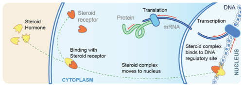
A hormone is a molecule released by a cell or a gland in one part of the body that exerts effects via specific receptors at other sites.

Key Points

The endocrine system relies on feedback mechanisms to control the hormone levels in the circulatory system.

Negative feedback systems prevent deviation from an ideal mean to maintain homeostasis.

Positive feedback systems facilitate deviation from the mean.

Key Terms

negative feedback: A system that prevents deviation from a mean value.

positive feedback: A system that promotes deviation from a mean value.

The physiological activity of a hormone depends largely on its concentration within the circulatory system. The effects of too high or too low a concentration of hormones can be damaging—this level must be tightly controlled.

The endocrine system relies on feedback systems to regulate hormone production and secretion.

Negative Feedback

Most endocrine glands are under negative feedback control that acts to maintain homeostasis, i.e., prevent deviation from an ideal value. A key example of a negative feedback system is the regulation of the thyroid hormone thyroxine, which regulates numerous key metabolic processes.

Briefly, neurons in the hypothalamus secrete a thyroid-releasing hormone that stimulates cells in the anterior pituitary to secrete thyroid-stimulating hormone. The thyroid-stimulating hormone then stimulates the release of thyroxine from the thyroid gland.

When the blood concentration of thyroxine rises above the ideal value as detected by sensory neurons, the hypothalamus is signaled to stop thyroid-releasing hormone production, which eventually lowers the levels of thyroxine in the blood. When these drop below the ideal value the hypothalamus is signaled to begin secreting thyroid-releasing hormone again.

Positive Feedback

Positive feedback mechanisms control self-perpetuating events, that is, they encourage deviation from the mean. Positive feedback systems are much less common although they do exist. A key example occurs during childbirth.

The hormone oxytocin is produced by the posterior pituitary that stimulates and enhances contractions during labor. During birth, as the baby moves through the birth canal, pressure receptors within the cervix signal the hypothalamus to stimulate the pituitary to secrete oxytocin.

Oxytocin travels to the uterus through the bloodstream, stimulating the muscles in the uterine wall to contract which in turn increases the activation of the pressure receptors and stimulates the further release of oxytocin. The strength of muscle contractions intensifies until the baby is born and the stimulation of the pressure receptors is removed, which stops the release of oxytocin.

The Endocrine system (along with the nervous system) controls and regulates the complex activities of the body. The Endocrine system regulates the activities of the body by secreting complex chemical substances (hormones) into the bloodstream. These secretions come from a variety of glands that control various organs of the body. The key functions are:

  • To regulate the metabolic functions of the body.
  • To regulate the rate of chemical reactions in various cells.
  • To influence the ability of substances to transport themselves through cell membranes.

About Hormones

  • Hormones are secreted from the glands of the endocrine system, they are specific in that each hormone causes a response in a specific target organ or group of cells, rather than on the body as a whole. Exocrine hormones are secreted via a duct into the blood and usually affect a distant organ or tissue. Endocrine hormones are secreted within the tissue (rather than via a duct) and enter the bloodstream via capillaries.

Hormones can be grouped into three main types

  • amines, these are simple molecules
    • proteins and peptides which are made from chains of amino acids
    • steroids which are derived from cholesterol.
  • Glands – discharge hormones directly into the bloodstream. They have built-in feedback mechanisms that maintain a proper balance of hormones, and prevent excess hormone secretion. Low concentrations of a hormone will often trigger the gland to secrete. Once the concentrations of the hormone in the blood rise this may cause the gland to stop secreting until once again hormone concentrations fall. This feedback mechanism (which is characteristic of most glands) causes a cycle of hormone secretions.
  •  Disposal of waste – Once hormones have served their function on their target organs/tissues they are destroyed. They are either destroyed by the liver or the actual tissues of the target organs. They are then removed by the kidneys.

The Pituitary Gland

This is known as the “master gland” because it exerts control over all of the other glands of the endocrine system. Despite its importance, the pituitary gland is no larger than a small pea. The Pituitary gland is made up of two separate glands: the Anterior lobe which is an outgrowth of the pharynx, and the

  • The posterior lobe is an outgrowth of the brain composed of neural (nerve) tissue.
  • The Anterior Lobe of the pituitary plays the ‘master’ role secreting six major hormones that affect most of the body, including the other Endocrine glands:
  • ACTH (Adrenocorticotrophic hormone) stimulates the adrenal glands to secrete their hormones.
  • HGH (Human growth hormone) also known as the somatotrophic hormone is responsible for the growth of long bones, muscles, and viscera.
  • TSH (Thyroid-stimulating hormone) influences the structure of the thyroid and causes it to secrete thyroid hormone.
  • FSH (Follicle-stimulating hormone) stimulates female egg production or male sperm production.
  • PRL (Prolactin) in females causes the corpus luteum the area around the mature follicle to produce two important hormones: Oestrogen and Progesterone. During pregnancy, PRL is also responsible for the development of the glandular tissues of the breast which produce milk.
  • LH (Luteinizing hormone) works in conjunction with FSH in females to cause ovulation and prepares the uterus for pregnancy, in males the testes to secrete testosterone.
  • The Posterior Lobe of the Pituitary Gland (or neurohypophysis) stores and releases hormones secreted by the hypothalamus section of the brain including:
  • ADH (Antidiuretic hormone) stimulates the smooth muscles, blood vessels, and intestine. ADH increases the kidney’s permeability to water allowing the body to re-absorb water that would otherwise escape in urine.
  • OT (Oxytocin) stimulates the smooth muscles of the uterus during pregnancy, causing it to contract during labor. It also stimulates the lacteals (milk ducts) in the breast.

The Thyroid gland

The thyroid is a butterfly-shaped gland which is located at the base of the throat. It has two lobes separated in the middle by a strip of tissue (the isthmus). The Thyroid itself secretes three main hormones:

  • Thyroxine contains iodine which is essential for the body’s normal growth and metabolism. Thyroxine helps control body size, regulating not only the growth of tissues but also the differentiation or specialization of tissues.
  • Triiodothyronine has similar functions to thyroxine.
  • Calcitonin causes a decrease in the concentration of calcium in the blood. Calcitonin works with secretions from the parathyroid glands to maintain the balance of calcium necessary for the body to function.

People who have surgery to remove the thyroid gland (thyroidectomy) for cancer or other thyroid problems usually need to take thyroxine supplements in order to maintain normal weight and body functions.

The Parathyroids

There are four Parathyroid glands which are small and rounded, arranged in two pairs usually located above and below the thyroid. Each Parathyroid is small, yellow, and smooth, sometimes they imbed themselves in the thyroid itself.

Parathyroid hormone increases the blood concentrations of calcium and phosphorous, working to balance the Calcitonin which is secreted by the thyroid to maintain the body’s balance of calcium.

The Pancreas

The pancreas is a long, narrow, lobed gland located behind the stomach. The Pancreas has two types of cells: exocrine and endocrine cells. The exocrine cells secrete Pancreatic juices which are used in the duodenum as an important part in the digestive system. The endocrine cells are arranged in clusters throughout the Pancreas, these known as Islets of Langerhans. There are three types of endocrine cells; alpha cells which secrete glucagon, beta cells which secrete insulin, and delta cells which inhibit the secretion of glucagon and insulin:

  • Glucagon increases the blood glucose level by stimulating the liver causing convert Glycogen into Glucose (sugar).
  • Insulin increases the permeability of the cell to glucose, which the cells use for energy. By promoting the utilization of glucose by the tissue cells, insulin causes a decrease in the concentration of glucose in the blood. Insulin also promotes the storage of glycogen in the liver.

The Adrenal Glands

The adrenal glands resemble small caps perched on top of each kidney. The Adrenal is actually a combination of two glands the adrenal cortex and the adrenal medulla.

  • The adrenal cortex is essential for life, as opposed to the adrenal medulla which is important but not indispensable. The anterior pituitary controls the adrenal cortex by secreting the hormone ACTH. All of the secretions of the adrenal cortex are known as steroids, many of which can now be manufactured synthetically. The adrenal cortex is made up of three layers associated with three classes of hormones:
  • Mineralocorticoids are produced by the outer layer of the adrenal cortex, the most important of which is aldosterone. Aldosterone promotes the retention of sodium (Na+) and the excretion of potassium (K+). This helps to maintain both the electrolyte and water content of the body.
  • Glucocorticoids are produced by the middle cortex. These affect almost every cell in the body regulating the metabolism of fats, proteins, and carbohydrates. Cortisone is one such glucocorticoid.
  • Gonadal hormones are produced by the inner cortex, there are roughly even amounts of two types of hormones secreted: Androgen (male) and Estrogen (female). The adrenal gland is not the only gland to secrete sex hormones.
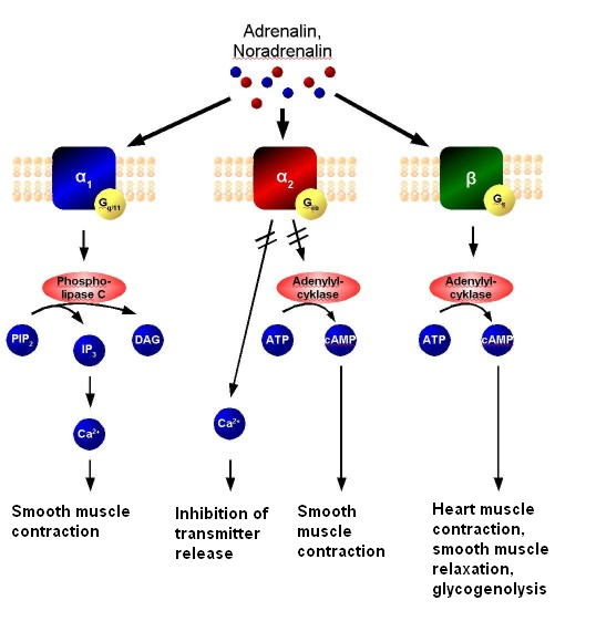
  • The Adrenal Medulla is the inner part of the adrenal gland. The hormones secreted affect the structures in the body that are under the control of the sympathetic nervous system, aiding the body to deal with stressful situations such as fright, attack or pursuit. They are both associated with an increased heartbeat, higher blood pressure, and higher blood glucose levels, thus preparing the body for quick action.
  • Adrenalin (or epinephrine) affects both alpha and beta receptors in the nervous system.
  • Noradrenalin (Norepinephrine) affects only the alpha receptors of the nervous system.

The Gonads

The gonads consist of ovaries in the female and testes in the male. These glands produce hormones important in the development and functioning of the reproductive organs. they are under the control of the pituitary gland and produce secondary sexual traits.

  • Male testes are egg-shaped glands located in the sac-like scrotum, and serve two main functions: (i) The production of sperm cells, and (ii) The secretion of testosterone. Testosterone is the masculizing hormone inducing male secondary sexual characteristics after puberty.
  • Female ovaries are two almond-shaped glands on each side of the uterus. They have three main functions; (i) Containing immature ova (eggs), (ii) secretion of estrogen and (ii) secretion of progesterone.
  • Estrogen is secreted by the adrenal cortex as well as the ovaries and is present in the blood of all females from puberty through to menopause. estrogen acts on the structure of the reproductive organs, especially during the menstrual cycle. This induces and maintains female secondary sexual characteristics. Progesterone works on the uterus to prepare it for the implantation of a fertilized ovum (egg). It causes the development of the breasts and is essential for the complete development of the maternal proportion of the placenta.
  • Giantism too much HGH is secreted before puberty.
  • Dwarfism is caused by a lack of HGH before puberty.
  • Diabetes Mellitus is a condition with under-secretion of insulin, causing the cells to lose their permeability to glucose preventing them from getting sugar needed for energy. Sugar remains in the blood and often the body will try and remove this leading to high sugar content in the urine, causing polyuria (passing of large volumes of urine) and polydipsia (excessive thirst).

Roots, suffixes, and prefixes

Most medical terms are comprised of a root word plus a suffix (word ending) and/or a prefix (beginning of the word). Here are some examples related to the Endocrine System.

component meaning example
A-, AN- without, lack muscular atrophy = ‘wasting away’ of muscles
ADEN- gland adenoma = tumor with gland-like structure
END-, Endo- within endocrine = secreting within
EXO- away from exocrine = secreting outwardly or away from
GLYCO-, GLUCO- sugar, sweet hyperglycemia = excessive blood sugar levels
PARA- near, beside parathyroid = beside the thyroid
POLY- much, many polyadenitis = inflammation of many glands
THYROID- thyroid hypoplasia = defective growth of the thyroid
-CRIME to secrete endocrine = endo (within) crime (secrete)
-TROPHY growth hypertrophy = excessive growth of an organ or part
-MAGALY enlargement hepatomegaly = enlarged liver with hepatitis
-PHYSIS growth a growth or outcropping (as opposed to a trophy where something is physically growing)

References

ByRx Harun

Mechanisms of Hormone Action – All About You Can Know

Mechanisms of Hormone Action/Hormones are the messenger molecules of the endocrine system. Endocrine hormones travel throughout the body in the blood. However, each hormone affects only certain cells, called target cells. A target cell is the type of cell on which a hormone has an effect. A target cell is affected by a particular hormone because it has receptor proteins that are specific to that hormone. A hormone travels through the bloodstream until it finds a target cell with a matching receptor it can bind to. When the hormone binds to a receptor, it causes a change within the cell. Exactly how this works depends on whether the hormone is a steroid hormone or a non-steroid hormone.

 Mechanisms of Hormone Action

Protein and peptide hormones, catecholamines like epinephrine, and eicosanoids such as prostaglandins find their receptors decorating the plasma membrane of target cells.

The binding of the hormone to the receptor initiates a series of events which leads to the generation of so-called second messengers within the cell (the hormone is the first messenger). The second messengers then trigger a series of molecular interactions that alter the physiologic state of the cell. Another term used to describe this entire process is signal transduction.

Structure of Cell Surface Receptors

Cell surface receptors are integral membrane proteins and, as such, have regions that contribute to three basic domains:

  • Extracellular domains: Some of the residues exposed to the outside of the cell interact with and bind the hormone – another term for these regions is the ligand-binding domain.
  • Transmembrane domains: Hydrophobic stretches of amino acids are “comfortable” in the lipid bilayer and serve to anchor the receptor in the membrane.
  • Cytoplasmic or intracellular domains: Tails or loops of the receptor that are within the cytoplasm react to hormone binding by interacting in some way with other molecules, leading to the generation of second messengers. Cytoplasmic residues of the receptor are thus the effector region of the molecule.

Several distinctive variations in receptor structure have been identified. As depicted below, some receptors are simple, single-pass proteins; many growth factor receptors take this form. Others, such as the receptor for insulin, have more than one subunit. Another class, which includes the beta-adrenergic receptor, is threaded through the membrane seven times.

Receptor molecules are neither isolated by themselves nor fixed in one location of the plasma membrane. In some cases, other integral membrane proteins interact with the receptor to modulate its activity. Some types of receptors cluster together in the membrane after binding hormones. Finally, as elaborated below, the interaction of the hormone-bound receptor with other membrane or cytoplasmic proteins is the key to the generation of second messengers and transduction of the hormonal signal.

Second Messenger Systems

Consider what would happen if, late at night, you noticed a building on fire. Hopefully, you would dial 911 or a similar emergency number. You would inform the dispatcher of the fire, and the dispatcher would, in turn, contact and “activate” a number of firemen. The firefighters would then rapidly go to work pouring water on the fire, setting up roadblocks, and the like. They would also probably activate other “players”, such as police and fire investigators that would come in later to try and determine the cause of the fire. Importantly, once the fire is out (or the building totally destroyed), the firemen go back to the station and to sleep.

The community response to a fire is, at least in some ways, analogous to a second messenger system involved in a hormone’s action. In the scenario described, you are the “first messenger”, the dispatcher is the “receptor”, the firefighters are the “second messengers”.

Currently, four-second messenger systems are recognized in cells, as summarized in the table below. Note that not only do multiple hormones utilize the same second messenger system, but a single hormone can utilize more than one system. Understanding how cells integrate signals from several hormones into a coherent biological response remains a challenge.

Second Messenger Examples of Hormones Which Utilize This System
Cyclic AMP Epinephrine and norepinephrine, glucagon, luteinizing hormone, follicle-stimulating hormone, thyroid-stimulating hormone, calcitonin, parathyroid hormone, antidiuretic hormone
Protein kinase activity Insulin, growth hormone, prolactin, oxytocin, erythropoietin, several growth factors
Calcium and/or phosphoinositides Epinephrine and norepinephrine, angiotensin II, antidiuretic hormone, gonadotropin-releasing hormone, thyroid-releasing hormone.
Cyclic GMP Atrial natriuretic hormone, nitric oxide

In all cases, the seemingly small signal generated by hormone binding its receptor is amplified within the cell into a cascade of actions that changes the cell’s physiologic state. Presented below are two examples of second messenger systems commonly used by hormones. The examples used are of glucagon and insulin, both of which ultimately work through a molecular switch involving protein phosphorylation. Be aware that in both cases, a very complex system is being simplified considerably.

Cyclic AMP Second Messenger Systems

Cyclic adenosine monophosphate (cAMP) is a nucleotide generated from ATP through the action of the enzyme adenylate cyclase. The intracellular concentration of cAMP is increased or decreased by a variety of hormones and such fluctuations affect a variety of cellular processes. One prominent and important effect of elevated concentrations of cAMP is the activation of a cAMP-dependent protein kinase called protein kinase A.

Protein kinase A is nominally in a catalytically inactive state but becomes active when it binds cAMP. Upon activation, protein kinase A phosphorylates a number of other proteins, many of which are themselves enzymes that are either activated or suppressed by being phosphorylated. Such changes in enzymatic activity within the cell clearly alter its state.

Now, let’s put this information together to understand the mechanism of action of a hormone-like glucagon

  • Glucagon binds its receptor in the plasma membrane of target cells (e.g. hepatocytes).
  • Bound receptor interacts with and, through a set of G proteins, turns on adenylate cyclase, which is also an integral membrane protein.
  • Activated adenylate cyclase begins to convert ATP to cyclic AMP, resulting in an elevated intracellular concentration of cAMP.
  • High levels of cAMP in the cytosol make it probable that protein kinase A will be bound by cAMP and therefore catalytically active.
  • Active protein kinase A “runs around the cell” adding phosphates to other enzymes, thereby changing their conformation and modulating their catalytic activity – – – abracadabra, the cell has been changed!
  • Levels of cAMP decrease due to destruction by cAMP-phosphodiesterase and the inactivation of adenylate cyclase.

In the above example, the hormone’s action was to modify the activity of pre-existing components in the cell. Elevations in cAMP also have important effects on the transcription of certain genes.

Tyrosine Kinase Second Messenger Systems

The receptors for several protein hormones are themselves protein kinases which are switched on by the binding of hormones. The kinase activity associated with such receptors results in the phosphorylation of tyrosine residues on other proteins. Insulin is an example of a hormone whose receptor is a tyrosine kinase.

The hormone binds to domains exposed on the cell’s surface, resulting in a conformational change that activates kinase domains located in the cytoplasmic regions of the receptor. In many cases, the receptor phosphorylates itself as part of the kinase activation process. The activated receptor phosphorylates a variety of intracellular targets, many of which are enzymes that become activated or are inactivated upon phosphorylations was seen with cAMP second messenger systems, activation of receptor tyrosine kinases leads to rapid modulation in a number of target proteins within the cell. Interestingly, some of the targets of receptor kinases are protein phosphatases which, upon activation by receptor tyrosine kinase, become competent to remove phosphates from other proteins and alter their activity. Again, a seemingly small change due to hormone binding is amplified into a multitude of effects within the cell.
In some cases, the binding of the hormone to a surface receptor induces a tyrosine kinase cascade even though the receptor is not itself a tyrosine kinase. The growth hormone receptor is one example of such a system – the interaction of growth hormone with its receptor leads to activation of cytoplasmic tyrosine kinases, with results conceptually similar to that seen with receptor kinases.

Steroid Hormones

Steroid hormones are made of lipids, such as phospholipids and cholesterol. They are fat-soluble, so they can diffuse across the plasma membrane of target cells and bind with receptors in the cytoplasm of the cell. The steroid hormone and receptor form a complex that moves into the nucleus and influences the expression of genes, essentially acting as a transcription factor. Examples of steroid hormones include cortisol and sex hormones.

Asteroid hormone crosses the plasma membrane of a target cell and binds with a receptor inside the cell.

Non-Steroid Hormones

Non-steroid hormones are made of amino acids. They are not fat-soluble, so they cannot diffuse across the plasma membrane of target cells. Instead, a non-steroid hormone binds to a receptor on the cell membrane. The binding of the hormone triggers an enzyme inside the cell membrane. The enzyme activates another molecule, called the second messenger, which influences processes inside the cell. Most endocrine hormones are non-steroid hormones, including insulin and thyroid hormones.

Direct Gene Activation and the Second-Messenger System

Nuclear receptors function as transcription factors because they can bind to DNA and regulate gene expression.

Key Points

Receptors that can directly influence gene expression are termed nuclear receptors.
Type I nuclear receptors (found in cytosol) are modified to translocate to the nucleus upon hormone binding.

Type II nuclear receptors remain in the nucleus where they often create a complex with co-repressor proteins, which are released upon hormone binding.

Secondary messengers relay signals from receptors on the cell surface to the target molecules.

The secondary messenger systems bind hormones to a receptor that causes a cascade of changes that leads to actions.

Key Terms

nuclear receptor: A class of proteins found within cells that are responsible for sensing steroid and thyroid hormones and certain other molecules, as well as influencing gene expression upon activation.

secondary messenger: Molecules that relay signals from receptors on the cell surface to target molecules inside the cell, in the cytoplasm or nucleus.

hormone response element: A short sequence of DNA within the promoter of a gene that is able to bind a specific hormone-receptor complex and therefore regulate gene expression.

Hormones can alter cell activity by binding with a receptor. Receptors can either directly influence gene expression and thus cell activity, or induce a secondary signaling cascade that will in turn influence cell activity.

Direct Gene Activation

Receptors that can directly influence gene expression are termed nuclear receptors. Located within the cytosol or nucleus, nuclear receptors are the target of steroid and thyroid hormones that are able to pass through the cell membrane. Nuclear receptors can bind directly to DNA to regulate specific gene expressions and are, therefore, classified as transcription factors.

Nuclear receptors can be classified into two broad classes according to their mechanism of action and their sub-cellular distribution in the absence of ligand. Type I nuclear receptors are located in the cytosol. Upon binding to a hormone the receptor and hormone translocate into the nucleus, and bind to specific sequences of DNA known as hormone response elements (HREs).

Type II receptors are retained in the nucleus. In the absence of ligand, type II nuclear receptors often form a complex with co-repressor proteins. Hormone binding to the nuclear receptor results in dissociation of the co-repressor and the recruitment of co-activator proteins.

This figure depicts the mechanism of a class I nuclear receptor (NR) that, in the absence of ligand, is located in the cytosol. Hormone binding to the NR triggers translocation to the nucleus, where the NR binds to a specific sequence of DNA known as a hormone response element (HRE).

Lipid soluble hormones directly regulate gene expression: This figure depicts the mechanism of a class I nuclear receptor (NR) that, in the absence of ligand, is located in the cytosol. Hormone binding to the NR triggers translocation to the nucleus, where the NR binds to a specific sequence of DNA known as a hormone response element (HRE).

Secondary Messengers

For lipophobic hormones that cannot pass the cellular membrane, activity is mediated and amplified within a cell by the action of second messenger mechanisms (molecules that relay signals from receptors on the cell surface to target molecules inside the cell in the cytoplasm or nucleus).

Most hormone receptors are G protein-coupled receptors. Upon hormone binding, the receptor undergoes a conformational change and exposes a binding site for a G-protein. The G-protein is bound to the inner membrane of the cell and consists of three sub-units: alpha, beta, and gamma.

Upon binding to the receptor, it releases a GTP molecule, at which point the alpha sub-unit of the G-protein breaks free from the beta and gamma sub-units and is able to move along the inner membrane until it contacts another membrane-bound protein: the primary effector.

The primary effector then has an action, which creates a signal that can diffuse within the cell. This signal is called the secondary messenger. The secondary messenger may then activate a secondary effector, whose effects depend on the particular secondary messenger system.

This is a general schematic diagram of second messenger generation following the activation of membrane-bound receptors. 1. The agonist activates the membrane-bound receptor. 2. G-protein is activated and produces an effector. 3. The effector stimulates a second messenger synthesis. 4. The second messenger activates an intercellular process.

Second messenger mechanisms: General schematic of second messenger generation following activation of membrane-bound receptors. 1. The agonist activates the membrane-bound receptor. 2. G-protein is activated and produces an effector. 3. The effector stimulates a second messenger synthesis. 4. The second messenger activates an intercellular process.

Target Cell Specificity

Hormones target a limited number of cells (based on the presence of a specific receptor) as they circulate in the bloodstream.

Key Points

Target cells are cells that are receptive to a secreted hormone.

Target cell activation is
dependent on three factors; the hormone levels in the blood, the receptor levels on the target cell, and hormone–receptor affinity.

Key Terms

target cell: A cell that is receptive to a secreted hormone.

EXAMPLES

An XY fetus will develop along a female pathway if the target cells fail to respond to androgen. This androgen insensitivity occurs when the receptors on the target cells are unable to accept the hormone due to an impairment in receptor shape.

In endocrinology, target cells can refer to the cells where hormones have an effect. Target cells are capable of responding to hormones because they display receptors to which the circulating hormone can bind. In this way, hormones only affect a limited number of cells even though they are transported in the bloodstream throughout the body.

Target cell activation is dependent on three factors:

  • The levels of hormones in the blood.
  • The relative number of hormone receptors on the target cell.
  • The hormone–receptor affinity.

Modulation of these factors can control target cell response. For example, after receptor stimulation, the signaling target cell often sends feedback to the hormone-secreting tissue to down-regulate hormone expression.

Additionally, the target cell can up or down-regulate receptor expression to make it more or less sensitive to the same hormone. Finally, hormone–receptor affinity can be altered by the expression of associated inhibitory or co-activating factors.

In some instances, alterations of receptor structure due to a genetic mutation can lead to a reduction in hormone–receptor affinity, as in the case of androgen insensitivity.

Onset, Duration, and Half-Life of Hormone Activity

A hormone’s half-life and duration of activity are limited and vary from hormone to hormone.

Key Points

The hormone receptors are dynamic structures that vary in number and sensitivity, that depend on the levels of the stimulating hormone.

The blood levels of hormones reflect a balance between secretion and degradation/excretion by the liver and kidneys.

The biological half- life of a hormone is the time it takes for the hormone to lose half of its physiological activity.

The duration of hormone activity refers to the duration of altered cellular behavior triggered by hormone binding.

Key Terms

hormone receptor: A molecule that binds to a specific hormone that triggers alterations in cell activity.

half-life: The time it takes for a substance (drug, radioactive nuclide, or other) to lose half of its pharmacological, physiological, or radiological activity.

EXAMPLES

Vitamin D is a hormone that has a half-life of one to two months. If one obtains vitamin D solely through sun (UVB) exposure during the summer months, serum vitamin D levels will be critically low by late winter. This is one reason why current recommendations are to take vitamin D supplements in order to maintain serum vitamin D levels throughout the year.

The number of hormone molecules available for complex formation is usually the key factor that determines the level at which signal transduction pathways are activated. The number of hormone molecules that are available is determined by the concentration of circulating hormones.

Half-Life

The blood levels of hormones reflect a balance between synthesis/secretion and degradation/excretion. The liver and kidneys are the major organs that degrade hormones with breakdown products excreted in urine and feces.

A hormone’s half-life and duration of activity are limited and vary from hormone to hormone. For instance, the biological half-life of luteinizing hormone is 20 minutes, which is shorter than that of a follicle-stimulating hormone (three to four hours), and of human chorionic gonadotropin (24 hours).

A biological half-life or elimination half-life is the time it takes for a substance such as a hormone or a drug to lose half of its pharmacologic or physiologic activity. In a medical context, the half-life may also describe the time it takes for the blood plasma concentration of a substance to halve (plasma half-life) its steady state.

The relationship between the biological and plasma half-lives of a substance can be complex, due to factors including their accumulation in tissues, active metabolites, and receptor interactions.

Duration

The duration of hormone activity refers to the duration of events that were stimulated by hormone-receptor binding. While typically relatively short and measured in minutes or hours, certain events, such as the onset of puberty, are much longer-lasting.

This image depicts the levels of certain hormones during the menstrual cycle, as they correspond to follicular growth and ovulation. Once ovulation starts, the hormone progesterone overtakes the follicle-stimulating hormone, the hormone estrogen, and the luteinizing hormone that were present earlier.

Hormone levels during the menstrual cycle: This image depicts the levels of certain hormones during the menstrual cycle (B), as they correspond to follicular growth and ovulation (A). 1. Follicle-stimulating hormone 2. Estrogen 3. Luteinizing hormone 4. Progesterone.

Mechanism

Growth Hormone

Growth Hormone has a direct and indirect mechanism of action. The direct effect of growth hormone involves growth hormone directly binding to its receptors on target cells to stimulate a response. The indirect effect is mediated by the action of insulin-like growth factor-1(IGF-1), which is secreted by the liver hepatocytes in response to growth hormone. Insulin-like growth factor-1 binds to its receptor, IGF-1R, on the cellular surface and activates a tyrosine kinase-mediated intracellular signaling pathway that phosphorylates various proteins intracellularly leading to increased anabolism, cellular replication and division, and metabolism.

Prolactin

Prolactin initiates its effect by binding to the prolactin receptor found on various tissues across the body, including but not limited to mammillary glands, ovaries, skeletal muscle, uterus, and thymus. Upon the binding of prolactin to its receptor, Jannu kinase 2, a tyrosine kinase is activated that furthermore initiates the JAK-STAT pathway.

FSH & LH

Both LH & FSH bind to G protein-coupled receptors. Upon binding to the receptor, adenylyl cyclase, an enzyme is activated, which goes on to produce cyclic-AMP. The intracellular concentrations of cyclic-AMP rise, which further activates a kinase molecule called protein kinase A. Protein kinase A primarily functions to phosphorylate specific intracellular proteins that then subsequently complete the physiological actions of FSH and LH.

Adrenocorticotropic Hormone

ACTH interacts with G protein-coupled receptors found on the extracellular membranes of the zona fasciculata and zona reticularis of the adrenal cortex. cAMP is the secondary messenger system. Activation of the g-couple receptor activates adenylyl cyclase, thus increase cAMP production and subsequent activation of Protein Kinase A.

Thyroid Stimulating Hormone (TSH)

TSH binds and activates the TSH receptor (TSHR) found on the basolateral surface of thyroid follicle cells. This binding site is a G-protein coupled receptor (GPCR), which couples to both Gs and Gq G-proteins, and hence activating both the cAMP pathway (via Gsa) and the phosphoinositol/calcium (IP/Ca2+; via Gq) second messenger signaling cascades. The Gs pathway activates iodide uptake,  increases thyroid hormone production, and enhances gland growth and differentiation. The Gq pathway is rate-limiting for hormone production by stimulating iodide organification.

Vasopressin

Two regulating receptors, the subfornical organ and the organum vasculum in the hypothalamus, since water deprivation and signal for ADH secretion. A small concentration of vasopressin is sufficient to generate water conservation in the renal tubules. The renal tubules are divided into the proximal, descending, ascending, distal regions, and the collecting duct. The most ADH-dependent segment of the renal tubule is the collecting duct, which has ADH receptors on its basolateral side for ADH to bind and stimulate the Gs protein. The Gs protein stimulates adenylyl cyclase, which further converts ATP into cAMP. High levels of cAMP cause the phosphorylation of protein kinase A, subsequently opening water channels known as aquaporins to allow passage of water from the luminal side to the basolateral side.

Oxytocin

Oxytocin binds to its extracellular receptor present in the myometrium of the uterus, which then activates the Gq protein further leading to activation of phospholipase C. The phospholipase C functions to break down the phosphoinositol diphosphate into two components, Inositol triphosphate (IP3) which will release calcium from the sarcoplasmic reticulum and diacylglycerol (DAG) which will activate protein kinase C. The protein kinase C phosphorylates proteins specifically on the cell membrane to allow calcium entry from the extracellular space. The increased intracellular calcium generates enough energy to cause the contraction of the uterus.

During lactation, when the newborn suckles, it transmits signals to the central nervous system to release oxytocin, a process known as the “milk letdown reflex.” The oxytocin binds to the breast myoepithelial cell receptors and initiates the same Gq cascade similar to uterine contraction, and ejects milk into the baby’s oral cavity.

References

ByRx Harun

Hormone Action – Mechanisms of Hormone Action

Hormones are released into the bloodstream through which they travel to target sites. … Water-soluble hormones bind to a receptor protein on the plasma membrane of the cell. Receptor stimulation results in a change in cell activity, which may send feedback to the original hormone-producing cel

Mechanisms of Hormone Action

A hormone is a secreted chemical messenger that enables the communication between cells and tissues throughout the body.

Key PointsHormones are released into the bloodstream through which they travel to target sites.

The target cell has receptors specific to a given hormone and will be activated by either a lipid-soluble (permeable to the plasma membrane) or water-soluble hormone (binds to a cell-surface receptor).

Lipid-soluble hormones diffuse through the plasma membrane to enter the target cell and bind to a receptor protein.

Water-soluble hormones bind to a receptor protein on the plasma membrane of the cell.

Receptor stimulation results in a change in cell activity, which may send feedback to the original hormone-producing cell.

Key Terms

Water-soluble hormone: A lipophobic hormone that binds to a receptor on, or within, the plasma membrane, to initiate an intracellular signaling cascade.

hormone: A molecule released by a cell or a gland in one part of the body that sends out messages affecting cells in other parts of the organism.

Lipid-soluble hormone: A lipophilic hormone that passes through the plasma membrane of a cell, binds to an intracellular receptor and changes gene expression.

A hormone is a chemical messenger that enables the communication between cells. Hormones are secreted by the glands of the endocrine system and they serve to maintain homeostasis and to regulate numerous other systems and processes, including reproduction and development.

Hormone Signaling

The glands of the endocrine system secrete hormones directly into the extracellular environment. The hormones then diffuse to the bloodstream via capillaries and are transported to the target cells through the circulatory system. This allows hormones to affect tissues and organs far from the site of production or to apply systemic effects to the whole body.

Hormone-producing cells are typically specialized and reside within a particular endocrine gland, such as thyrocytes in the thyroid gland. Hormones exit their cell of origin through the process of exocytosis or by other means of membrane transport.

Cellular recipients of a particular hormonal signal may be one of several cell types that reside within a number of different tissues. This is so in the case of insulin, which triggers a diverse range of systemic physiological effects. Different tissue types may also respond differently to the same hormonal signal. As a result, hormonal signaling is elaborate and hard to dissect.

Hormones activate target cells by diffusing through the plasma membrane of the target cells (lipid-soluble hormones) to bind a receptor protein within the cytoplasm of the cell, or by binding a specific receptor protein in the cell membrane of the target cell (water-soluble proteins). In both cases, the hormone complex will activate a chain of molecular events within the cell that will result in the activation of gene expression in the nucleus.

The reaction of the target cells may then be recognized by the original hormone-producing cells, leading to a down-regulation in hormone production. This is an example of a homeostatic negative feedback loop.

This is a diagram that shows how lipid-soluble hormones, such as estrogen, activate the hormone receptors and bind themselves to a cell. The diagram shows a steroid hormone passing through the cytoplasm, where it binds to a steroid receptor, and then into the nucleus where it activates mRNA transcriptions.

Lipid-soluble hormone receptor activation: Nuclear hormone receptors are activated by a lipid-soluble hormone such as estrogen, binding to them inside the cell. Lipid-soluble hormones can cross the plasma membrane.

Steps of Hormonal Signaling

  • Biosynthesis of a particular hormone in a particular tissue.
  • Storage and secretion of the hormone.
  • Transport of the hormone to the target cells, tissues, or organs.
  • Recognition of the hormone by an associated cell membrane or an intracellular receptor protein.
  • Relay and amplification of the received hormonal signal via a signal transduction process.
  • Potential feedback to a hormone-producing cell.

This diagram shows how water-soluble hormones, such as epinephrine, bind to a cell-surface localized receptor, initiating a signaling cascade using intracellular second messengers.

Water-soluble hormone receptor activation: Water-soluble hormones, such as epinephrine, bind to a cell-surface localized receptor, initiating a signaling cascade using intracellular second messengers.

Hormone Classes

Hormones are typically divided into three classes:

Peptide: Hormones that are modified amino acids or short (peptide) or long (protein) chains of amino acids. Additionally, they can contain carbohydrate moieties.

Lipid: Steroid hormones that contain lipids synthesized from cholesterol and eicosanoids that contain lipids synthesized from the fatty acid chains of phospholipids found in the plasma membrane.

Monoamine: Hormones derived from aromatic amino acids such as
phenylalanine, tyrosine, and tryptophan.

Hormone Receptors

Hormones activate a cellular response in the target cell by binding to a specific receptor in the target cell.

Key Points

For water-soluble proteins, the receptor will be at the plasma membrane of the cell.

The ligand-bound receptor will trigger a cascade of secondary messengers inside the cell.

For lipid-soluble hormones, the receptor is typically located within the cytoplasm or nucleus of the cell.

The binding of the hormone allows the receptor to influence transcription in the nucleus, either alone or in association with other transcription factors.

The number of hormone molecules is usually the key factor for determining hormone action and it is determined by the concentration of circulating hormones, which in turn is influenced by the rate and level of secretion.

Another limiting factor for hormone action is the effective concentration of hormone-bound receptor complexes that are formed within the cell. This is determined by the number of hormone/receptor molecules available for complex formation and the binding affinity between the hormone and receptor.

Key Terms

secondary messenger: These are molecules that relay signals from receptors on the cell surface to target molecules inside the cell, in the cytoplasm, or the nucleus.

A hormone receptor is a molecule that binds to a specific hormone. Receptors for peptide hormones tend to be found on the plasma membrane of cells, whereas receptors for lipid-soluble hormones are usually found within the cytoplasm.

Upon hormone binding, the receptor can initiate multiple signaling pathways that ultimately lead to changes in the behavior of the target cells.

The hormone activity within a target cell is dependent on the effective concentration of hormone-receptor complexes that are formed. The number of these complexes is in turn regulated by the number of hormone or receptor molecules available, and the binding affinity between hormone and receptor.

Lipophobic Hormones

Many hormones are composed of polypeptides—such as thyroid-stimulating hormones, follicle-stimulating hormones, luteinizing hormones, and insulin. These molecules are not lipid-soluble and therefore cannot diffuse through cell membranes.

The receptors for these hormones need to be localized to the cells’ plasma membranes. Following an interaction with the hormones, a cascade of secondary effects within the cytoplasm of the cell is triggered, often involving the addition or removal of phosphate groups to cytoplasmic proteins, changes in ion channel permeability, or an increase in the concentrations of intracellular molecules that may act as secondary messengers, such as cyclic AMP.

Lipophilic Hormones

Lipophilic hormones—such as steroid or thyroid hormones—are able to pass through the cell and nuclear membrane; therefore receptors for these hormones do not need to be, although they sometimes are, located in the cell membrane.

The majority of lipophilic hormone receptors are transcription factors that are either located in the cytosol and move to the cell nucleus upon activation or remain in the nucleus waiting for the steroid hormone to enter and activate them.
Upon binding by the hormone the receptor undergoes a conformational change, and the receptor together with the bound hormone influence transcription, either alone or in association with other transcription factors.

This is a diagram of a hormone passing into a nuclear envelope, binding to a receptor, then leaving the nucelar envelope with changed cell function. The example is of a thyroid hormone receptor (TR) heterodimerized to the RXR. In the absence of a ligand, the TR is bound to a corepressor protein. Ligand binding to the TR causes a dissociation of co-repressor and recruitment of co-activator proteins, which in turn recruit additional proteins (such as RNA polymerase) that are responsible for the transcription of downstream DNA into RNA, and eventually into protein that results in a change in cell function.

Example hormone receptor: The thyroid hormone receptor (TR) heterodimerized to the RXR. In the absence of a ligand, the TR is bound to a corepressor protein. Ligand binding to the TR causes dissociation of co-repressor and recruitment of co-activator proteins, which in turn recruit additional proteins (such as RNA polymerase) that are responsible for the transcription of downstream DNA into RNA, and eventually into protein that results in a change in cell function.

Chemistry of Hormones

There are three classes of hormones: peptide hormones, lipid hormones, and monoamine hormones.

Key Points

Peptide hormones are comprised of short (peptides) and long ( proteins ) chains of amino acids. They are water-soluble but cannot pass through the plasma membrane alone.

Glycoprotein hormones have a carbohydrate moiety attached to the protein.

Lipid hormones include steroid and eicosanoid hormones. They are lipid-soluble and can pass through the plasma membrane.

Steroid hormones are derived from cholesterol and eicosanoid hormones from fatty acids that compose the plasma membrane.

The third class of hormones is the monoamines that are derived from aromatic amino acids like phenylalanine, tyrosine, and tryptophan.

A hormone is a chemical released by a cell or a gland in one part of the body that sends out messages that affect cells in other parts of the organism.

There are three classes of hormones:

  1. Peptide hormones
  2. Lipid-derived hormones
  3. Monoamine hormones

Peptide Hormones

Peptide hormones consist of short chains of amino acids, such as vasopressin, that are secreted by the pituitary gland and regulate osmotic balance; or long chains, such as insulin, that are secreted by the pancreas, which regulates glucose metabolism.

Some peptide hormones contain carbohydrate side chains and are termed glycoproteins, such as the follicle-stimulating hormone. All peptide hormones are hydrophilic and are therefore unable to cross the plasma membrane alone.

This is a color illustration of the molecular structure of a peptide hormone.

Peptide hormone: Representation of the molecular structure of a peptide hormone.

Lipid-Derived Hormones

Lipid and phospholipid-derived hormones are produced from lipids such as linoleic acid and arachidonic acid. Steroid hormones, which form the majority of lipid hormones, are derived from carbohydrates; for example, testosterone is produced primarily in the testes and plays a key role in the development of the male reproductive system.

Eicosanoids are also lipid hormones that are derived from fatty acids in the plasma membrane. Unlike other hormones, eicosanoids are not stored in the cell—they are synthesized as required. Both are lipophilic and can cross the plasma membrane.

Monoamine Hormones

Monoamine hormones are derived from single aromatic amino acids like phenylalanine, tyrosine, and tryptophan. For example, the tryptophan-derived melatonin that is secreted by the pineal gland regulates sleep patterns.

Transport of Hormones

Hormones synthesized by the endocrine glands are transported throughout the body by the bloodstream.

Key Points

Hormones are typically secreted into the systemic circulation. However, some are secreted into portal systems that allow for direct hormone targeting.

Hormones can exist freely in the systemic circulation, but the majority are bound with transport proteins.

Transport proteins hold hormones inactive in systemic circulation and create a reservoir within the circulation that facilitates an even distribution of hormones throughout the tissue or organ.

Key Terms

transport protein: A protein that binds with a hormone in systemic circulation that facilitates its efficient transport.

The endocrine system is a system of ductless glands that secrete hormones directly into the circulatory system to be carried long distances to other target organs that regulate key body and organ functions.

Some endocrine glands secrete into a portal system rather than the systemic circulation that allows for the direct targeting of hormones. For example, hormones secreted by the pancreas pass into the hepatic portal vein that transports them directly to the liver.

Once within the circulatory system, a small proportion of hormones circulate freely, however, the majority are bound with a transport protein. Mainly produced in the liver, these transport proteins are hormone specific, such as the sex hormone-binding globulin that binds with the sex hormones.

When bound with a transport protein hormones are typically inactive, and their release is often triggered in regions of low hormone concentration or can be controlled by other factors. Therefore, transport proteins can act as a reservoir within the circulatory system and help ensure an even distribution of hormones within an organ or tissue.

reference


ByRx Harun

Endocrine System – Anatomy, Types, Functions

The endocrine system is the collection of glands that produce hormones that regulate metabolism, growth and development, tissue function, sexual function, reproduction, sleep, and mood, among other things.

The endocrine system is a messenger system comprising feedback loops of the hormones released by internal glands of an organism directly into the circulatory system, regulating distant target organs. In invertebrates, the hypothalamus is the neural control center for all endocrine systems. In humans, the major endocrine glands are the thyroid gland and the adrenal glands. The study of the endocrine system and its disorders is known as endocrinology.

Structure

Major endocrine systems

The human endocrine system consists of several systems that operate via feedback loops. Several important feedback systems are mediated via the hypothalamus and pituitary.[rx]

  • TRH – TSH – T3/T4
  • GnRH – LH/FSH – sex hormones
  • CRH – ACTH – cortisol
  • Renin – angiotensin – aldosterone
  • leptin vs. insulin

Glands

Endocrine glands are glands of the endocrine system that secrete their products, hormones, directly into interstitial spaces and then absorbed into the blood rather than through a duct. The major glands of the endocrine system include the pineal gland, pituitary gland, pancreas, ovaries, testes, thyroid gland, parathyroid gland, hypothalamus, and adrenal glands. The hypothalamus and pituitary gland are neuroendocrine organs.

The hypothalamus and the anterior pituitary are two out of the three endocrine glands that are important in cell signaling. They are both part of the HPA axis which is known to play a role in cell signaling in the nervous system.

Hypothalamus: The hypothalamus is a key regulator of the autonomic nervous system. The endocrine system has three sets of endocrine outputs[rx] which include the magnocellular system, the parvocellular system, and autonomic intervention. The magnocellular is involved in the expression of oxytocin or vasopressin. The parvocellular is involved in controlling the secretion of hormones from the anterior pituitary.

Anterior Pituitary: The main role of the anterior pituitary gland is to produce and secret tropic hormones.[rx] Some examples of tropic hormones secreted by the anterior pituitary gland include TSH, ACTH, GH, LH, and FSH.

Cells

There are many types of cells that make up the endocrine system and these cells typically make up larger tissues and organs that function within and outside of the endocrine system.

  • Hypothalamus
  • Anterior pituitary gland
  • Pineal gland
  • Posterior pituitary gland
    • The posterior pituitary gland is a section of the pituitary gland. This organ secretes hormones such as antidiuretic hormone (ADH) and oxytocin. ADH functions to help the body to retain water; this is important in maintaining a homeostatic balance between blood solutions and water. Oxytocin functions to induce uterine contractions, stimulate lactation, and allows for ejaculation.[5][6]
  • Thyroid gland
    • follicular cells of the thyroid gland produce and secrete T3 and T4 in response to elevated levels of TRH, produced by the hypothalamus, and subsequently elevated levels of TSH, produced by the anterior pituitary gland, which further regulates the metabolic activity and rate of all cells, including cell growth and tissue differentiation.
  • Parathyroid gland
    • Epithelial cells of the parathyroid glands are richly supplied with blood from the inferior and superior thyroid arteries and secrete parathyroid hormone (PTH). PTH acts on bone, the kidneys, and the GI tract to increase calcium reabsorption and phosphate excretion. In addition, PTH stimulates the conversion of Vitamin D to its most active variant, 1,25-dihydroxyvitamin D3, which further stimulates calcium absorption in the GI tract.[rx]
  • Adrenal glands
    • Adrenal cortex
    • Adrenal medulla
  • Pancreas
    • Alpha cells
      • The alpha cells of the pancreas secrete hormones to maintain homeostatic blood sugar. Insulin is produced and excreted to lower blood sugar to normal levels. Glucagon, another hormone produced by alpha cells, is secreted in response to low blood sugar levels; glucagon stimulates glycogen stores in the liver to release sugar into the bloodstream to raise blood sugar to normal levels.[7]
    • Beta cells
    • Delta cells
    • F Cells
  • Ovaries
    • Granulosa cells
  • Testis
    • Leydig cells

The endocrine system is a system of ductless glands that secrete hormones—chemical messengers that are carried for long distances.

Key Points

The endocrine system is a system of ductless glands that secrete hormones directly into the circulatory system to be carried long distances to other target organs that regulate key body and organ functions.

The major endocrine glands include the pituitary, pineal, ovaries, testes, thyroid, hypothalamus, and adrenal glands.

Key Terms

hormone: A molecule released by a cell or a gland in one part of the body that sends out messages affecting cells in other parts of the organism.

endocrine system: The system of ductless glands that secretes hormones directly into the circulatory system.

The Endocrine System

The endocrine system is a system of ductless glands that secretes hormones directly into the circulatory system to be carried long distances to other target organs regulating key body and organ functions. For example, the pineal gland, located at the base of the brain, secretes the hormone melatonin, responsible for regulating sleep patterns.

Endocrine glands are typically well vascularized and the cells comprising the tissue are typically rich in intracellular vacuoles or granules that store hormones prior to release. Endocrine signaling is typically slow to initiate but is prolonged in response; this provides a counterpoint to the more rapid and short-lived nervous system signals.

The endocrine system is in contrast to the exocrine system, which features ducted glands that secrete substances onto an epithelial surface; for example, a sweat gland. Additionally, the endocrine system is differentiated from shorter distance signaling such as autocrine (a cell affecting itself), juxtacrine (a cell affecting its direct neighbors), and paracrine (a cell affecting other nearby cells) signaling.

Key Endocrine Glands

The major endocrine glands include the pituitary, pineal, ovaries, testes, thyroid, hypothalamus and adrenal glands, additionally other tissues such as the kidney and liver also display secondary adrenal functions.

This is a drawing of the head and neck that shows the locations of the endocrine systems. The endocrine systems found in the head and neck include the hypothalamus, pineal, pituitary and thyroid glands.

Endocrine glands of the head and neck: The endocrine systems found in the head and neck include the hypothalamus, pineal, pituitary, and thyroid glands.

Hormones Produced by the Major Hormone-Producing (i.e., Endocrine) Glands and Their Primary Functions

Endocrine Gland Hormone Primary Hormone Function
Hypothalamus Corticotropin-releasing hormone (CRH) Stimulates the pituitary to release adrenocorticotropic hormone (ACTH)
Gonadotropin-releasing hormone (GnRH) Stimulates the pituitary to release luteinizing hormone (LH) and follicle-stimulating hormone (FSH)
Thyrotropin-releasing hormone (TRH) Stimulates the pituitary to release thyroid-stimulating hormone (TSH)
Growth hormone-releasing hormone (GHRH) Stimulates the release of growth hormone (GH) from the pituitary
Somatostatin Inhibits the release of GH from the pituitary
Dopamine Inhibits the release of prolactin from the pituitary
Anterior pituitary gland ACTH Stimulates the release of hormones from the adrenal cortex
LH In women, stimulates the production of sex hormones (i.e., estrogens) in the ovaries as well as during ovulation; in men, stimulates testosterone production in the testes
FSH In women, stimulates follicle development; in men, stimulates sperm production
TSH Stimulates the release of thyroid hormone
GH Promotes the body’s growth and development
Prolactin Controls milk production (i.e., lactation)
Posterior pituitary gland1 Vasopressin Helps control the body’s water and electrolyte levels
Oxytocin Promotes uterine contraction during labor and activates milk ejection in nursing women
Adrenal cortex Cortisol Helps control carbohydrate, protein, and lipid metabolism; protects against stress
Aldosterone Helps control the body’s water and electrolyte regulation
Testes Testosterone Stimulates development of the male reproductive organs, sperm production, and protein anabolism
Ovaries Estrogen (produced by the follicle) Stimulates development of the female reproductive organs
Progesterone (produced by the corpus luteum) Prepares uterus for pregnancy and mammary glands for lactation
Thyroid gland Thyroid hormone (i.e., thyroxine [T4] and triiodothyronine [T3]) Controls metabolic processes in all cells
Calcitonin Helps control calcium metabolism (i.e., lowers calcium levels in the blood)
Parathyroid gland Parathyroid hormone (PTH) Helps control calcium metabolism (i.e., increases calcium levels in the blood)
Pancreas Insulin Helps control carbohydrate metabolism (i.e., lowers blood sugar levels)
Glucagon Helps control carbohydrate metabolism (i.e., increases blood sugar levels)
1These hormones are produced in the hypothalamus but stored in and released from the posterior pituitary gland.

Comparing the Nervous and Endocrine Systems

The nervous system and endocrine system both use chemical messengers to signal cells, but each has a different transmission speed.

Key Points

The nervous system can respond quickly to stimuli, through the use of action potentials and neurotransmitters.

Responses to nervous system stimulation are typically quick but short-lived.

The endocrine system responds to stimulation by secreting hormones into the circulatory system that travel to the target tissue.

Responses to endocrine system stimulation are typically slow but long-lasting.

Key Terms

hormone: A molecule released by a cell or a gland in one part of the body that sends out messages affecting cells in other parts of the organism.

neurotransmitters: Endogenous chemicals that transmit signals from a neuron to a target cell across a synapse.

The body must maintain a constant internal environment, through a process termed homeostasis, while also being able to respond and adapt to external events. The nervous and endocrine systems both work to bring about this adaptation, but their response patterns are different. The nervous system and the endocrine system use chemical messengers to signal cells, but the speed at which these messages are transmitted and the length of their effects differs.

Nervous System

The nervous system responds rapidly to stimuli by sending electrical action potentials along neurons, which in turn transmit these action potentials to their target cells using neurotransmitters, the chemical messenger of the nervous system. The response to stimuli by the nervous system is near-instantaneous, although the effects are often short-lived. An example is the recoil mechanism of an arm when touching something hot.

Endocrine System

The endocrine system relies on hormones to elicit responses from target cells. These hormones are synthesized in specialized glands at a distance from their target and travel through the bloodstream or inter-cellular fluid. Upon reaching their target, hormones can induce cellular responses at a protein or genetic level.

This process takes significantly longer than that of the nervous system, as endocrine hormones must first be synthesized, transported to their target cell, and enter or signal the cell. However, although hormones act more slowly than a nervous impulse, their effects are typically longer lasting.

Additionally, the target cells can respond to minute quantities of hormones and are sensitive to subtle changes in hormone concentration. For example, the growth hormones secreted by the pituitary gland are responsible for sustained growth during childhood.

The hypothalamic hormones are released into blood vessels that connect the hypothalamus and the pituitary gland (i.e., the hypothalamic-hypophyseal portal system). Because they generally promote or inhibit the release of hormones from the pituitary gland, hypothalamic hormones are commonly called releasing or inhibiting hormones. The major releasing and inhibiting hormones include the following

  • Corticotropin-releasing hormone (CRH) – which is part of the hormone system regulating carbohydrate, protein, and fat metabolism as well as sodium and water balance in the body

  • Gonadotropin-releasing hormone (GnRH), which helps control sexual and reproductive functions, including pregnancy and lactation (i.e., milk production)

  • Thyrotropin-releasing hormone (TRH), which is part of the hormone system controlling the metabolic processes of all cells and which contributes to the hormonal regulation of lactation

  • Growth hormone-releasing hormone (GHRH), which is an essential component of the system promoting the organism’s growth

  • Somatostatin, which also affects bone and muscle growth but has the opposite effect as that of GHRH

  • Dopamine, a substance that functions primarily as a neurotransmitter but also has some hormonal effects, such as repressing lactation until it is needed after childbirth.

THE MAIN HORMONE-PRODUCING GLANDS ARE:

  • Hypothalamus: The hypothalamus is responsible for body temperature, hunger, moods, and the release of hormones from other glands; and also controls thirst, sleep, and sex drive.
  • Pituitary: Considered the “master control gland,” the pituitary gland controls other glands and makes the hormones that trigger growth.
  • Parathyroid: This gland controls the amount of calcium in the body.
  • Pancreas: This gland produces insulin that helps control blood sugar levels
  • Thyroid: The thyroid produces hormones associated with calorie burning and heart rate.
  • Adrenal: Adrenal glands produce the hormones that control sex drive and cortisol, the stress hormone.
  • Pineal: This gland produces melatonin which affects sleep.
  • Ovaries: Only in women, the ovaries secrete estrogen, testosterone, and progesterone, the female sex hormones.
  • Testes: Only in men, the testes produce the male sex hormone, testosterone, and produce sperm.

References

ByRx Harun

Neurotransmitters and Receptors

Neurotransmitters and Receptors/Neurotransmitters are chemical messengers that transmit a signal from a neuron across the synapse to a target cell, which can be a different neuron, muscle cell, or gland cell. Neurotransmitters are chemical substances made by the neuron specifically to transmit a message.[rx]

Neurotransmitters are released from synaptic vesicles in synapses into the synaptic cleft, where they are received by neurotransmitter receptors on the target cell. Many neurotransmitters are synthesized from simple and plentiful precursors such as amino acids, which are readily available and only require a small number of biosynthetic steps for conversion. Neurotransmitters are essential to the function of complex neural systems. The exact number of unique neurotransmitters in humans is unknown, but more than 500 have been identified.[rx][rx][rx]

Neurotransmitter, also called chemical transmitter or chemical messenger, any of a group of chemical agents released by neurons (nerve cells) to stimulate neighboring neurons or muscle or gland cells, thus allowing impulses to be passed from one cell to the next throughout the nervous system. The following is an overview of neurotransmitter action and types; for more information, see the nervous system.

Cholinergic Neurons and Receptors

Acetylcholine is a neurotransmitter in the central and peripheral nervous systems that affect plasticity, arousal, and reward.

Key Points

The neurotransmitter acetylcholine (ACh) is the only neurotransmitter used in the motor division of the somatic nervous system and the principal neurotransmitter at autonomic ganglia.

In the CNS, the neurons that release and respond to ACh comprise the cholinergic system, which causes anti-excitatory effects.

ACh plays a role in synaptic plasticity, including learning and short-term memory.

ACh may bind either muscarinic or nicotinic receptors.

ACh is synthesized in cholinergic neurons (such as those in the nucleus basalis of Meynert) from choline and acetyl-CoA using an enzyme called choline acetyltransferase.

Key Terms

choline acetyltransferase: Abbreviated as ChAT, this is an enzyme that is synthesized within the body of a neuron. It is then transferred to the nerve terminal via axoplasmic flow. The role of choline acetyltransferase is to join Acetyl-CoA to choline, resulting in the formation of the neurotransmitter acetylcholine.

autonomic ganglia: Clusters of neuronal cell bodies and their dendrites that are a junction between the autonomic nerves originating from the central nervous system and the autonomic nerves innervating their target organs in the periphery.

nicotinic receptors: Also called nAChRs, these are cholinergic receptors that form ligand-gated ion channels in the plasma membranes of certain neurons and on the postsynaptic side of the neuromuscular junction.

Acetylcholine

Acetylcholine (ACh) is an organic, polyatomic ion that acts as a neurotransmitter in both the peripheral nervous system (PNS) and central nervous system (CNS) in many organisms, including humans. Acetylcholine is one of many neurotransmitters in the autonomic nervous system (ANS) and the only neurotransmitter used in the motor division of the somatic nervous system (sensory neurons use glutamate and various peptides at their synapses ).

Acetylcholine is also the principal neurotransmitter in all autonomic ganglia. In cardiac tissue, acetylcholine neurotransmission has an inhibitory effect, which lowers heart rate. However, acetylcholine also behaves as an excitatory neurotransmitter at neuromuscular junctions in skeletal muscle.

The chemical structure of acetylcholine is depicted. 

Acetylcholine: The chemical structure of acetylcholine is depicted.

Acetylcholine was first identified in 1914 by Henry Hallett Dale for its actions on heart tissue. It was confirmed as a neurotransmitter by Otto Loewi, who initially gave it the name Vagusstoff because it was released from the vagus nerve. They jointly received the 1936 Nobel Prize in physiology or medicine for their work. Acetylcholine was also the first neurotransmitter to be identified.

Functions

This is a drawing of a human M2 muscarinic acetylcholine receptor that is bound to ACh. 

Muscarinic acetylcholine receptor M2: This human M2 muscarinic acetylcholine receptor is bound to an antagonist (ACh).

Acetylcholine has functions both in the peripheral nervous system (PNS) and in the central nervous system (CNS) as a neuromodulator. In the peripheral nervous system, acetylcholine activates muscles and is a major neurotransmitter in the autonomic nervous system. In the central nervous system, acetylcholine and its associated neurons form the cholinergic system.

When acetylcholine binds to acetylcholine receptors on skeletal muscle fibers, it opens ligand-gated sodium channels in the cell membrane. Sodium ions then enter the muscle cell, initiating a sequence of steps that finally produce muscle contraction. Although acetylcholine induces the contraction of skeletal muscle, it acts via a different type of receptor to inhibit the contraction of cardiac muscle fibers.

In the autonomic nervous system, acetylcholine is released in the following sites: all pre-and post-ganglionic parasympathetic neurons, all pre-ganglionic sympathetic neurons, some post-ganglionic sympathetic fibers, and in the pseudo motor neurons to sweat glands.

In the central nervous system, ACh has a variety of effects as a neuromodulator for plasticity, arousal, and reward. ACh has an important role in the enhancement of sensory perceptions when we wake up and in sustaining attention.

Damage to the cholinergic (acetylcholine-producing) system in the brain has been shown to be plausibly associated with the memory deficits associated with Alzheimer’s disease. ACh has also been shown to promote REM sleep.

In the cerebral cortex, tonic ACh inhibits layer 4 neurons, the main targets of thalamocortical inputs while exciting pyramidal cells in layers 2/3 and 5. This filters out weak sensory inputs in layer 4 and amplifies inputs that reach the layers 2/3 and layer 5 excitatory microcircuits.

As a result, these layer-specific effects of ACh might function to improve the signal-to-noise ratio of cortical processing. At the same time, acetylcholine acts through nicotinic receptors to excite certain groups of inhibitory interneurons in the cortex that further dampen cortical activity.

This is a drawing of of two nicotinic acetylcholine receptors, a heteromeric receptor and a homomeric receptor. These different subtypes of nicotinic acetylcholine receptors have alpha and beta subunits. The acetylcholine binding sites are indicated by ACh. 

Nicotinic acetylcholine receptors: These schematics describe the heteromeric and homomeric nature of nAChRs. The heteromeric receptors found in the central nervous system are made up of 2 α and 3 β subunits with the binding site at the interface of α and adjacent subunits. Homomeric receptors contain 5 identical subunits and have 5 binding sites located at the interfaces between adjacent subunits.

One well-supported function of ACh in the cortex is increased responsiveness to sensory stimuli, a form of attention. Phasic increases of ACh during visual, auditory, and somatosensory stimulus presentations have been found to increase the firing rate of neurons in the corresponding primary sensory cortices.

When cholinergic neurons in the basal forebrain are lesioned, animals’ ability to detect visual signals was robustly and persistently impaired. In that same study, an animals’ ability to correctly reject non-target trials was not impaired, further supporting the interpretation that phasic ACh facilitates responsiveness to stimuli.

ACh has been implicated in reporting expected uncertainty in the environment, based both on the suggested functions listed above and results recorded while subjects perform a behavioral cuing task. Reaction time differences between correctly cued trials and incorrectly cued trials, called the cue validity, was found to vary inversely with ACh levels in primates with pharmacologically and surgically altered levels of ACh. The result was also found in Alzheimer’s disease patients and smokers after nicotine (an ACh agonist) consumption.

Creation of ACh

Acetylcholine is synthesized in certain neurons by the enzyme choline acetyltransferase from the compounds choline and acetyl-CoA. Cholinergic neurons are capable of producing ACh.

An example of a central cholinergic area is the nucleus basalis of Meynert in the basal forebrain. The enzyme acetylcholinesterase converts acetylcholine into the inactive metabolites choline and acetate. This enzyme is abundant in the synaptic cleft, and its role in rapidly clearing free acetylcholine from the synapse is essential for proper muscle function.

Certain neurotoxins work by inhibiting acetylcholinesterase, leading to excess acetylcholine at the neuromuscular junction. This results in paralysis of the muscles needed for breathing and stops the beating of the heart.

Adrenergic Neurons and Receptors

Adrenergic receptors are molecules that bind catecholamines. Their activation leads to overall stimulatory and sympathomimetic responses.

Key Points

Adrenergic receptors consist of two main groups, α and β, multiple subgroups (α1, α2, β1, β2, β3), and several subtypes of the α2 subgroup (α2A, α2B, α2C).

Epinephrine binds both α and β adrenergic receptors to cause vasoconstriction and vasodilation.

When activated, the α1 receptor triggers smooth muscle contraction in blood vessels in the skin, gastrointestinal tract, kidney, and brain, among other areas.

When activated, the α2 receptor triggers inhibition of insulin and the induction of glucagon release in the pancreas, contraction of GI tract sphincters, and increased thrombocyte aggregation.

When activated, the α2 receptor triggers inhibition of insulin and induction of glucagon release in the pancreas, contraction of GI tract sphincters, and increased thrombocyte aggregation.

Key Terms

adrenoreceptor: These are a class of G protein-coupled receptors that are targets of the catecholamines, especially norepinephrine (noradrenaline) and epinephrine (adrenaline). Many cells possess these receptors, and the binding of a catecholamine to the receptor will generally stimulate the sympathetic nervous system.

G protein-coupled receptors: These comprise a large protein family of transmembrane receptors that sense molecules outside the cell and activate inside signal transduction pathways and, ultimately, cellular responses. Any adrenergic effects on cells are generally mediated by G protein-coupled receptors.

adrenergic receptor: Any of several sites in the surface membranes of cells innervated by adrenergic neurons.

The adrenergic receptors (or adrenoceptors) are a class of metabotropic G protein-coupled receptors that are targets of the catecholamines, especially norepinephrine or noradrenaline, and epinephrine ( adrenaline ). Although dopamine is a catecholamine, its receptors are in a different category.

Many cells possess these receptors, and the binding of an agonist will generally cause a sympathetic (or sympathomimetic) response (e.g., the fight-or-flight response). For instance, the heart rate will increase, pupils will dilate, energy will be mobilized, and blood flow will be diverted from non-essential organs to skeletal muscle.

This is a drawing of the 2D structure of adrenaline (epinephrine). 

Adrenaline (epinephrine): The 2D structure of adrenaline (epinephrine) is illustrated.

This is a drawing of the 2D structure of noradrenaline (norepinephrine). 

Noradrenaline (norepinephrine): The 2D structure of noradrenaline (norepinephrine) is illustrated here.

There are two main groups of adrenergic receptors, α, and β, with several subtypes. α receptors have the subtypes α1 (a Gq coupled receptor) and α2 (a Gi-coupled receptor). Phenylephrine is a selective agonist of the α receptor.

β-receptors have the subtypes β1, β2, and β3. All three are linked to Gs proteins (although β2 also couples to Gi), which in turn are linked to adenylate cyclase. Agonist binding thus causes a rise in the intracellular concentration of the second messenger cAMP. Downstream effectors of cAMP include the cAMP-dependent protein, kinase (PKA), which mediates some of the intracellular events following hormone binding. Isoprenaline is a nonselective agonist.

Adrenaline or noradrenaline is a receptor-ligand to α1, α2, or β-adrenergic receptors (the pathway is shown in the following diagram).

  • α1 couples to Gq, which results in increased intracellular Ca2+ that results in smooth muscle contraction.
  • α2, on the other hand, couples to Gi, which causes a decrease of cAMP activity, that results in smooth muscle contraction.
  • β receptors couple to Gs, and increases intracellular cAMP activity, resulting in heart muscle contraction, smooth muscle relaxation, and glycogenolysis.
This schematic shows the mechanism of adrenergic receptors. Adrenaline and noradrenaline are ligands to α1, α2, or β-adrenergic receptors. α1 receptors couple to Gq, resulting in increased intracellular Ca2+ and causing smooth muscle contraction. α2 receptors couple to Gi, causing a decrease in cAMP activity and resulting in smooth muscle contraction. β receptors couple to Gs, increasing intracellular cAMP activity and resulting in heart muscle contraction, smooth muscle relaxation and glycogenolysis. 

Adrenergic signal transduction: This schematic shows the mechanism of adrenergic receptors. Adrenaline and noradrenaline are ligands to α1, α2, or β-adrenergic receptors. α1-receptors couple to Gq, resulting in increased intracellular Ca2+ and causing smooth muscle contraction. α2 receptors couple to Gi, causing a decrease in cAMP activity and resulting in smooth muscle contraction. β-receptors couple to Gs, increasing intracellular cAMP activity and resulting in heart muscle contraction, smooth muscle relaxation, and glycogenolysis.

Adrenaline (epinephrine) reacts with both α- and β-adrenoceptors, causing vasoconstriction and vasodilation, respectively. Although α receptors are less sensitive to epinephrine, when activated, they override the vasodilation mediated by β-adrenoceptors. The result is that high levels of circulating epinephrine cause vasoconstriction. At lower levels of circulating epinephrine, β-adrenoceptor stimulation dominates, producing overall vasodilation.

Smooth muscle behavior is variable depending on anatomical location. One important note is the differential effects of increased cAMP in smooth muscle compared to cardiac muscle. Increased cAMP will promote relaxation in smooth muscle while promoting increased contractility and pulse rate in cardiac muscle.

α-receptors have several functions in common, but also individual effects. Common (or still unspecified) effects include: vasoconstriction of cardiac arteries (coronary artery), vasoconstriction of veins, and decreased motility of smooth muscle in the gastrointestinal tract.

α1-adrenergic receptors are members of the G protein-coupled receptor superfamily. On activation, a heterotrimeric G protein, Gq, activates phospholipase C (PLC).

The PLC cleaves phosphatidylinositol 4,5-bisphosphate (PIP2), which in turn causes an increase in inositol triphosphate (IP3) and diacylglycerol (DAG). The former interacts with calcium channels of the endoplasmic and sarcoplasmic reticulum, thus changing the calcium content in a cell. This triggers all other effects.

Specific actions of the α1-receptor mainly involve smooth muscle contraction. It causes vasoconstriction in many blood vessels, including those of the skin, gastrointestinal system, kidney (renal artery), and brain. Other areas of smooth muscle contraction are:

  • Ureter.
  • Vas deferens.
  • Hair (arrector pili muscles).
  • Uterus (when pregnant).
  • Urethral sphincter.
  • Bronchioles (although minor to the relaxing effect of β2 receptor on bronchioles).
  • Blood vessels of the ciliary body (stimulation causes mydriasis).

Further effects include glycogenolysis and gluconeogenesis from adipose tissue and the liver, as well as secretion from sweat glands, and Na+ reabsorption from the kidney. Antagonists may be used in hypertension.

There are 3 highly homologous subtypes of α2 receptors: α2A, α2Β, and α2C.

α2-Receptor Effects

  • Inhibition of insulin release in the pancreas.
  • Induction of glucagon release from the pancreas.
  • Contraction of sphincters of the gastrointestinal tract.
  • Negative feedback in the neuronal synapses—presynaptic inhibition of noradrenaline release in CNS.

β1-Receptor Effects

  • Increases cardiac output, by raising heart rate (positive chronotropic effect), increasing impulse conduction (positive dromotropic effect), and increasing contraction (positive inotropic effect), thus increasing the volume expelled with each beat (increased ejection fraction).
  • Increases renin secretion from the juxtaglomerular cells of the kidney.
  • Increases ghrelin secretion from the stomach.

β2-Receptor Effects

  • Smooths muscle relaxation, e.g., in bronchi and the GI tract (decreased motility).
  • Lipolysis in adipose tissue.
  • Anabolism in skeletal muscle.
  • Relaxes a non-pregnant uterus.
  • Dilates arteries to skeletal muscle.
  • Glycogenolysis and gluconeogenesis.
  • Stimulates insulin secretion.
  • Contracts the sphincters of the GI tract.
  • Thickens secretions from the salivary glands.
  • Inhibits histamine release from mast cells.
  • Increases renin secretion from the kidney.
  • Relaxation of bronchioles (salbutamol, a beta-2 agonist, relieves bronchiole constriction).

Agonists, Antagonists, and Drugs

Drugs affecting cholinergic neurotransmission may block, hinder, or mimic the action of acetylcholine and alter post-synaptic transmission.

Key Points

Acetylcholine receptor agonists and antagonists have either direct effects on the receptors or act indirectly by affecting the enzyme acetylcholinesterase.

Agents targeting ACh receptors may target either the nicotinic or muscarinic receptors for ACh.

Atropine, an antagonist for muscarinic ACh receptors, lowers the parasympathetic activity of muscles and glands in the parasympathetic nervous system.

Neostigmine is an indirect ACh receptor agonist that inhibits acetylcholinesterase, preventing the breakdown of acetylcholine. It is used in the treatment of myasthenia gravis and to reverse the effects of neuromuscular blockers used for anesthesia.

Phenylephrine, marketed as a substitute for Sudafed for decongestant purposes, is an α1- adrenergic receptor agonist.

Beta-blockers, as their name suggests, block the action of epinephrine and norepinephrine on β-adrenergic receptors and are used for the management of cardiac arrhythmias, cardio-protection after a heart attack, and hypertension.

Key Terms

acetylcholinesterase: An enzyme that catalyzes the breakdown of the neurotransmitter acetylcholine.

beta-blockers: Also called beta-adrenergic blocking agents, beta-adrenergic antagonists, beta-adrenoreceptor antagonists, or beta antagonists, these are a class of drugs used for various indications. As beta-adrenergic receptor antagonists, they diminish the effects of epinephrine (adrenaline) and other stress hormones.

atropine: An alkaloid extracted from the plant deadly nightshade (Atropa belladonna) and other sources. It is used as a drug in medicine for its paralytic effects (e.g., in surgery to relax muscles, in dentistry to dry the mouth, in ophthalmology to dilate the pupils), though overdoses are fatal.

Blocking, hindering, or mimicking the action of acetylcholine has many uses in medicine. Drugs that act on the acetylcholine system are either agonists to the receptors that stimulate the system, or antagonists that inhibit it.

Acetylcholine receptor agonists and antagonists can have a direct effect on the receptors or exert their effects indirectly. For example, by affecting the enzyme acetylcholinesterase the receptor-ligand is degraded. Agonists increase the level of receptor activation, antagonists reduce it.

Acetylcholine in the ANS

The vagus (parasympathetic) nerves that innervate the heart release acetylcholine (ACh) as their primary neurotransmitter. ACh binds to muscarinic receptors (M2) that are found principally on cells comprising the sinoatrial (SA) and atrioventricular (AV) nodes.

Muscarinic receptors are coupled to the Gi-protein; therefore, vagal activation decreases cAMP. Gi-protein activation also leads to the activation of KACh channels that increase potassium efflux and hyperpolarizes the cells.

Increases in vagal activity to the SA node decrease the firing rate of the pacemaker cells by decreasing the slope of the pacemaker potential and decreasing heart rate. By hyperpolarizing the cells, vagal activation increases the cell’s threshold for firing, which contributes to the reduction of the firing rate.

Similar electrophysiological effects also occur at the atrioventricular AV node. However, in this tissue, these changes are manifested as a reduction in impulse conduction velocity through the AV node. In the resting state, there is a large degree of vagal tone on the heart, which is responsible for low, resting heart rates.

There is also some vagal innervation of the atrial muscle, and to a much lesser extent, the ventricular muscle. Vagus activation, therefore, results in modest reductions in atrial contractility (inotropy) and even smaller decreases in ventricular contractility.

Muscarinic Antagonists

The 2D chemical structure of atropine is illustrated here. 

Atropine: The 2D chemical structure of atropine is illustrated here.

Muscarinic receptor antagonists bind to muscarinic receptors, thereby preventing ACh from binding to and activating the receptor. By blocking the actions of ACh, muscarinic receptor antagonists very effectively block the effects of vagal nerve activity on the heart. By doing so, they increase heart rate and conduction velocity.

Atropine is a naturally occurring tropane alkaloid extracted from deadly nightshade (Atropa belladonna), Jimson weed (Datura stramonium), mandrake (Mandragora officinarum), and other plants of the family Solanaceae. Atropine’s pharmacological effects are due to its ability to bind to muscarinic acetylcholine receptors. It is an anti-muscarinic agent.

Working as a nonselective muscarinic acetylcholinergic antagonist, atropine increases firing of the sinoatrial node (SA) and conduction through the atrioventricular node (AV) of the heart, opposes the actions of the vagus nerve, blocks acetylcholine receptor sites, and decreases bronchial secretions. In overdoses, atropine is poisonous.

Nicotinic Agonists

A nicotinic agonist is a drug that mimics, in one way or another, the action of acetylcholine (ACh) at nicotinic acetylcholine receptors (nAChRs). Nicotinic acetylcholine receptors are receptors found in the central nervous system, the peripheral nervous system, and skeletal muscles.

They are ligand-gated ion channels with binding sites for acetylcholine as well as other agonists. When agonists bind to a receptor it stabilizes the open state of the ion channel allowing an influx of cations.

This is a drawing of of two nicotinic acetylcholine receptors, a heteromeric receptor and a homomeric receptor. These different subtypes of nicotinic acetylcholine receptors have alpha and beta subunits. The acetylcholine binding sites are indicated by ACh. 

Nicotinic acetylcholine receptors: NAchR are cholinergic receptors that form ligand-gated ion channels in the plasma membranes of certain neurons and on the postsynaptic side of the neuromuscular junction.

The development of nicotinic acetylcholine receptor agonists began in the early nineties after the discovery of nicotine’s positive effects on animal memory. Nicotinic antagonists are mainly used for peripheral muscle paralysis in surgery, the classical agent of this type being tubocurarine, but some centrally acting compounds such as bupropion, mecamylamine, and 18-methoxycoronaridine block nicotinic acetylcholine receptors in the brain and have been proposed for treating drug addiction.

The nicotinic acetylcholine receptor agonists are gaining increasing attention as drug candidates for multiple central nervous system disorders such as Alzheimer’s disease, schizophrenia, attention-deficit hyperactivity disorder (ADHD), and nicotine addiction. In 2009 there were at least five drugs on the market that affect the nicotinic acetylcholine receptors.

Most indirect-acting ACh receptor agonists work by inhibiting the enzyme acetylcholinesterase. The resulting accumulation of acetylcholine causes continuous stimulation of the muscles, glands, and central nervous system.

They are examples of enzyme inhibitors, and increase the action of acetylcholine by delaying degradation; some have been used as nerve agents (sarin and VX nerve gas) or pesticides (organophosphates and carbamates). In clinical use, they are administered to reverse the action of muscle relaxants, to treat myasthenia gravis, and to treat symptoms of Alzheimer’s disease (rivastigmine increases cholinergic activity in the brain).

Beta Receptor Antagonists

Beta-blockers (sometimes written as β-blockers) or beta-adrenergic blocking agents, beta-adrenergic antagonists, beta-adrenoreceptor antagonists, or beta antagonists, are a class of drugs used for various indications. They are particularly used for the management of cardiac arrhythmias, cardiac protection after myocardial infarction (heart attack), and hypertension.

As beta-adrenergic receptor antagonists, they diminish the effects of epinephrine (adrenaline) and other stress hormones. Beta-blockers block the action of endogenous catecholamines —epinephrine (adrenaline) and norepinephrine (noradrenaline) in particular—on β-adrenergic receptors, part of the sympathetic nervous system that mediates the fight-or-flight response.

The basis of autonomic pharmacology reflects the physiology of the sympathetic nervous system (SNS) and the parasympathetic nervous system (PSNS) to regulate involuntary reactions to stresses on multiorgan systems within the body. When a pathologic process is present that affects the homeostasis achieved between the SNS and PSNS in this process, either of these branches can become overactive while the other is excessively inhibited. This break in homeostasis results in various clinical manifestations that can range in severity from simply presenting rhinorrhea symptomology to fatal presentations like cardiovascular collapse. For a wide range of presentations and severity of pathologies, the agents classified in autonomic pharmacology are indicated to re-establish the homeostasis that the human body attempts to produce via the autonomic nervous system (ANS).

Within autonomic pharmacology, there are four specific categories of drugs based on how they affect the ANS

  • Cholinomimetics/cholinesterase antagonists
  • Anticholinergics
  • Adrenoreceptor agonists/sympathomimetics
  • Adrenoreceptor antagonists

The clinical indications of medications from each of the four categories are listed below. Important to note is that this is not a complete list due to the vastness of this topic; the drugs included are representative of each category.

 FDA-labeled indications

Cholinomimetics/Cholinesterase antagonists

  • Bethanechol – postoperative and neurogenic ileus and urinary retention
  • Pilocarpine – glaucoma and alleviating the symptoms of Sjogren’s syndrome
  • Nicotine – found in smoking cessation regimens
  • Cholinesterase inhibitors (neostigmine, edrophonium, pyridostigmine, physostigmine) – the diagnosis and treatment of myasthenia gravis, maintenance treatment of Alzheimer disease, and specifically neostigmine used commonly with glycopyrrolate to reverse neuromuscular blockade in postoperative anesthesia practice

Anticholinergics:

  • Atropine – used in ACLS guidelines to correct bradyarrhythmias and in ophthalmic surgery as a retinal dilator
  • Ipratropium and tiotropium – correct acute exacerbations of bronchospasm (asthma, COPD), as well as exacerbation prophylaxis for those conditions
  • Scopolamine – prevents motion sickness and postoperative nausea/vomiting
  • Oxybutynin – urge incontinence and postoperative bladder spasm
  • Dicyclomine, glycopyrrolate – can be used for reducing diarrhea output in irritable bowel syndrome; glycopyrrolate can also be added to cholinesterase reversal of neuromuscular blockades in postoperative anesthesia care to prevent bronchospasm and is currently undergoing investigation as an adjunct treatment in COPD

Adrenoreceptor agonists/Sympathomimetics:

  • Clonidine – used as an antihypertensive
  • Dobutamine, phenylephrine, epinephrine – used to correct severe hypotension in cardiogenic shock and acute heart failure exacerbation; epinephrine specifically also used in ACLS guidelines for non-shockable heart rhythms in cardiac arrest and rapid reversal of fatal anaphylactic reactions
  • Albuterol – fast-acting bronchodilator used in acute asthma exacerbations
  • Fenoldopam – corrects hypertension
  • Bromocriptine – involved in the maintenance of Parkinson disease and conditions involving prolactinoma

Adrenoreceptor antagonists:

  • Phenoxybenzamine, phentolamine – used to correct high catecholamine states
  • Prazosin, doxazosin, terazosin, tamsulosin – indicated to correct urinary retention in benign prostatic hyperplasia
  • Beta-blockers (propranolol, metoprolol, labetalol, etc.) – indicated for many cardiovascular conditions since they are in the classification of class II antiarrhythmics; these agents are used to manage tachyarrhythmias, hypertension, angina, heart failure, and migraine prophylaxis

Mechanism of Action

As with the homeostasis established via processes performed by the SNS and PSNS, drugs from each of the four categories listed above also work inversely to each other. The primary mechanism of action for most of these agents are to serve as either agonists or antagonists of specific receptors within these systems. The receptors with their locations and physiologic actions are listed below.

For adrenoreceptors stimulated by norepinephrine (synapses) and epinephrine (endocrine), involved in SNS processes:

  • Alpha-1 (A1) – located mostly in postsynaptic effector cells found in smooth muscle; effects mediated by IP3/DAG path, include mydriasis due to contraction of radial muscles, constriction of arteries and veins, urinary retention due to internal/external urethral sphincter contraction, and a decrease in renin release from renal juxtaglomerular cells
  • Alpha-2 (A2) – located in presynaptic adrenergic terminals found in lipocytes and smooth muscle; effects mediated by decreasing cAMP, including a decrease in norepinephrine release, stimulates platelet aggregation and decreases insulin secretion
  • Beta-1 (B1) – located in postsynaptic effector cells in the SA node of the heart, lipocytes, brain, juxtaglomerular apparatus of renal tubules, and the ciliary body epithelium; effects mediated by increasing cAMP, including increased heart rate and the conduction velocity through the cardiac nodes, also increases renin release from renal juxtaglomerular cells
  • Beta-2 (B2) – located in postsynaptic effector cells in smooth muscle and cardiac myocytes; effects mediated by increasing cAMP, include vasodilation, bronchiole dilation, increased insulin secretion, and uterine relaxation
  • Beta-3 (B3) – located in postsynaptic effector cells in lipocytes and myocardium; similar effects to beta-1 receptors mediated by increasing cAMP

For choline receptors stimulated by acetylcholine, most involved in PSNS processes

  • Muscarinic-1 (M1) – important to note is the only choline receptor involved in an SNS process, located in sweat glands of the skin; effects mediated by IP3/DAG path, include glandular contraction and increased secretion
  • Muscarinic-2 (M2) – located in SA and AV nodes and myocardium; effects mediated by decreasing cAMP, include decreasing heart rate and myocardial conduction velocity
  • Muscarinic-3 (M3) – located in the smooth muscle of various organ systems; effects mediated by IP3/DAG path, include contraction of the ciliary muscle causing miosis, contraction of bronchioles, increased bronchiole secretions, increased GI motility, detrusor muscle contraction, and internal/external urethral sphincter relaxation
  • Muscarinic-4 (M4) and Muscarinic (M5) – located primarily in the CNS, e.g., forebrain and substania nigra, respectively
  • Nicotinic-N (NN) – located in postsynaptic dendrites of both sympathetic and parasympathetic postganglionic neurons; effects mediated by Na+/K+ depolarization, include increased neurotransmission
  • Nicotinic-M (NM) – located in neuromuscular endplates of skeletal muscle; effects mediated by Na+/K+ depolarization, include skeletal muscle contraction

For dopamine receptors, most involved in both SNS and PSNS processes:

  • Dopamine 1-5 (D1-5) – located in the CNS, except for Dopamine-1 receptors, which also appear in renal vasculature; effects mediated by cAMP path, include renal artery vasodilation, increased renal blood flow, and modulation of neuroendocrine signaling

In terms of the four categories mentioned, each is an agonist and/or antagonist of the receptors listed. Cholinomimetics have agonist activity at muscarinic receptors augmenting PSNS activity to achieve the desired effects of increasing GI motility and decreasing intraocular pressure. Whereas the other agents mentioned work directly on receptors as agonists/antagonists, the subcategory of drugs that also achieve similar effects to cholinomimetics is the cholinesterase antagonists. These agents inhibit acetylcholinesterase enzymes within the synaptic cleft to increase the concentration of acetylcholine, resulting in increased PSNS neurotransmission and facilitating skeletal muscle contraction. Inversely, the anticholinergic agents work to inhibit PSNS activity, the main mechanism of action involving antagonism of muscarinic receptors resulting in increased heart rate and conduction velocity and stimulate bronchodilation.

Within the SNS system, adrenoreceptor agonists/sympathomimetics work at alpha and beta receptors to potentiate SNS activity to achieve higher cardiac output and fast bronchodilation. Inversely, adrenoreceptor antagonists are also active at alpha and beta receptors in decreasing SNS neurotransmission to reduce heart rate, dampen high catecholamine states, and increase urinary smooth muscle relaxation.

Administration

Most agents are available as IV, IM, SC, PO formulations. Some agents can also be given topically as eye drops, specific to ophthalmologic surgery requiring extended pupillary dilation and the medical treatment of open-angle and closed-angle glaucoma.

Adverse Effects

Due to the various effects of the ANS on cardiovascular, pulmonary, gastrointestinal, and genitourinary systems, the general theme of reactions to these medications involves effects on these organ systems. The various reactions to each of the categories of agents include:

  • Cholinomimetics/cholinesterase inhibitors – nausea, vomiting, diarrhea, urinary urgency, excessive salivation, sweating, cutaneous vasodilation, bronchial constriction
  • Anticholinergics – tachycardia, urinary retention, xerostomia (dry mouth), constipation, increased intraocular pressure
  • Adrenoreceptor agonists/sympathomimetics – tremor, tachycardia, hypertension, urinary retention, piloerection
  • Adrenoreceptor antagonists – bradycardia, bronchospasm, hypotension

Contraindications

Based on the adverse reaction profiles of each category, several significant contraindications can be elucidated:

  • Cholinomimetics/Cholinesterase inhibitors – relative contraindications in asthma/COPD, bradycardia, volume-depleted/hypotension, cardiogenic shock, sepsis, reduced ejection fraction heart failure
  • Anticholinergics – relative contraindications in glaucoma especially angle-closure, older men with benign prostatic hyperplasia, and peptic ulcer disease; atropine specifically not recommended for children, especially infants who are sensitive to its hyperthermic effects
  • Adrenoreceptor agonists/Sympathomimetics – relative contraindications in patients with a previous/current history of tachycardia or hypokalemia, hypertension, urinary retention, gastroparesis; for clonidine specifically in elderly who are more prone to fall from orthostatic hypotension, and epinephrine in those with angle-closure glaucoma
  • Adrenoreceptor antagonists – relative contraindications for alpha-blockers in orthostatic hypotension, tachycardia, myocardial ischemia; for beta-blockers asthma/COPD for the nonselective agents, bradycardia, hypotension

Toxicity

Toxic profiles of the four categories described are mostly involved in overdose, exhibiting the same effects that are augmented so that the benefits no longer outweigh the risks. The primary reversal strategy for these situations typically is to discontinue the offending agent and treat the resultant symptoms. Several agents of each category have toxic effects which require more specific reversal methods as listed:

  • Cholinesterase inhibitors (neostigmine, pyridostigmine, physostigmine) – formerly, high doses of these agents were used in chemical warfare would present as miosis, bronchial constriction, vomiting and diarrhea, and progress to convulsions, coma, and finally death; this toxicity profile remains the same and can be reversed with pralidoxime with adjunctive parenteral atropine and benzodiazepines for possible seizure activity
  • Atropine – can cause vision disturbances when in excess resulting in prolonged mydriasis and cycloplegia, can also exacerbate closed-angle glaucoma by increasing intraocular pressure; reversal generally is to discontinue; however, physostigmine has utility in extreme cases such as severe elevation of body temperature and rapid supraventricular tachycardia
  • Clonidine – can cause xerostomia and sedation; though currently there is no approved reversal, studies are currently investigating the use of naloxone as a reversal agent
  • Beta-blockers – besides severe hypotension and bradycardia, tremors and bronchospasm are worrisome in the event of overdose; glucagon serves as the reversal agent

Enhancing Healthcare Team Outcomes

Healthcare professionals who prescribe medications that work on the autonomic system must be fully aware of the side effects of these agents. Requisite close monitoring of vital signs, including blood pressure, heart rate, respiratory rate, oxygen saturation, and temperature is strongly recommended when attempting to reestablish autonomic homeostasis with ANS agents. Several common conditions which require autonomic pharmacological correction need specific monitoring:

  • Glaucoma – ocular telemetry sensors can help to continuously monitor intraocular pressure.
  • Shock – requires several monitoring functions as listed:

    • Maintaining a MAP of 65 and above
    • MAP measurements via an arterial line
    • Pulse pressure variation to guide fluid therapy
    • Bedside echocardiography to assess chambers of the heart and looking for cardiogenic shock vs. obstructive shock (massive PE) and calculate cardiac output/ejection fraction
    • Pulse index continuous cardiac output (PiCCO) device which can serve to continuously monitor continuous cardiac output and assess fluid response
  • Asthma/COPD – pulmonary function testing is the standard to diagnose and monitor the severity of pulmonary obstruction; can also evaluate the effectiveness of inhaled autonomic agents in reversing obstructive processes
  • Arrhythmias – for acute monitoring 4-lead ECG and 12-lead EKG are standard for monitoring tachycardias or bradycardias; if extended monitoring is required extended continuous ambulatory rhythm monitors (ECAM) is the monitoring modality of choice

Physicians, nurses, and pharmacists need to work collaboratively when using medications that interact with the autonomic nervous system to make sure that the pharmacotherapy is safe and effective for each patient.

References

ByRx Harun

Functions of the Autonomic Nervous System

Functions of the Autonomic Nervous System/The autonomic nervous system (ANS), formerly the vegetative nervous system, is a division of the peripheral nervous system that supplies smooth muscle and glands, and thus influences the function of internal organs.[rx] The autonomic nervous system is a control system that acts largely unconsciously and regulates bodily functions, such as the heart rate, digestion, respiratory rate, pupillary response, urination, and sexual arousal.[rx] This system is the primary mechanism in control of the fight-or-flight response.

The autonomic nervous system is regulated by integrated reflexes through the brainstem to the spinal cord and organs. Autonomic functions include control of respiration, cardiac regulation (the cardiac control center), vasomotor activity (the vasomotor center), and certain reflex actions such as coughing, sneezing, swallowing, and vomiting. Those are then subdivided into other areas and are also linked to autonomic subsystems and the peripheral nervous system. The hypothalamus, just above the brain stem, acts as an integrator for autonomic functions, receiving autonomic regulatory input from the limbic system.[rx]

Structure

The autonomic nervous system is divided into the sympathetic nervous system and parasympathetic nervous system. The sympathetic division emerges from the spinal cord in the thoracic and lumbar areas, terminating around L2-3. The parasympathetic division has craniosacral “outflow”, meaning that the neurons begin at the cranial nerves (specifically the oculomotor nerve, facial nerve, glossopharyngeal nerve, and vagus nerve) and sacral (S2-S4) spinal cord.

The autonomic nervous system is unique in that it requires a sequential two-neuron efferent pathway; the preganglionic neuron must first synapse onto a postganglionic neuron before innervating the target organ. The preganglionic, or first, neuron will begin at the “outflow” and will synapse at the postganglionic, or second, neuron’s cell body. The postganglionic neuron will then synapse at the target organ.

Sympathetic division

The sympathetic nervous system consists of cells with bodies in the lateral grey column from T1 to L2/3. These cell bodies are “GVE” (general visceral efferent) neurons and are the preganglionic neurons. There are several locations upon which preganglionic neurons can synapse for their postganglionic neurons:

  • Paravertebral ganglia (3) of the sympathetic chain (these run on either side of the vertebral bodies)
  • cervical ganglia (3)
  • thoracic ganglia (12) and rostral lumbar ganglia (2 or 3)
  • caudal lumbar ganglia and sacral ganglia
  • Prevertebral ganglia (celiac ganglion, aorticorenal ganglion, superior mesenteric ganglion, inferior mesenteric ganglion)
  • Chromaffin cells of the adrenal medulla (this is the one exception to the two-neuron pathway rule: the synapse is directly efferent onto the target cell bodies)

These ganglia provide the postganglionic neurons from which innervation of target organs follows. Examples of splanchnic (visceral) nerves are:

  • Cervical cardiac nerves and thoracic visceral nerves, which synapse in the sympathetic chain
  • Thoracic splanchnic nerves (greater, lesser, least), which synapse in the prevertebral ganglia
  • Lumbar splanchnic nerves, which synapse in the prevertebral ganglia
  • Sacral splanchnic nerves, which synapse in the inferior hypogastric plexus

These all contain afferent (sensory) nerves as well, known as GVA (general visceral afferent) neurons.

Parasympathetic division

The parasympathetic nervous system consists of cells with bodies in one of two locations: the brainstem (Cranial Nerves III, VII, IX, X) or the sacral spinal cord (S2, S3, S4). These are the preganglionic neurons, which synapse with postganglionic neurons in these locations:

  • Parasympathetic ganglia of the head: Ciliary (Cranial nerve III), Submandibular (Cranial nerve VII), Pterygopalatine (Cranial nerve VII), and Otic (Cranial nerve IX)
  • In or near the wall of an organ innervated by the Vagus (Cranial nerve X) or Sacral nerves (S2, S3, S4)

These ganglia provide the postganglionic neurons from which innervations of target organs follow. Examples are:

  • The postganglionic parasympathetic splanchnic (visceral) nerves
  • The vagus nerve, which passes through the thorax and abdominal regions innervating, among other organs, the heart, lungs, liver, and stomach

Sympathetic Responses

The sympathetic division of the autonomic nervous system maintains internal organ homeostasis and initiates the stress response.

Key Points

The fibers from the sympathetic nervous system (SNS) innervate the tissues in almost every organ system.

The SNS is best known for mediating the neuronal and hormonal response to stress known as the fight-or-flight response, also known as a sympathoadrenal response.

The catecholamine hormones adrenaline and noradrenaline are secreted by the adrenal medulla and facilitate physical activity and mobilize the body to respond to threatening environments.

The primary neurotransmitter of SNS postganglionic fibers is noradrenaline, also called norepinephrine.

Key Terms

sympathetic nervous system (SNS): One of the three parts of the autonomic nervous system, along with the enteric and parasympathetic systems. Its general action is to mobilize the body’s nervous system fight-or-flight response; it is also constantly active at a basal level to maintain homeostasis.

sympathoadrenal response: Also called the fight-or-flight response, this activates the secretion of adrenaline (epinephrine) and, to a lesser extent, noradrenaline (norepinephrine).

stress response: This halts or slows down various processes, such as sexual responses and digestive systems, to focus on the stressor situation; this usually causes negative effects like constipation, anorexia, difficulty urinating, and difficulty maintaining sexual arousal.

EXAMPLES

Physiological changes induced by the sympathetic nervous system include accelerating the heart rate, widening bronchial passages, decreasing motility of the large intestine, dilating the pupils, and causing perspiration.

Sympathetic Nervous System Physiology

Alongside the other two components of the autonomic nervous system, the sympathetic nervous system aids in the control of most of the body’s internal organs. Stress—as in the hyperarousal of the flight-or-fight response—is thought to counteract the parasympathetic system, which generally works to promote maintenance of the body at rest.

This is an anatomical drawing showing a schematic profile of a person. The sympathetic nervous system is seen to extend from the thoracic to lumbar vertebrae and has connections with the thoracic, abdominal aortic, and pelvic plexuses.

Sympathetic nervous system: The sympathetic nervous system extends from the thoracic to lumbar vertebrae and has connections with the thoracic, abdominal aortic, and pelvic plexuses.

The sympathetic nervous system is responsible for regulating many homeostatic mechanisms in living organisms. Fibers from the SNS innervate tissues in almost every organ system and provide physiological regulation over diverse body processes including pupil diameter, gut motility (movement), and urinary output.

The SNS is perhaps best known for mediating the neuronal and hormonal stress response commonly known as the fight-or-flight response, also known as the sympathoadrenal response of the body. This occurs as the preganglionic sympathetic fibers that end in the adrenal medulla secrete acetylcholine, which activates the secretion of adrenaline (epinephrine), and to a lesser extent noradrenaline (norepinephrine).

Therefore, this response is mediated directly via impulses transmitted through the sympathetic nervous system, and also indirectly via catecholamines that are secreted from the adrenal medulla, and acts primarily on the cardiovascular system.

Messages travel through the SNS in a bidirectional flow. Efferent messages can trigger simultaneous changes in different parts of the body.

For example, the sympathetic nervous system can accelerate heart rate, widen bronchial passages, decrease motility of the large intestine, constrict blood vessels, increase peristalsis in the esophagus, cause pupillary dilation, piloerection (goosebumps) and perspiration (sweating), and raise blood pressure.

Afferent messages carry sensations such as heat, cold, or pain. Some evolutionary theorists suggest that the sympathetic nervous system operated in early organisms to maintain survival since the sympathetic nervous system is responsible for priming the body for action. One example of this priming is in the moments before waking, in which sympathetic outflow spontaneously increases in preparation for activity.

The Fight-or-Flight Response

The fight-or-flight response was first described by Walter Bradford Cannon. His theory states that animals react to threats with a general discharge of the sympathetic nervous system, priming the animal for fighting or fleeing. This response was later recognized as the first stage of a general adaptation syndrome that regulates stress responses among vertebrates and other organisms.

Catecholamine hormones, such as adrenaline or noradrenaline, facilitate the immediate physical reactions associated with a preparation for violent muscular action. These include the following:

  • Acceleration of heart and lung action.
  • Paling or flushing, or alternating between both.
  • Inhibition of stomach and upper-intestinal action to the point where digestion slows down or stops.
  • General effect on the sphincters of the body.
  • Constriction of blood vessels in many parts of the body.
  • Liberation of nutrients (particularly fat and glucose) for muscular action.
  • Dilation of blood vessels for muscles.
  • Inhibition of the lacrimal gland (responsible for tear production) and salivation.
  • Dilation of the pupil (mydriasis).
  • Relaxation of the bladder.
  • Inhibition of erection.
  • Auditory exclusion (loss of hearing).
  • Tunnel vision (loss of peripheral vision).
  • Disinhibition of spinal reflexes; and shaking.

In prehistoric times, the human fight-or-flight response manifested fight as aggressive, combative behavior and flight as fleeing potentially threatening situations, such as being confronted by a predator.

In current times, these responses persist, but fight-and-flight responses have assumed a wider range of behaviors. For example, the fight response may be manifested in angry, argumentative behavior, and the flight response may be manifested through social withdrawal, substance abuse, and even television viewing.

Males and females tend to deal with stressful situations differently. Males are more likely to respond to an emergency situation with aggression (fight), while females are more likely to flee (flight), turn to others for help, or attempt to defuse the situation (tend and befriend). During stressful times, a mother is especially likely to show protective responses toward her offspring and affiliate with others for shared social responses to threats.

Parasympathetic Responses

The parasympathetic nervous system regulates organ and gland functions during rest and is considered a slowly activated, dampening system.

Key Points

Body functions stimulated by the parasympathetic nervous system (PSNS) include sexual arousal, salivation, lacrimation, urination, digestion, and defecation.

The PSNS primarily uses acetylcholine as its neurotransmitter.

Peptides (such as cholecystokinin) may also act on the PSNS as neurotransmitters.

Key Terms

acetylcholine: An organic, polyatomic cation (often abbreviated ACh) that acts as a neurotransmitter in both the peripheral nervous system (PNS) and central nervous system (CNS) in many organisms, including humans.

parasympathetic nervous system: One of the divisions of the autonomic nervous system, based between the brain and the spinal cord, that slows the heart and relaxes muscles.

lacrimation: Shedding tears; crying.

The Parasympathetic Nervous System

image

Nerve innervation of the autonomic nervous system: The parasympathetic nervous system, shown in blue, is a division of the autonomic nervous system.

The parasympathetic nervous system (PSNS, or occasionally PNS) is one of the two main divisions of the autonomic nervous system (ANS). The autonomic nervous system (ANS, or visceral nervous system, or involuntary nervous system) is the part of the peripheral nervous system that acts as a control system, functioning largely below the level of consciousness and controlling visceral functions.

The ANS is responsible for regulating the internal organs and glands, which occurs unconsciously. Its roles include stimulation of rest-and-digest activities that occur when the body is at rest, including sexual arousal, salivation, lacrimation (tears), urination, digestion, and defecation.

Its action is described as being complementary to that of one of the other main branches of the ANS, the sympathetic nervous system, which is responsible for stimulating activities associated with the fight-or-flight response.

The sympathetic and parasympathetic divisions typically function in opposition to each other. This natural opposition is better understood as complementary in nature rather than antagonistic.

The sympathetic nervous system can be considered a quick response, mobilizing system; and the parasympathetic system is a more slowly activated, dampening system.

Parasympathetic Nervous System Functions

A useful acronym to summarize the functions of the parasympathetic nervous system is SLUDD (salivation, lacrimation, urination, digestion, and defecation). The parasympathetic nervous system may also be known as the parasympathetic division.

The parasympathetic nervous system uses chiefly acetylcholine (ACh) as its neurotransmitter, although peptides (such as cholecystokinin) may act on the PSNS as neurotransmitters. The ACh acts on two types of receptors, the muscarinic and nicotinic cholinergic receptors.

Most transmission occurs in two stages. When stimulated, the preganglionic nerve releases ACh at the ganglion, which acts on nicotinic receptors of the postganglionic neurons. The postganglionic nerve then releases ACh to stimulate the muscarinic receptors of the target organ.

This is a drawing of of two nicotinic acetylcholine receptors, a heteromeric receptor and a homomeric receptor. These different subtypes of nicotinic acetylcholine receptors have alpha and beta subunits. The acetylcholine binding sites are indicated by ACh.

Nicotinic acetylcholine receptors: Two different subtypes of nicotinic acetylcholine receptors with alpha and beta subunits are shown. The acetylcholine binding sites are indicated by ACh.

Autonomic Interactions

The sympathetic and parasympathetic autonomic nervous systems cooperatively modulate internal physiology to maintain homeostasis.

Key Points

The sympathetic and parasympathetic divisions typically function in opposition to each other, with one division exciting, triggering, or activating a response that is countered by the alternate system, which serves to relax, decrease, or negatively modulate a process.

The sympathetic division typically functions in actions requiring quick responses. The parasympathetic division functions with actions that do not require immediate reaction. The sympathetic division initiates the fight-or-flight response and the parasympathetic initiates the rest-and-digest or feed-and-breed responses.

The sympathetic and parasympathetic nervous systems are important for modulating many vital functions, including respiration and cardiac contractility. For example, the activities of both the sympathetic and parasympathetic systems maintain adequate blood pressure, vagal tone, and heart rate.

Key Terms

feed-and-breed: The parasympathetic nervous system is often colloquially described as the feed-and-breed or rest-and-digest portion of the autonomic nervous system.

fight or flight: All the coordinated physiological responses that the sympathetic nervous system initiates in response to stress or other emergency situations.

vital function: A measure of various physiological states that life depends on, such as recording body temperature, pulse rate (or heart rate), blood pressure, and respiratory rate.

EXAMPLES

Some processes that are modulated by the sympathetic and parasympathetic systems but that are not easily labeled as fight or rest include the maintenance of blood pressure when standing and the maintenance of regular heart rhythms.

Sympathetic and parasympathetic divisions typically function in opposition to each other. However, this opposition is better termed complementary in nature rather than antagonistic. For an analogy, one may think of the sympathetic division as the accelerator and the parasympathetic division as the brake.

The sympathetic division typically functions in actions requiring quick responses. The parasympathetic division functions with actions that do not require immediate reaction. Consider sympathetic as fight or flight and parasympathetic as rest and digest or feed and breed.

This diagram shows the subdivisions of the autonomic nervous system: the sympathetic and parasympathetic nervous systems.

The subdivisions of the autonomic nervous system: In the autonomic nervous system, preganglionic neurons connect the CNS to the ganglion.

However, many instances of sympathetic and parasympathetic activity cannot be ascribed to fight or rest situations. For example, standing up from a reclining or sitting position would entail an unsustainable drop in blood pressure if not for a compensatory increase in the arterial sympathetic tonus.

Another example is the constant, second-to-second modulation of heart rate by sympathetic and parasympathetic influences, as a function of the respiratory cycles. More generally, these two systems should be seen as permanently modulating vital functions, usually in an antagonistic fashion, to achieve homeostasis. Some typical actions of the sympathetic and parasympathetic systems are listed below.

The SNS promotes a fight-or-flight response, corresponds with arousal and energy generation, and performs the following functions:

  • Inhibits digestion.
  • Diverts blood flow away from the gastrointestinal (GI) tract and skin via vasoconstriction.
  • Blood flow to skeletal muscles and the lungs is enhanced (by as much as 1,200% in the case of skeletal muscles).
  • Dilates bronchioles of the lung, which allows for greater alveolar oxygen exchange.
  • Increases heart rate and the contractility of cardiac cells (myocytes), thereby providing a mechanism for the enhanced blood flow to skeletal muscles.
  • Dilates pupils and relaxes the ciliary muscle to the lens, allowing more light to enter the eye and far vision.
  • Provides vasodilation for the coronary vessels of the heart.
  • Constricts all the intestinal sphincters and the urinary sphincter.
  • Inhibits peristalsis.
  • Stimulates orgasm.

Conversely, the PSNS promotes a rest-and-digest response, and promotes the following functions:

  • Dilates blood vessels leading to the GI tract, increasing blood flow.
  • Constricts the bronchiolar diameter when the need for oxygen has diminished.
  • Causes constriction of the pupil and contraction of the ciliary muscle to the lens, allowing for closer vision.
  • Stimulates salivary gland secretion, and accelerates peristalsis.
  • Stimulates sexual arousal.

Control of Autonomic Nervous System Function

The medulla oblongata, in the lower half of the brainstem, is the control center of the autonomic nervous system.

Key Points

The medulla contains the cardiac, respiratory, and vasomotor centers.

The ANS is classically divided into two subdivisions, the sympathetic division and the parasympathetic division.

As a rule, the SNS functions in actions that require quick responses, while the PSNS is initiated in actions that don’t require an immediate response.

Key Terms

fight or flight: This theory states that animals react to threats with a general discharge of the sympathetic nervous system, priming the animal for fighting or fleeing.

The autonomic nervous system (ANS) is the part of the peripheral nervous system that controls involuntary functions that are critical for survival. The ANS participates in the regulation of heart rate, digestion, respiratory rate, pupil dilation, and sexual arousal, among other bodily processes.

Within the brain, the ANS is located in the medulla oblongata in the lower brainstem. The medulla’s main functions are to control the cardiac, respiratory, and vasomotor centers, to mediate autonomic, involuntary functions, such as breathing, heart rate, and blood pressure, and to regulate reflex actions such as coughing, sneezing, vomiting, and swallowing.

This is a drawing of the brain and spinal cord. It shows, from top to bottom, the positions of the pineal gland, pituitary gland, pons, cerebellum, and the medulla oblongata.

The brain stem with pituitary and pineal glands: The medulla is a subregion of the brainstem and is a major control center for the autonomic nervous system.

The hypothalamus acts to integrate autonomic functions and receives autonomic regulatory feedback from the limbic system to do so. The ANS is classically divided into two subdivisions, the sympathetic division and the parasympathetic division.

The sympathetic division of the ANS is often referred to as the sympathetic nervous system (SNS). The SNS provides a noradrenergic drive to the ANS. It is often referred to as a quick response mobilizing system that initiates the body’s fight-or-flight response.

PSNS input to the ANS is responsible for the stimulation of feed-and-breed and rest-and-digest responses, as opposed to the fight-or-flight response initiated by the SNS. The parasympathetic division of the ANS (PSNS) acts to complement and modulate the drive provided by SNS neurotransmission within the ANS.

As a rule, the SNS functions in actions requiring quick responses while the PSNS is initiated in actions that don’t require an immediate response.

Innervation

Autonomic nerves travel to organs throughout the body. Most organs receive parasympathetic supply by the vagus nerve and sympathetic supply by splanchnic nerves. The sensory part of the latter reaches the spinal column at certain spinal segments. Pain in any internal organ is perceived as referred pain, more specifically as pain from the dermatome corresponding to the spinal segment.[rx]

Autonomic nervous supply to organs in the human body 
Organ Nerves[rx] Spinal column origin[rx]
stomach
  • PS: anterior and posterior vagal trunks
  • S: greater splanchnic nerves
T5, T6, T7, T8, T9, sometimes T10
duodenum
  • PS: vagus nerves
  • S: greater splanchnic nerves
T5, T6, T7, T8, T9, sometimes T10
jejunum and ileum
  • PS: posterior vagal trunks
  • S: greater splanchnic nerves
T5, T6, T7, T8, T9
spleen
  • S: greater splanchnic nerves
T6, T7, T8
gallbladder and liver
  • PS: vagus nerve
  • S: celiac plexus
  • right phrenic nerve
T6, T7, T8, T9
colon
  • PS: vagus nerves and pelvic splanchnic nerves
  • S: lesser and least splanchnic nerves
  • T10, T11, T12 (proximal colon)
  • L1, L2, L3, (distal colon)
pancreatic head
  • PS: vagus nerves
  • S: thoracic splanchnic nerves
T8, T9
appendix
  • nerves to superior mesenteric plexus
T10
kidneys and ureters
  • PS: vagus nerve
  • S: thoracic and lumbar splanchnic nerves
T11, T12

References

ByRx Harun

Structure and Functions of the Autonomic Nervous System

Structure and Functions of the Autonomic Nervous System/The autonomic nervous system (ANS), formerly the vegetative nervous system, is a division of the peripheral nervous system that supplies smooth muscle and glands, and thus influences the function of internal organs.[rx] The autonomic nervous system is a control system that acts largely unconsciously and regulates bodily functions, such as the heart rate, digestion, respiratory rate, pupillary response, urination, and sexual arousal.[rx] This system is the primary mechanism in control of the fight-or-flight response.

Preganglionic Neurons

In the autonomic nervous system (ANS), nerve fibers that connect the central nervous system to ganglia are known as preganglionic fibers.

Key Points

All preganglionic fibers of the ANS are cholinergic —meaning they have acetylcholine as their neurotransmitter and are myelinated for faster transmission.

Differences between sympathetic and parasympathetic preganglionic fibers include that sympathetic preganglionic fiber tend to be shorter than parasympathetic fibers and sympathetic fibers tend to form more synapses than parasympathetic fibers.

The parasympathetic division (craniosacral outflow) consists of cell bodies from one of two locations: the brainstem (cranial nerves III, VII, IX, X) or the sacral spinal cord (S2, S3, S4).

The sympathetic division (thoracolumbar outflow) consists of cell bodies in the lateral horn of the spinal cord (intermediolateral cell columns) from T1 to L2. These cell bodies are GVE (general visceral efferent ) neurons and are the preganglionic neurons.

Key Terms

cholinergic: Pertaining to, activated by, producing, or having the same function as acetylcholine.

postsynaptic neuron: The nerve cell that bears receptors for neurotransmitters released into the synaptic cleft by the presynaptic neuron.

preganglionic fiber: In the autonomic nervous system, fibers from the CNS to the ganglion are known as preganglionic fibers.

ganglion: A cluster of interconnecting nerve cells outside the brain.

preganglionic neuron: The nerve fibers that supply a ganglion.

Preganglionic Neuron Properties

In the autonomic nervous system (ANS), fibers from the central nervous system to the ganglion are known as preganglionic fibers. All preganglionic fibers, whether they are in the sympathetic nervous system (SNS) or in the parasympathetic nervous system (PSNS), are cholinergic—that is, these fibers use acetylcholine as their neurotransmitter—and are myelinated.

The ANS is unique in that it requires a sequential two-neuron efferent pathway; the preganglionic neuron must first cross a synapse onto a postganglionic neuron before innervating the target organ. The preganglionic, or first neuron will begin at the outflow and will cross a synapse at the postganglionic, or second neuron’s cell body. The postganglionic neuron will then cross a synapse at the target organ.

Sympathetic preganglionic fibers tend to be shorter than parasympathetic preganglionic fibers because sympathetic ganglia are often closer to the spinal cord while parasympathetic preganglionic fibers tend to project to and synapse with the postganglionic fiber close to the target organ.

Outflow Sites

Properties of the SNS and PSNS preganglionic neurons also differ with respect to the spinal cord exit points. The sympathetic division has thoracolumbar outflow, meaning that the neurons begin at the thoracic and lumbar (T1–L2) portions of the spinal cord. The parasympathetic division has craniosacral outflow, meaning that the neurons begin at the cranial nerves (CN3, CN7, CN9, CN10) and sacral (S2–S4) spinal cord.

The sympathetic division (thoracolumbar outflow) consists of cell bodies in the lateral horn of the spinal cord (intermediolateral cell columns) from T1 to L2. These cell bodies are GVE (general visceral efferent) neurons and are the preganglionic neurons. There are several locations where preganglionic neurons create synapses with their postganglionic neurons:

  • The paravertebral ganglia of the sympathetic chain (these run on either side of the vertebral bodies), cervical ganglia, thoracic ganglia, rostral lumbar ganglia, caudal lumbar ganglia, and pelvic ganglia.
  • The prevertebral ganglia celiac ganglion, aorticorenal ganglion, superior mesenteric ganglion, inferior mesenteric ganglion.
  • The chromaffin cells of the adrenal medulla. This is the one exception to the two-neuron pathway rule: they create a synapse directly onto the target cell bodies.

The parasympathetic division (craniosacral outflow) consists of cell bodies from one of two locations: the brainstem (cranial nerves III, VII, IX, X) or the sacral spinal cord (S2, S3, S4).

These are the preganglionic neurons that synapse with the postganglionic neurons in these locations:

This is a diagram of the parasympathetic division of the head. It depicts craniosacral outflow, meaning that the neurons begin at the cranial nerves (CN3, CN7, CN9, CN10) and the sacral (S2-S4) spinal cord. The pre- and post-ganglionic fibers and targets are depicted for the eyes, nose, and mouth.

Parasympathetic ganglia of the head: The parasympathetic division has craniosacral outflow, meaning that the neurons begin at the cranial nerves (CN3, CN7, CN9, CN10) and the sacral (S2–S4) spinal cord. Pre- and post-ganglionic fibers and targets are depicted.

  • Parasympathetic ganglia of the head (ciliary (CN III)).
  • Submandibular (CN VII).
  • Pterygopalatine (CN VII).
  • Otic (CN IX)).
  • In or near the wall of an organ innervated by the vagus (CN X) or sacral nerves (S2, S3, S4).

Divergence

Another major difference between the two ANS systems is divergence or the number of postsynaptic fibers a single preganglionic fiber creates a synapse with. Whereas in the parasympathetic division there is a divergence factor of roughly 1:4, in the sympathetic division there can be a divergence of up to 1:20.

The site of synapse formation and this divergence for both the sympathetic and parasympathetic preganglionic neurons do, however, occur within ganglia situated within the peripheral nervous system.

Autonomic Ganglia

Autonomic ganglia are clusters of neuron cell bodies that transmit sensory signals from the periphery to the integration centers in the CNS.

Key Points

Autonomic ganglia can be classified as either sympathetic ganglia and parasympathetic ganglia.

A dorsal root ganglion (or spinal ganglion) is a nodule on a dorsal root of the spine that contains the cell bodies of nerve cells ( neurons ) that carry signals from sensory organs to the appropriate integration center.

Sympathetic ganglia deliver information to the body about stress and impending danger, and are responsible for the familiar fight-or-flight response. They contain approximately 20,000–30,000 nerve cell bodies and are located close to and on either side of the spinal cord in long chains.

The axons of dorsal root ganglion neurons are known as afferents. In the peripheral nervous system, afferents refer to the axons that relay sensory information into the central nervous system (i.e., the brain and the spinal cord).

Key Terms

sympathetic ganglion: The ganglia of the sympathetic nervous system. They deliver information to the body about stress and impending danger, and are responsible for the familiar fight-or-flight response.

dorsal root ganglia: A dorsal root ganglion (or spinal ganglion) is a nodule on a dorsal root of the spine that contains the cell bodies of nerve cells (neurons) that carry signals from sensory organs toward the appropriate integration center.

parasympathetic ganglion: The autonomic ganglia of the parasympathetic nervous system. Most are small terminal ganglia or intramural ganglia, so named because they lie near or within (respectively) the organs they innervate.

Autonomic ganglia are clusters of neuronal cell bodies and their dendrites. They are essentially a junction between autonomic nerves originating from the central nervous system and autonomic nerves innervating their target organs in the periphery.

The dorsal root ganglia lie along the vertebral column by the spine and develop in the embryo from neural crest cells, not neural tube. Therefore, the spinal ganglia can be regarded as a gray matter of the spinal cord that became translocated to the periphery.

The two main categories are sympathetic ganglia and parasympathetic ganglia. An example of a parasympathetic ganglion is the ciliary ganglion, involved in pupil constriction and accommodation. A depiction of all the parasympathetic ganglia in the head and neck is shown in the following illustration.

This is a diagram of the ciliary ganglion. The pathways of the ciliary ganglion include sympathetic neurons (shown in red), parasympathetic neurons (shown in green), and sensory neurons (shown in blue).

Ciliary ganglion: The pathways of the ciliary ganglion include sympathetic neurons (red), parasympathetic neurons (green), and sensory neurons (blue).

This diagram shows the parasympathetic ganglia of the head (shown as red circles located between the organs and the nerves). These ganglia help supply all parasympathetic innervation to the head and neck.

Parasympathetic ganglia of the head: Parasympathetic ganglia of the head (shown as red circles) help supply all parasympathetic innervation to the head and neck.

The sympathetic connections of the ciliary and superior cervical ganglia are shown in this digram. The postganglionic fibers travel from the ganglion to the effector organ (an eye in this case).

Anatomy of an autonomic ganglion: The sympathetic connections of the ciliary and superior cervical ganglia are shown in this diagram. The postganglionic fibers travel from the ganglion to the effector organ.

Dorsal Root Ganglia

A dorsal root ganglion (or spinal ganglion) is a nodule on a dorsal root of the spine that contains the cell bodies of nerve cells (neurons) that carry signals from the sensory organs towards the appropriate integration center.

Nerves that carry signals towards the brain are known as afferent nerves. The axons of dorsal root ganglion neurons are known as afferents. In the peripheral nervous system, afferents refer to the axons that relay sensory information into the central nervous system (i.e., the brain and the spinal cord).

These neurons are of the pseudo-unipolar type, meaning they have an axon with two branches that act as a single axon, often referred to as a distal process and a proximal process.

Unlike the majority of neurons found in the central nervous system, an action potential in a dorsal root ganglion neuron may initiate in the distal process in the periphery, bypass the cell body, and continue to propagate along the proximal process until reaching the synaptic terminal in the dorsal horn of the spinal cord.

The distal section of the axon may either be a bare nerve ending or encapsulated by a structure that helps relay specific information to a nerve. The nerve endings of dorsal root ganglion neurons have a variety of sensory receptors that are activated by mechanical, thermal, chemical, and noxious stimuli.

In these sensory neurons, a group of ion channels thought to be responsible for somatosensory transduction have been identified. For example, a Meissner’s corpuscle or Pacinian corpuscle may encapsulate the nerve ending, rendering the distal process sensitive to mechanical stimulation, such as stroking or vibration, respectively.

Sympathetic Ganglia

Sympathetic ganglia are the ganglia of the sympathetic nervous system. They deliver information to the body about stress and impending danger and are responsible for the familiar fight-or-flight response. They contain approximately 20,000–30,000 nerve cell bodies and are located close to and on either side of the spinal cord in long chains.

Sympathetic ganglia are the tissue from which neuroblastoma tumors arise. The bilaterally symmetric sympathetic chain ganglia —also called the paravertebral ganglia —are located just ventral and lateral to the spinal cord. The chain extends from the upper neck down to the coccyx, forming the unpaired coccygeal ganglion.

Preganglionic nerves from the spinal cord create a synapse end at one of the chain ganglia, and the postganglionic fiber extends to an effector, typically a visceral organ in the thoracic cavity. There are usually 21 or 23 pairs of these ganglia: three in the cervical region, 12 in the thoracic region, four in the lumbar region, four in the sacral region, and a single, unpaired ganglion lying in front of the coccyx called the ganglion impair.

Neurons of the collateral ganglia also called the prevertebral ganglia, receive input from the splanchnic nerves and innervate organs of the abdominal and pelvic region. These include the celiac ganglia, superior mesenteric ganglia, and inferior mesenteric ganglia.

Parasympathetic Ganglia

Parasympathetic ganglia are the autonomic ganglia of the parasympathetic nervous system. Most are small terminal ganglia or intramural ganglia, so named because they lie near or within (respectively) the organs they innervate. The exceptions are the four paired parasympathetic ganglia of the head and neck.

Efferent parasympathetic nerve signals are carried from the central nervous system to their targets by a system of two neurons. The first neuron in this pathway is referred to as the preganglionic or presynaptic neuron. Its cell body sits in the central nervous system and its axon usually extends to a ganglion somewhere else in the body, where it synapses with the dendrites of the second neuron in the chain.

This second neuron is referred to as the postganglionic or postsynaptic neuron. The axons of presynaptic parasympathetic neurons are usually long. They extend from the CNS into a ganglion that is either very close to or embedded in their target organ. As a result, the postsynaptic parasympathetic nerve fibers are very short.

Postganglionic Neurons

In the autonomic nervous system, fibers from the ganglion to the effector organ are called postganglionic fibers.

Key Points

Postganglionic fibers in the sympathetic division are adrenergic and use norepinephrine (also called noradrenaline) as a neurotransmitter. By contrast, postganglionic fibers in the parasympathetic division are cholinergic and use acetylcholine as a neurotransmitter.

In the sympathetic nervous system, the postganglionic neurons of sweat glands release acetylcholine for the activation of muscarinic receptors.

Chromaffin cells of the adrenal medulla are analogous to post-ganglionic neurons; the adrenal medulla develops in tandem with the sympathetic nervous system and acts as a modified sympathetic ganglion.

In the sympathetic nervous system, presynaptic nerves ‘ axons terminate in either the paravertebral ganglia or prevertebral ganglia. In all cases, the axon enters the paravertebral ganglion at the level of its originating spinal nerve.

Key Terms

postganglionic fiber: In the autonomic nervous system, these are the fibers that run from the ganglion to the effector organ.

cholinergic: Pertaining to, activated by, producing, or having the same function as acetylcholine.

adrenergic: Containing or releasing adrenaline.

postganglionic neuron: A nerve cell that is located distal or posterior to a ganglion.

In the autonomic nervous system, fibers from the ganglion to the effector organ are called postganglionic fibers. The post-ganglionic neurons are directly responsible for changes in the activity of the target organ via biochemical modulation and neurotransmitter release.

The neurotransmitters used by postganglionic fibers differ. In the parasympathetic division, they are cholinergic and use acetylcholine as their neurotransmitter. In the sympathetic division, most are adrenergic, meaning they use norepinephrine as their neurotransmitter.

This is a diagram of the postganglionic nerve fibers. On the left is the sympathetic division and on the right is the parasympathetic division. Preganglionic fibers are seen coming from both divisions towards the center of the diagram, where various organs are innervated by the fibers.

Postganglionic nerve fibers: In the autonomic nervous system, preganglionic fibers (shown in light blue) carry information from the CNS to the ganglion.

The Sympathetic Fibers

At the synapses within the ganglia, the preganglionic neurons release acetylcholine, a neurotransmitter that activates nicotinic acetylcholine receptors on postganglionic neurons. In response to this stimulus, postganglionic neurons—with two important exceptions—release norepinephrine, which activates adrenergic receptors on the peripheral target tissues. The activation of target tissue receptors causes the effects associated with the sympathetic system.

The two exceptions mentioned above are the postganglionic neurons of sweat glands and the chromaffin cells of the adrenal medulla. The postganglionic neurons of sweat glands release acetylcholine for the activation of muscarinic receptors. The chromaffin cells of the adrenal medulla are analogous to post-ganglionic neurons—the adrenal medulla develops in tandem with the sympathetic nervous system and acts as a modified sympathetic ganglion. Within this endocrine gland, the pre-ganglionic neurons create synapses with chromaffin cells and stimulate the chromaffin cells to release norepinephrine and epinephrine directly into the blood.

Presynaptic nerves’ axons terminate in either the paravertebral ganglia or prevertebral ganglia. In all cases, the axon enters the paravertebral ganglion at the level of its originating spinal nerve.

After this, it can then either create a synapse in this ganglion, ascend to a more superior ganglion, or descend to a more inferior paravertebral ganglion and make a synapse there, or it can descend to a prevertebral ganglion and create a synapse there with the postsynaptic cell. The postsynaptic cell then goes on to innervate the targeted end effector (i.e., gland, smooth muscle, etc.).

Because paravertebral and prevertebral ganglia are relatively close to the spinal cord, presynaptic neurons are generally much shorter than their postsynaptic counterparts, which must extend throughout the body to reach their destinations.

The Parasympathetic Fibers

The axons of presynaptic parasympathetic neurons are usually long. They extend from the CNS into a ganglion that is either very close to or embedded in their target organ. As a result, the postsynaptic parasympathetic nerve fibers are very short.

In the cranium, preganglionic fibers (cranial nerves III, VII, and IX) usually arise from specific nuclei in the central nervous system (CNS) and create a synapse at one of four parasympathetic ganglia: ciliary, pterygopalatine, otic, or submandibular.

From these four ganglia the postsynaptic fibers complete their journey to target tissues via cranial nerve V (the trigeminal ganglion with its ophthalmic, maxillary, and mandibular branches).

The vagus nerve does not participate in these cranial ganglia, as most of its fibers are destined for a broad array of ganglia on or near the thoracic viscera (esophagus, trachea, heart, lungs) and the abdominal viscera (stomach, pancreas, liver, kidneys). It travels all the way down to the midgut/hindgut junction, which occurs just before the splenic flexure of the transverse colon.

The pelvic splanchnic efferent preganglionic nerve cell bodies reside in the lateral gray horn of the spinal cord at the S2–S4 spinal levels. Their axons continue away from the CNS to synapse at an autonomic ganglion close to the organ of innervation. This differs from the sympathetic nervous system, where synapses between pre- and post-ganglionic efferent nerves in general occur at ganglia that are farther away from the target organ.

The parasympathetic nervous system uses acetylcholine (ACh) as its chief neurotransmitter, although peptides (such as cholecystokinin) may act on the PSNS as a neurotransmitter. The ACh acts on two types of receptors, the muscarinic and nicotinic cholinergic receptors.

Most transmissions occur in two stages: When stimulated, the preganglionic nerve releases ACh at the ganglion, which acts on the nicotinic receptors of the postganglionic neurons. The postganglionic nerve then releases ACh to stimulate the muscarinic receptors of the target organ.

Autonomic Plexuses

Autonomic plexuses are formed from sympathetic and parasympathetic fibers that innervate and regulate the overall activity of visceral organs.

Key Points

The autonomic plexuses include the cardiac plexus, the pulmonary plexus, the esophageal plexus, the abdominal aortic plexus, and the superior and inferior hypogastric plexuses.

Autonomic plexuses are formed from sympathetic postganglionic axons, parasympathetic preganglionic axons, and some visceral sensory axons.

Plexuses provide a complex innervation pattern to the target organs since most organs are innervated by both divisions of the autonomic nervous system.

Key Terms

autonomic plexus: Any of the extensive networks of nerve fibers and cell bodies associated with the autonomic nervous system that are found in the thorax, abdomen, and pelvis, and that contain sympathetic, parasympathetic, and visceral afferent fibers.

abdominal aortic plexus: This is formed by branches derived, on either side, from the celiac plexus and ganglia, and receives filaments from some of the lumbar ganglia. It is situated upon the sides and front of the aorta, between the origins of the superior and inferior mesenteric arteries.

pulmonary plexus: An autonomic plexus formed from the pulmonary branches of the vagus nerve and the sympathetic trunk. It supplies the bronchial tree and the visceral pleura.

Autonomic plexuses are formed from sympathetic postganglionic axons, parasympathetic preganglionic axons, and some visceral sensory axons. The nerves in each plexus are close to each other, as in the plexuses of the somatic nervous system, but typically do not interact or synpase together.

This is a drawing of a cross section of the sympathetic trunk. It shows both the celiac and the hypogastric plexus.

Sympathetic trunk: This section of the sympathetic trunk shows both the celiac and the hypogastric plexus.

Instead, they provide a complex innervation pattern to the target organs, since most organs are innervated by both divisions of the autonomic nervous system. The autonomic plexuses include the cardiac plexus, the pulmonary plexus, the esophageal plexus, and the abdominal aortic plexus, and the superior and inferior hypogastric plexuses.

Plexuses

Cardiac

The cardiac plexus is a plexus of nerves situated at the base of the heart that innervates the heart. The superficial part of the cardiac plexus lies beneath the arch of the aorta, in front of the right pulmonary artery. It is formed by the superior cardiac branch of the left sympathetic trunk and the lower superior cervical cardiac branch of the left vagus nerve. A small ganglion, the cardiac ganglion of Wrisberg, is occasionally found connected with these nerves at their point of junction.

Pulmonary

The pulmonary plexus is an autonomic plexus formed from pulmonary branches of vagus nerve and the sympathetic trunk. It supplies the bronchial tree and the visceral pleura.

Esophageal

The esophageal plexus is formed by nerve fibers from two sources: the branches of the vagus nerve and the visceral branches of the sympathetic trunk. The esophageal plexus and the cardiac plexus contain the same types of fibers and are both considered thoracic autonomic plexus(es).

Abdominal

The abdominal aortic plexus is formed by branches derived, on either side, from the celiac plexus and ganglia, and receives filaments from some of the lumbar ganglia. It is situated on the sides and front of the aorta, between the origins of the superior and inferior mesenteric arteries.

From this plexus arise parts of the spermatic, the inferior mesenteric, and the hypogastric plexuses; it also distributes filaments to the inferior vena cava.

Superior Hypogastric Plexus

The superior hypogastric plexus (in older texts, hypogastric plexus or presacral nerve) is a plexus of nerves situated on the vertebral bodies below the bifurcation of the abdominal aorta.

Inferior Hypogastric Plexus

The inferior hypogastric plexus (pelvic plexus in some texts) is a plexus of nerves that supplies the viscera of the pelvic cavity. The inferior hypogastric plexus is a paired structure, with each situated on the side of the rectum in the male, and at the sides of the rectum and vagina in the female.

Parasympathetic (Craniosacral) Division

Parasympathetic ganglia are the autonomic ganglia of the parasympathetic nervous system that lie near or within the organs they innervate.

Key Points

Each PSNS ganglion has three roots: a motor root, a sympathetic root, and a sensory root, as well as a number of existing branches.

Most are small terminal ganglia or intramural ganglia, so named because they lie near or within the organs they innervate.

The parasympathetic system is referred to as having craniosacral outflow because of the location of PSNS fiber origins.

Key Terms

lacrimal gland: One of a pair of almond-shaped glands, one for each eye, that secrete the aqueous layer of the tear film.

sympathetic root: This carries postsynaptic sympathetic fibers that traverse the ganglion without crossing a synapse.

motor root: This carries presynaptic parasympathetic nerve fibers that terminate in the ganglion and create synapses for the postsynaptic fibers to travel to their target organs.

sensory root: The proximal end of a dorsal afferent nerve that is attached to the spinal cord.

parasympathetic: Relating to the part of the autonomic nervous system that inhibits or opposes the effects of the sympathetic nervous system.

EXAMPLES

Nerves that supply parasympathetic fibers to the parasympathetic ganglia of the head include the oculomotor nerve (ciliary ganglion); the facial nerve (pterygopalatine ganglion, submandibular ganglion); the glossopharyngeal nerve (otic ganglion); the vagus nerve (no named ganglion); and the pelvic splanchnic nerves (no named ganglion).

Parasympathetic ganglia are the autonomic ganglia of the parasympathetic nervous system, blue fibers). Most are small terminal ganglia or intramural ganglia, so named because they lie near or within (respectively) the organs they innervate.

The exceptions are the four paired parasympathetic ganglia of the head and neck. These paired ganglia supply all parasympathetic innervation to the head and neck: ciliary ganglion (spincter pupillae, ciliary muscle), pterygopalatine ganglion (lacrimal gland, glands of nasal cavity), submandibular ganglion (submandibular and sublingual glands), and otic ganglion (parotid gland).

This is a diagram that shows the nerve innervation of the autonomic nervous system. The sympathetic fibers are shown as red lines in their places on the spinal cord. The parasympathetic nervous system, a division of the autonomic nervous system, is shown as blue lines that connect a particular organ to the spinal cord.

Nerve innervation of the autonomic nervous system: The parasympathetic nervous system, shown in blue, is a division of the autonomic nervous system.

Each has three roots entering the ganglion (motor, sympathetic, and sensory roots) and a variable number of exiting branches.

  • The motor root carries presynaptic parasympathetic nerve fibers (general visceral efferent fibers) that terminate in the ganglion by creating a synapse for the postsynaptic fibers traveling to target organs.
  • The sympathetic root carries postsynaptic sympathetic fibers (general visceral efferent fibers) that traverse the ganglion without creating a synapse.
  • The sensory root carries general sensory fibers (general somatic afferent fibers) that also do not create a synapse in the ganglion.

Some ganglia also carry special sensory fibers (special visceral afferent) for taste sensation.

The nerves that supply parasympathetic fibers to the parasympathetic ganglia of the head include the oculomotor nerve (ciliary ganglion), the facial nerve (pterygopalatine ganglion, submandibular ganglion), the glossopharyngeal nerve (otic ganglion), the vagus nerve, and the pelvic splanchnic nerves.

Because of its location, the parasympathetic system is commonly referred to as having craniosacral outflow, in contrast to the sympathetic nervous system, which is said to have thoracolumbar outflow.

This is a diagram of the parasympathetic ganglia of the head. It shows how the cranial and sacral nerves innervate the eyes, nose, and mouth.

Parasympathetic ganglia of the head: The parasympathetic division has craniosacral outflow, meaning that the neurons begin at the cranial nerves (CN3, CN7, CN9, CN10) and the sacral spinal cord (S2–S4). Pre- and post-ganglionic fibers and targets are depicted.

Sympathetic Nervous System

Sympathetic ganglia are the ganglia of the sympathetic nervous system that initiate fight-or-flight, stress-mediated responses.

Key Points

The bilaterally symmetric sympathetic chain ganglia, also called the paravertebral ganglia, extend from the upper neck down to the coccyx, lateral and slightly ventral to the vertebral column.

Most sympathetic ganglia are bilaterally symmetric, but an example of an unpaired ganglion, called the ganglion impar is present in front of the coccyx.

The sympathetic nervous system is said to have thoracolumbar outflow based on the proximity of the ganglia to the corresponding thoracic and lumbar vertebrae.

Key Terms

paravertebral ganglia: Located along the length of the sympathetic trunk, these ganglia are designated as cervical, thoracic, lumbar, and sacral and, except in the neck, closely correspond in number to the vertebrae.

fight or flight: All the coordinated physiological responses that the sympathetic nervous system initiates in response to stress or other emergency situations.

sympathetic chain ganglia: Also called the paravertebral ganglia, these are located just ventral and lateral to the spinal cord. The chain extends from the upper neck down to the coccyx, forming the unpaired coccygeal ganglion.

This is a diagram of how the nerves innervate the autonomic nervous system. The spinal cord is shown with the ganglia of the sympathetic nervous system (the red lines in the diagram) linked to their vertebral position and the organs they innervate. The parasympathetic nervous system, shown as blue lines, is a division of the autonomic nervous system, and is also linked to its vertebral positions and the organs it innervates.

Nerve innervation of the autonomic nervous system: The parasympathetic nervous system, shown in blue, is a division of the autonomic nervous system.

The sympathetic ganglia are the ganglia of the sympathetic nervous system (the red lines in the diagram below). They deliver information to the body about stress and impending danger and are responsible for the familiar fight-or-flight response.

This response is also known as the sympathetic-adrenal response because the pre-ganglionic sympathetic fibers that end in the adrenal medulla—like all sympathetic fibers—secrete acetylcholine. This secretion activates the secretion of adrenaline (epinephrine) and to a lesser extent noradrenaline (norepinephrine) from the adrenal medulla.

Therefore, this response is mediated directly via impulses transmitted through the sympathetic nervous system, and indirectly via catecholamines secreted from the adrenal medulla, and acts primarily on the cardiovascular system.

An example of a sympathetic ganglion in a thoracic nerve is shown in. ganglia contain approximately 20000–30000 nerve cell bodies and are located close to and on either side of the spinal cord in long chains. Sympathetic ganglia are the tissue from which neuroblastoma tumors arise.

The bilaterally symmetric sympathetic chain ganglia, also called the paravertebral ganglia, are located just ventral and lateral to the spinal cord. The chain extends from the upper neck down to the coccyx, forming the unpaired coccygeal ganglion.

Preganglionic nerves from the spinal cord create a synapse at one end of the chain ganglia and the postganglionic fiber extends to an effector, typically a visceral organ, in the thoracic cavity. There are usually 21 or 23 pairs of these ganglia: 3 in the cervical region, 12 in the thoracic region, 4 in the lumbar region, 4 in the sacral region, and a single, unpaired ganglion lying in front of the coccyx called the ganglion impair.

This is a drawing of an intercostal nerve with the sympathetic ganglion identified near the nerve's posterior division.

Sympathetic ganglion: This intercostal nerve shows the sympathetic ganglion at the top left.

Neurons of the collateral ganglia also called the prevertebral ganglia, receive input from the splanchnic nerves and innervate organs of the abdominal and pelvic region. These include the celiac ganglia, the superior mesenteric ganglia, and the inferior mesenteric ganglia.

The sympathetic nervous system is said to have thoracolumbar outflow based on its location.

Autonomic Reflexes

Autonomic reflexes are unconscious motor reflexes relayed from the organs and glands to the CNS through visceral afferent signaling.

Key Points

While the unconscious reflex arcs are normally undetectable, in certain instances they may trigger pain, typically masked as referred pain.

The sympathetic nervous system is a quick-response, mobilizing system while the parasympathetic system is a more slowly activated, dampening system—but there are exceptions, such as in sexual arousal and orgasm where both systems play a role.

Within the brain, the ANS is located in the medulla oblongata in the lower brainstem. The medulla’s major ANS functions include respiration, cardiac regulation, vasomotor activity, and certain reflex actions (such as coughing, sneezing, vomiting, and swallowing).

Key Terms

reflex arc: A neural pathway that controls an action reflex. There are two types of reflex arcs: the autonomic reflex arc that affects the inner organs, and the somatic reflex arc that affects muscles.

referred pain: Pain perceived at a location other than the site of the painful stimulus.

somatic: Part of, or relating to, the body of an organism.

EXAMPLES

An example of referred pain from an autonomic reflex arc occurs when the bowel is suddenly distended. In these cases, the body will interpret the afferent pain stimulus as somatic.

The Autonomic Nervous System

The autonomic nervous system (ANS, visceral nervous system, or involuntary nervous system) is the part of the peripheral nervous system that acts as a control system. It functions largely below the level of consciousness and controls visceral functions.

The ANS affects heart rate, digestion, respiratory rate, salivation, perspiration, pupillary dilation, micturition (urination), and sexual arousal. Most autonomic functions are involuntary but a number of ANS actions can work alongside some degree of conscious control. Everyday examples include breathing, swallowing, and sexual arousal, and in some cases functions such as heart rate.

Functions

Within the brain, the ANS is located in the medulla oblongata in the lower brainstem. The medulla’s major ANS functions include respiration (the respiratory control center, or RCC), cardiac regulation (the cardiac control center, or CCC), vasomotor activity (the vasomotor center or VMC), and certain reflex actions (such as coughing, sneezing, vomiting, and swallowing).

These then subdivide into other areas and are also linked to ANS subsystems and nervous systems external to the brain. The hypothalamus, just above the brain stem, acts as an integrator for autonomic functions, receiving ANS regulatory input from the limbic system to do so.

Classifications

The ANS is classically divided into two subsystems: the parasympathetic nervous system (PSNS) and sympathetic nervous system (SNS) that operate independently in some functions and interact co-operatively in others. In many cases, the two have opposite actions. When one activates a physiological response, the other inhibits it.

An older simplification of the sympathetic and parasympathetic nervous systems as excitatory and inhibitory was overturned due to the many exceptions found. A more modern characterization is that the sympathetic nervous system is a quick-response, mobilizing system and the parasympathetic is a more slowly activated, dampening system—but there are exceptions, such as in sexual arousal and orgasm where both play a role.

The enteric nervous system is also sometimes considered part of the autonomic nervous system, and sometimes considered an independent system.

The ANS is unique in that it requires a sequential two-neuron efferent pathway; the preganglionic neuron must first create a synapse to a postganglionic neuron before innervating the target organ. The preganglionic, or first neuron will begin at the outflow and will cross a synapse at the postganglionic, or second neuron’s cell body. The postganglionic neuron will then create a synapse at the target organ.

General visceral afferent sensations are mostly unconscious, visceral motor reflex sensations from hollow organs and glands that are transmitted to the CNS (see the following illustration for a depiction of a typical nerve fiber, including general visceral afferent fibers).

While the unconscious reflex arcs are normally undetectable, in certain instances they may send pain sensations to the CNS, masked as referred pain. If the peritoneal cavity becomes inflamed or if the bowel is suddenly distended, the body will interpret the afferent pain stimulus as somatic in origin. This pain is usually non-localized. The pain is usually referred to as dermatomes that are at the same spinal nerve level as the visceral afferent synapse.

Innervation

Autonomic nerves travel to organs throughout the body. Most organs receive parasympathetic supply by the vagus nerve and sympathetic supply by splanchnic nerves. The sensory part of the latter reaches the spinal column at certain spinal segments. Pain in any internal organ is perceived as referred pain, more specifically as pain from the dermatome corresponding to the spinal segment.[rx]

Autonomic nervous supply to organs in the human body 
Organ Nerves[rx] Spinal column origin[rx]
stomach
  • PS: anterior and posterior vagal trunks
  • S: greater splanchnic nerves
T5, T6, T7, T8, T9, sometimes T10
duodenum
  • PS: vagus nerves
  • S: greater splanchnic nerves
T5, T6, T7, T8, T9, sometimes T10
jejunum and ileum
  • PS: posterior vagal trunks
  • S: greater splanchnic nerves
T5, T6, T7, T8, T9
spleen
  • S: greater splanchnic nerves
T6, T7, T8
gallbladder and liver
  • PS: vagus nerve
  • S: celiac plexus
  • right phrenic nerve
T6, T7, T8, T9
colon
  • PS: vagus nerves and pelvic splanchnic nerves
  • S: lesser and least splanchnic nerves
  • T10, T11, T12 (proximal colon)
  • L1, L2, L3, (distal colon)
pancreatic head
  • PS: vagus nerves
  • S: thoracic splanchnic nerves
T8, T9
appendix
  • nerves to superior mesenteric plexus
T10
kidneys and ureters
  • PS: vagus nerve
  • S: thoracic and lumbar splanchnic nerves
T11, T12

Function

Function of the autonomic nervous system [rx]

Sympathetic and parasympathetic divisions typically function in opposition to each other. But this opposition is better termed complementary in nature rather than antagonistic. For an analogy, one may think of the sympathetic division as the accelerator and the parasympathetic division as the brake. The sympathetic division typically functions in actions requiring quick responses. The parasympathetic division functions with actions that do not require immediate reaction. The sympathetic system is often considered the “fight or flight” system, while the parasympathetic system is often considered the “rest and digest” or “feed and breed” system.

However, many instances of sympathetic and parasympathetic activity cannot be ascribed to “fight” or “rest” situations. For example, standing up from a reclining or sitting position would entail an unsustainable drop in blood pressure if not for a compensatory increase in the arterial sympathetic tonus. Another example is the constant, second-to-second, modulation of heart rate by sympathetic and parasympathetic influences, as a function of the respiratory cycles. In general, these two systems should be seen as permanently modulating vital functions, in usually antagonistic fashion, to achieve homeostasis. Higher organisms maintain their integrity via homeostasis which relies on negative feedback regulation which, in turn, typically depends on the autonomic nervous system.[rx] Some typical actions of the sympathetic and parasympathetic nervous systems are listed below.[15]

 
Target organ/system Parasympathetic Sympathetic
Digestive system Increase peristalsis and amount of secretion by digestive glands Decrease activity of digestive system
Liver No effect Causes glucose to be released to blood
Lungs Constricts bronchioles Dilates bronchioles
Urinary bladder/ Urethra Relaxes sphincter Constricts sphincter
Kidneys No effects Decrease urine output
Heart Decreases rate Increase rate
Blood vessels No effect on most blood vessels Constricts blood vessels in viscera; increase BP
Salivary and Lacrimal glands Stimulates; increases production of saliva and tears Inhibits; result in dry mouth and dry eyes
Eye (iris) Stimulates constrictor muscles; constrict pupils Stimulate dilator muscle; dilates pupils
Eye (ciliary muscles) Stimulates to increase bulging of lens for close vision Inhibits; decrease bulging of lens; prepares for distant vision
Adrenal Medulla No effect Stimulate medulla cells to secrete epinephrine and norepinephrine
Sweat gland of skin No effect Stimulate to produce perspiration

Sympathetic nervous system

Promotes a fight-or-flight response, corresponds with arousal and energy generation, and inhibits digestion

  • Diverts blood flow away from the gastrointestinal (GI) tract and skin via vasoconstriction
  • Blood flow to skeletal muscles and the lungs is enhanced (by as much as 1200% in the case of skeletal muscles)
  • Dilates bronchioles of the lung through circulating epinephrine, which allows for greater alveolar oxygen exchange
  • Increases heart rate and the contractility of cardiac cells (myocytes), thereby providing a mechanism for enhanced blood flow to skeletal muscles
  • Dilates pupils and relaxes the ciliary muscle to the lens, allowing more light to enter the eye and enhances far vision
  • Provides vasodilation for the coronary vessels of the heart
  • Constricts all the intestinal sphincters and the urinary sphincter
  • Inhibits peristalsis
  • Stimulates orgasm

Parasympathetic nervous system

The parasympathetic nervous system has been said to promote a “rest and digest” response, promote calming of the nerves return to regular function, and enhancing digestion. Functions of nerves within the parasympathetic nervous system include

  • Dilating blood vessels leading to the GI tract, increasing the blood flow.
  • Constricting the bronchiolar diameter when the need for oxygen has diminished
  • Dedicated cardiac branches of the vagus and thoracic spinal accessory nerves impart parasympathetic control of the heart (myocardium)
  • Constriction of the pupil and contraction of the ciliary muscles, facilitating accommodation and allowing for closer vision
  • Stimulating salivary gland secretion, and accelerates peristalsis, mediating digestion of food and, indirectly, the absorption of nutrients
  • Sexual. Nerves of the peripheral nervous system are involved in the erection of genital tissues via the pelvic splanchnic nerves 2–4. They are also responsible for stimulating sexual arousal.

Enteric nervous system

The enteric nervous system is the intrinsic nervous system of the gastrointestinal system. It has been described as “the Second Brain of the Human Body”.[rx] Its functions include:

  • Sensing chemical and mechanical changes in the gut
  • Regulating secretions in the gut
  • Controlling peristalsis and some other movements

Neurotransmitters

A flow diagram showing the process of stimulation of the adrenal medulla that makes it release adrenaline, that further acts on adrenoreceptors, indirectly mediating or mimicking sympathetic activity.

At the effector organs, sympathetic ganglionic neurons release noradrenaline (norepinephrine), along with other transmitters such as ATP, to act on adrenergic receptors, with the exception of the sweat glands and the adrenal medulla:

  • Acetylcholine is the preganglionic neurotransmitter for both divisions of the ANS, as well as the postganglionic neurotransmitter of parasympathetic neurons. Nerves that release acetylcholine are said to be cholinergic. In the parasympathetic system, ganglionic neurons use acetylcholine as a neurotransmitter to stimulate muscarinic receptors.
  • At the adrenal medulla, there is no postsynaptic neuron. Instead, the presynaptic neuron releases acetylcholine to act on nicotinic receptors. Stimulation of the adrenal medulla releases adrenaline (epinephrine) into the bloodstream, which acts on adrenoceptors, thereby indirectly mediating or mimicking sympathetic activity.

References

ByRx Harun

Autonomic Nervous System – Anatomy, Types, Functions

The autonomic nervous system is a component of the peripheral nervous system that regulates involuntary physiologic processes including heart rate, blood pressure, respiration, digestion, and sexual arousal. It contains three anatomically distinct divisions: sympathetic, parasympathetic, and enteric.

The autonomic nervous system (ANS) is made up of pathways of neurons that control various organ systems inside the body, using many diverse chemicals and signals to maintain homeostasis. It divides into the sympathetic and parasympathetic systems. The sympathetic component is better known as “fight or flight” and the parasympathetic component is “rest and digest.” It functions without conscious control throughout the lifespan of an organism to control cardiac muscle, smooth muscle, and exocrine and endocrine glands, which in turn regulate blood pressure, urination, bowel movements, and thermoregulation.[rx]

Organ Systems Involved

The autonomic nervous system exerts influence over the organ systems of the body to upregulate and downregulate various functions. The two aspects of the ANS operate as opposing functions that act to achieve homeostasis. This next section will discuss this in terms of its divisions, the sympathetic and parasympathetic nervous systems.

The sympathetic nervous system, also known as the “fight or flight” system, increases energy expenditure and inhibits digestion. The following changes take place on activation of the sympathetic nervous system:

  • Heart rate and cardiac muscle contractility increase
  • The ciliary muscle relaxes, and the pupil becomes dilated for the betterment of far vision.
  • Bronchodilation of the lungs
  • Decreased GI motility
  • Decreased urine output
  • Relaxation of the detrusor muscle of the bladder and contraction of urethral sphincters
  • Increased secretions from sweat glands
  • Increased blood flow to muscles because of relaxation of arterioles
  • Dilation of coronary arteries
  • Constriction of large arteries and large veins
  • Increased metabolism
  • Increased glucose production and mobilization by the liver
  • Increased lipolysis within fat tissue, ejaculation
  • Suppression of the immune system

These changes function to increase movement and strength. Stressful situations that threaten survival, exercise, or the moments just before wakening are just a few examples of increases in sympathetic nervous system activity.[4]

The parasympathetic nervous system, also known as “rest and digest,” can be thought of as functioning in opposition to the sympathetic nervous system. Its functions include:

  • A decrease in heart rate and contractility of cardiac muscle
  • Constriction of the ciliary muscle and the pupil for near vision
  • Increased secretion by lacrimal glands and salivary glands
  • Increased gut motility, bronchoconstriction of the lungs
  • Contraction of the detrusor muscle with the relaxation of urethral sphincters
  • Glycogen synthesis by the liver
  • Swelling of the clitoris and erection of the penis
  • Activation of the immune system

The parasympathetic nervous system does not appear to exert much control over vascular tone as the sympathetic nervous system does.[rx]

Comparing the Somatic and Autonomic Nervous Systems

The peripheral nervous system includes both a voluntary, somatic branch and an involuntary branch that regulates visceral functions.

Key Points

The somatic nervous system (SoNS) is the part of the peripheral nervous system associated with the voluntary control of body movements through the skeletal muscles and the mediation of involuntary reflex arcs.

The autonomic nervous system (ANS) is the part of the peripheral nervous system that controls visceral functions that occur below the level of consciousness.

The ANS can be subdivided into the parasympathetic nervous system (PSNS) and the sympathetic nervous system (SNS).

Key Terms

peripheral nervous system: Consists of the nerves and ganglia outside of the brain and spinal cord.

autonomic: Acting or occurring involuntarily, without conscious control.

somatic nervous system: The part of the peripheral nervous system that transmits signals from the central nervous system to skeletal muscles, and from receptors of external stimuli, thereby mediating sight, hearing, and touch.

EXAMPLES

Examples of body processes controlled by the ANS include heart rate, digestion, respiratory rate, salivation, perspiration, pupillary dilation, urination, and sexual arousal.

The peripheral nervous system (PNS) is divided into the somatic nervous system and the autonomic nervous system. The somatic nervous system (SoNS) is the part of the peripheral nervous system associated with the voluntary control of body movements via skeletal muscles.

The SoNS consists of efferent nerves responsible for stimulating muscle contraction, including all the non-sensory neurons connected with skeletal muscles and skin. The somatic nervous system controls all voluntary muscular systems within the body, and also mediates involuntary reflex arcs. The somatic nervous system consists of three parts:

This is a full body view of the human nervous system The major organs (brain, cerebellum, and spinal column) and nerves of the human nervous system are shown.

The human nervous system: The major organs and nerves of the human nervous system.

  1. Spinal nerves are peripheral nerves that carry motor commands and sensory information into the spinal cord.
  2. Cranial nerves are the nerve fibers that carry information into and out of the brain stem. They include information related to smell, vision, eyes, eye muscles, the mouth, taste, ears, the neck, shoulders, and the tongue.
  3. Association nerves integrate sensory input and motor output; these nerves number in the thousands.

The autonomic nervous system (ANS) is the part of the peripheral nervous system that acts as a control system, functioning largely below the level of consciousness and controlling visceral functions. The ANS affects heart rate, digestion, respiratory rate, salivation, perspiration, pupillary dilation, micturition (urination), and sexual arousal.

Whereas most of its actions are involuntary, some, such as breathing, work in tandem with the conscious mind. The ANS is classically divided into two subsystems: the parasympathetic nervous system (PSNS) and the sympathetic nervous system (SNS).

The enteric nervous system is sometimes considered part of the autonomic nervous system and sometimes considered an independent system.

Divisions of the Autonomic Nervous System

The autonomic nervous system (ANS) contains two subdivisions: the parasympathetic (PSNS) and sympathetic (SNS) nervous systems.

Key Points

The enteric nervous system is sometimes considered part of the autonomic nervous system, and sometimes considered an independent system.

Sympathetic and parasympathetic divisions have complementary roles: the sympathetic division functions in actions requiring quick responses (fight or flight) and the parasympathetic division regulates actions that do not require rapid responsiveness (rest and digest).

The SNS and PSNS can be seen as constantly modulating vital functions, in a usually antagonistic fashion, to achieve homeostasis. This includes both cardiovascular and respiratory functions.

Key Terms

autonomic: Acting or occurring involuntarily, without conscious control.

fight or flight: This theory states that animals react to threats with a general discharge of the sympathetic nervous system, priming the animal for fighting or fleeing.

vasoconstriction: The constriction (narrowing) of a blood vessel.

EXAMPLES

Example functions of the SNS include diverting blood flow away from the gastrointestinal (GI) tract and increasing heart rate. Example functions of the PSNS include dilating the blood vessels that lead to the GI tract and stimulating salivary gland secretion.

The autonomic nervous system (ANS) is classically divided into two subsystems: the parasympathetic nervous system (PSNS) and sympathetic nervous system (SNS). The enteric nervous system is sometimes considered part of the autonomic nervous system, and sometimes considered an independent system.

This is a diagram of the CNS and the subdivisions of the autonomic nervous system. In the autonomic nervous system, preganglionic neurons connect the CNS to the ganglion.

The subdivisions of the autonomic nervous system: In the autonomic nervous system, preganglionic neurons connect the CNS to the ganglion.

Sympathetic and parasympathetic divisions typically function in opposition to each other. This opposition is often viewed as complementary in nature rather than antagonistic. For an analogy, one may think of the sympathetic division as the accelerator and the parasympathetic division as the brake.

The sympathetic division typically functions in actions requiring quick responses. The parasympathetic division functions with actions that do not require immediate reaction. Many think of sympathetic as fight or flight and parasympathetic as rest and digest or feed and breed.

However, many instances of sympathetic and parasympathetic activity cannot be ascribed to fight or rest situations. For example, standing up from a reclining or sitting position would entail an unsustainable drop in blood pressure if not for a compensatory increase in the arterial sympathetic tonus.

Another example is the constant, second-to-second modulation of heart rate by sympathetic and parasympathetic influences as a function of the respiratory cycles. More generally, these two systems should be seen as permanently modulating vital functions, in usually antagonistic fashion, to achieve homeostasis.

Some functions of the SNS include diverting blood flow away from the gastrointestinal (GI) tract and skin via vasoconstriction, enhancing blood flow to skeletal muscles and the lungs, dilating the bronchioles of the lung to allow for greater oxygen exchange, and increasing heart rate.

The PSNS typically functions in contrast to the SNS by dilating the blood vessels leading to the GI tract, causing constriction of the pupil and contraction of the ciliary muscle to the lens to enable closer vision, and stimulating salivary gland secretion, in keeping with the rest and digest functions.

Functional Organization of the Vegetative/Autonomic Nervous System

The vegetative or autonomic nervous system (from Greek: autos = self; nomos = law) uses both sensory and efferent neurons, which especially control the function of the internal organs.

The characteristic feature of the autonomic system is that its efferent nerves emerge as medullated fibers from the brain and spinal cord, are interrupted in their course by a synapse in a peripheral ganglion, and are then relayed for distribution as fine, non-medullated fibers. In this respect, they differ from the cerebrospinal efferent nerves, which pass without interruption to their terminations.

The autonomic nervous system allows the higher brain centers (the cerebral cortex and the limbic system) to subconsciously control organs of the autonomic nervous system. It controls functions such as sexual arousal, urination, digestion, and cardiorespiratory functions.

Diagram showing the divisions of the nervous system

Autonomic nervous system divisions

The autonomic nervous system has 2 divisions based on anatomical, functional, and to a considerable extent, pharmacological grounds: the sympathetic and parasympathetic divisions. The 2 divisions have antagonistic effects on the internal organs they innervate.

Anatomically, the sympathetic nervous system has its motor cell stations in the lateral grey column of the thoracic and upper 2 lumbar segments of the spinal cord. The parasympathetic system is less neatly defined anatomically since it is divided into a cranial outflow, which passes along the cranial nerves 3, 7, 9, and 10, and a sacral outflow, with cell stations in the 2nd, 3rd, and sometimes 4th sacral segments of the cord.

Sensory neurons

The main input of the ANS particularly comes from autonomic sensory (viscerosensory) neurons, which are usually associated with interoceptors and act as sensory receptors in the blood vessels, the visceral organs, and the muscles. These neurons, which have the task to transduce information to the CNS, are usually located in the stomach and the lungs.

Unlike the signals that are triggered by a nice-smelling odor or a delicious-looking meal, the inner sensory signals are normally not experienced consciously, although the activation of the interoceptors can indeed advance into consciousness. Here, 2 typical examples would be the pain caused by damaged intestines or angina pectoris (chest pain) caused by inadequate perfusion of the myocardium.

Efferent neurons

On the other hand, efferent neurons forward the nerve impulses from the CNS to the target tissue (smooth muscle, heart muscle, or glands) and regulate visceral activities by an increase (excitation) or decrease (inhibiting).

Nerves of the sympathetic and parasympathetic nervous systems are responsible for these oppositional effects.

Sympathetic neurons accelerate the heartbeat, support the processes or effort of the body, and enable a ‘fight-or-flight’ reaction. This allows an improvement in performance and consequently, is stimulated in states of excitement, activity, and stress.

Parasympathetic neurons, in contrast, decelerate the heartbeat and induce a ‘rest-and-digest’ reaction. These neurons are responsible for relaxation, recreation, and inducing the regeneration of vital energy reserves.

Efferent responses are not controlled consciously, thus the activity of the ANS is unintentional. So-called ‘lie-detector tests’ are based on several autonomous reactions, since the pulse cannot be manipulated intentionally to half of the standard value. However, some people can alter their autonomic activities by applying adequate relaxing techniques.

Unlike the transmission of the impulse via the somatic motor neurons to the skeletal muscles, transmission to the visceral effectors involves 2 neurons.

Structure of the ANS/VNS

The 1st of the 2 motor neurons in every motoric signaling pathway is called a preganglionic neuron. Its soma is located in the brain or the spinal cord and its axon, on the other hand, leaves the CNS as a part of a cranial nerve or a spinal nerve.

Generally, the preganglionic neuron connects with an autonomic ganglion, where it forms a synapse with the second neuron of the signaling pathway, the postganglionic neuron. The soma and the dendrites of the postganglionic neurons are located in an autonomic ganglion, where they form synapses with 1 or more preganglionic neurons.

The ANS transduces nerve impulses from the preganglionic neurons to the autonomic neurons, where the signals are forwarded to the postganglionic neurons and then transmitted to the target tissue.

The following example deserves mention:

Spinal cord (CNS) → preganglionic neuron → autonomic ganglion → postganglionic neuron → heart (target tissue/effector)

Preganglionic neurons

Thoracolumbar part

The sympathetic part of the ANS is also called the thoracolumbar part since the somas of the preganglionic neurons are located in the lateral horn of the 12 thoracic segments, as well as the 1st 2 or 3 lumbar segments of the spinal cord.

Craniosacral part

On the other hand, the parasympathetic part of the ANS is also referred to as the craniosacral part because the somas of the parasympathetic, preganglionic neurons are located in the nuclei of 4 cranial nerves in the area of the brain stem, as well as in the lateral horns of the second to the fourth sacral segment of the spinal cord.

Autonomic ganglia

Autonomic ganglia are divided into 3 groups:

  • Sympathetic chain ganglia
  • Sympathetic prevertebral ganglia
  • Parasympathetic ganglia

Sympathetic ganglia are located where the synapses between preganglionic and postganglionic sympathetic neurons interact.

Sympathetic chain ganglia

Ganglia of the sympathetic trunk (paravertebral ganglia) are arranged in a vertical column on each side of the spine and reach from the base of the skull down to the coccygeal bone. For the most part, organs above the diaphragm are innervated by the postganglionic axons of the ganglia of the sympathetic trunk.

Sympathetic prevertebral ganglia

Sympathetic prevertebral ganglia are located on the ventral side of the spine close to the large abdominal arteries. On the other hand, postganglionic axons of prevertebral ganglia innervate organs inferior to the diaphragm. The 3 largest prevertebral ganglia are:

  • Coeliac ganglion (located right underneath the diaphragm)
  • Superior mesenteric ganglion (located in the epigastric region)
  • Inferior mesenteric ganglion (located in the umbilical region)

The branches of the sympathetic ganglionic chain have somatic and visceral distribution.

Somatic distribution

Each spinal nerve receives 1 or more grey rami from a sympathetic ganglion which distributes postganglionic non-medullated sympathetic fibers to the segmental skin area supplied by the spinal nerve. These fibers are vasoconstrictive to the skin arterioles, pseudo motor to sweat glands, and pilomotor to the cutaneous hairs.

Visceral distribution

Postganglionic fibers to the head and neck and to the thoracic viscera arise from the ganglion cells of the sympathetic chain. Those to the head ascend along the internal carotid and vertebral arteries, whereas those to the thoracic organs are distributed by the cardiac, pulmonary, and esophageal plexuses.

The abdominal and pelvic viscera are supplied by the postganglionic fibers which have their cell stations in more peripherally-placed prevertebral ganglia—the celiac, hypogastric, and pelvic plexuses—which receive their preganglionic fibers from the splanchnic nerves.

Parasympathetic Ganglia

Preganglionic axons of the parasympathetic nervous system form synapses with postganglionic neurons in terminal or intramural ganglia. For the most part, the ganglia are located near to or inside the wall of an organ.

Axons of preganglionic parasympathetic neurons are usually longer than most of the axons of preganglionic sympathetic neurons since they reach from the CNS all the way to an intramural ganglion of the innervated organ.

Afferent parasympathetic fibers

Visceral afferent fibers from the heart, lungs, and alimentary tract are conveyed in the vagus nerve. Sacral afferents are conveyed in the pelvic splanchnic nerves and are responsible for visceral pain experienced in the bladder, prostate, rectum, and uterus.

Although afferent fibers are conveyed in both sympathetic and parasympathetic nerves, they are completely independent of the autonomic system. They do not relay in the autonomic ganglia and have their cell stations, just like somatic sensory fibers, in the dorsal ganglia of the spinal and cranial nerves. They simply use the autonomic nerves as a convenient anatomical conveyor system from the periphery to the brain.

Postganglionic neurons

Axons of preganglionic sympathetic neurons can be connected to postganglionic neurons by the following 3 possibilities after they have headed to the ganglia of the sympathetic trunk:

  1. An axon can form a synapse with a postganglionic neuron directly in the first reached ganglion.
  2. An axon can ascend or descend to a higher or lower located ganglion before it is wired up with the postganglionic neuron, which runs vertically next to the sympathetic trunk.
  3. An axon can run through the sympathetic ganglion without forming a synapse and end in a prevertebral ganglion to be switched over to the postganglionic neuron.
Sympathetic Innervation

A single preganglionic sympathetic fiber has many branches, which is the reason why it can be connected with more than 20 or more postganglionic neurons over synapses. The postganglionic axons typically end in different target tissues.

In these ganglia, presynaptic neurons only transmit to 4–5 postsynaptic neurons. They all individually provide a visceral target tissue and, as a consequence, this target tissue can be controlled separately by parasympathetic fibers.

Parasympathetic Innervation

Autonomic plexuses

Axons and sympathetic and parasympathetic neurons form networks that are called autonomic plexuses. They run along large arteries and can be found in the thorax, the abdomen, and the pelvis. The large cardiac plexus in the thorax is in charge of the innervation of the heart and the pulmonary plexus for the bronchial tree.

The largest autonomic plexus is the celiac (solar) plexus, which passes onto the liver, gallbladder, stomach, pancreas, spleen, kidneys, adrenal cortex, testicles, and the ovaries.

Neurotransmitter and Receptors of the ANS/VNS

Neurotransmitters are assigned to receptors – integral membrane proteins that are located in the plasma membrane of the postsynaptic neuron or a cell of the target tissue.

We differentiate between cholinergic and adrenergic neurons.

Cholinergic neurons and receptors

The ANS includes the following cholinergic neurons:

  • All sympathetic and parasympathetic preganglionic neurons
  • Sympathetic postganglionic neurons for most of the sweat glands
  • All postganglionic parasympathetic neurons

Cholinergic neurons release the neurotransmitter acetylcholine (ACh) which is stored in synaptic vesicles and liberated by exocytosis. After that, it diffuses through the synaptic cleft and binds to specific cholinergic receptors.

Cholinergic receptors are further subdivided into the nicotinic and muscarinergic receptors that both bind to ACh.

Nicotinic receptors are embedded in the sympathetic and parasympathetic postganglionic neurons, as well as in the neuromuscular junction. They bear this term because nicotine simulates the action of ACh after binding to the receptors. In non-smokers, this substance is not traceable since nicotine is not a physiologically present substance in the human organism.

However, in the plasma membrane of all target tissues (the smooth muscle, myocardium, and the glands), the muscarinergic receptors are present, which are supplied by the parasympathetic postganglionic axons. An inhibition occurs in several receptors, whereas in others, an excitation occurs. Similarly, sweat glands have muscarinergic receptors which result in increased sweating.

Note: ACh is able to activate both types of cholinergic receptors, whereas nicotine is not able to activate muscarinergic receptors; muscarine is not able to activate nicotinic receptors.

ACh is quickly deactivated by the enzyme acetylcholine esterase and thus, the effects triggered by cholinergic neurons are short.

Adrenergic neurons and receptors

Noradrenaline (NA) is released in the ANS by adrenergic neurons. A great number of postganglionic sympathetic neurons are adrenergic. The NA is stored, just like ACh, in the synaptic vesicles, and released by exocytosis, which diffuses through the synaptic cleft and binds to specific adrenergic receptors of the postsynaptic membrane. The consequence is excitation or inhibition of the effector cell.

NA, as well as adrenaline, binds to the adrenergic receptors. The NA can be released as a neurotransmitter by sympathetic postganglionic neurons or as a hormone by the adrenal medulla, into the blood. Adrenaline is solely released as a hormone.

Also, the adrenergic receptors can again be subdivided into 2 subtypes which are innervated by most of the postganglionic sympathetic neurons. They are called alpha (α)-receptors and beta (β)-receptors, which are further subdivided according to their specific answers and the corresponding binding properties (α1, α2, β1, β2, etc.).

Broadly speaking, an activation of the α1- and β1-receptors induces an excitation, whereas α2- and β2-receptors yield an inhibition of the target tissue.

Note: NA stimulates α-receptors stronger than the β-receptors. There is no noticeable difference in the stimulation of α- and β-receptors with ADR. NA remains longer in the synaptic cleft than the ACh. Consequently, the effects triggered by the adrenergic neurons last longer than the actions caused by cholinergic neurons.

Autonomic reflexes

Answers that are triggered by the nerve impulses in an autonomic reflex arc are called autonomic reflexes. They play a key role in the following processes:

  • Blood pressure (i.e. by adjusting the heart rate)
  • Digestion (adjustment of motility and muscle tone in the GI tract)
  • Defecation
  • Urination (regulating the opening and closing of the sphincter)

The main control and integration center of the ANS is the hypothalamus, which receives sensory information about visceral functions (smell, taste, temperature etc.). Signals of the limbic system, which cohere with emotions, influence this process as well. The signals coming from the hypothalamus affect the autonomous centers in the brain stem, as well as in the spinal cord (medulla spinalis).

An autonomic reflex arc consists of the following components:

Receptor

  • The distal end of a sensory neuron is the receptor of an autonomic reflex arc, which reacts to a stimulus and triggers a nerve impulse. Usually, autonomic sensory receptors are associated with interoceptors.

Sensory neurons

  • The sensory neuron forwards nerve impulses to the CNS.

Integration center

  • The main integration centers for the autonomic reflexes are located in the hypothalamus and the brain stem. Some autonomic reflexes are situated in the integration centers of the spinal cord, which are mostly responsible for urination and defecation.
  • Connectivity: interneurons of the CNS forward signals from the sensory neurons to the motor neurons.

Motor neurons

  • Signals triggered by the integration centers leave the CNS, via motor neurons, towards the target tissue. Two motor neurons connect the CNS in an autonomic reflex arc with the effector. The impulse is transduced by the preganglionic neuron to an autonomic ganglion from where it is forwarded through the postganglionic neuron to the target tissue.

Target tissue (effector)

  • The effectors of the autonomic reflex are the smooth muscles, the heart muscle, or the glands.

Clinical Significance

Due to the extensive nature of the autonomic nervous system, it can be affected by a wide range of conditions. Some of these include

Inherited

  • Amyloidosis
  • Fabry disease
  • Hereditary sensory autonomic neuropathy
  • Porphyrias

Acquired

  • Diabetes mellitus
  • Uremic neuropathy/chronic liver diseases
  • Vitamin B12 deficiency
  • Toxin/drug-induced: alcohol, amiodarone, chemotherapy
  • Infections: Botulism, Chagas disease, HIV, leprosy, Lyme disease, tetanus
  • Autoimmune: Guillain-Barre, Lambert-Eaton myasthenic syndrome, rheumatoid arthritis, Sjogren, systemic lupus erythematosus
  • Neurological: multiple system atrophy/Shy-Drager syndrome, Parkinson disease, Lewy body dementia/
    Neoplasia: Brain tumors, paraneoplastic syndromes

Likewise, autonomic neuropathy can present in nearly any system. Orthostatic hypotension is the most common autonomic dysautonomia, but numerous other, less understood, findings may present

Cardiovascular
  • Fixed heart rate
  • Postural hypotension
  • Resting tachycardia
Gastrointestinal
  • Dysphagia
  • Gastroparesis; nausea, vomiting, abdominal fullness
  • Constipation
Genitourinary
  • Bladder atony
Pupillary
  • Absent/delayed light reflexes
  • Decreased pupil size
Sexual
  • Erectile dysfunction
  • Retrograde ejaculation
Sudomotor
Vasomotor
  • Cold extremities (due to loss of vasomotor responses)
  • Edema (due to loss of vasomotor tone and increased vascular permeability)

The most prevalent symptoms of orthostatic hypotension are lightheadedness, tunnel vision, and discomfort in the head, neck, or chest. It may present concomitantly with supine hypertension due to increased peripheral resistance, which induces natriuresis, exacerbating orthostatic hypotension. There are numerous other, more benign stimuli that may either lower blood pressure (standing, food, Valsalva, dehydration, exercise, hyperventilation, etc.) or raise blood pressure (lying supine, water ingestion, coffee, head-down tilt, hypoventilation, etc.).

References

ByRx Harun

Hearing – Anatomy, Structure, Functions

Hearing allows one to identify and recognize objects in the world based on the sound they produce, and hearing makes communication using sound possible. Sound is derived from objects that vibrate producing pressure variations in a sound-transmitting medium, such as air. A pressure wave is propagated outward from the vibrating source. When the pressure wave encounters another object, the vibration can be imparted to that object and the pressure wave will propagate in the medium of the object. The sound wave may also be reflected from the object or it may diffract around the object. Thus, a sound wave propagating outward from a vibrating object can reach the eardrum of a listener causing the eardrum to vibrate and initiate the process of hearing.

Sound

Sound waves, characterized by frequency and amplitude, are perceived uniquely by different organisms.

Key Points

Sound waves are mechanical pressure waves that must travel through a medium and cannot exist in a vacuum.

There are four main characteristics of a sound wave: frequency, wavelength, period, and amplitude.

Frequency is the number of waves per unit of time and is heard as pitch; high-frequency sounds are high-pitched, and low-frequency sounds are low-pitched.

Most humans can perceive sounds with frequencies between 30 and 20,000 Hz; other animals, such as dolphins, can detect sounds at far higher frequencies.

Amplitude, the dimension of a wave from peak to trough, is heard as volume; louder sounds have greater amplitudes than those of softer sounds.

Key Terms

frequency: characterized as a periodic vibration that is audible; property of sound that most determines pitch and is measured in hertz

amplitude: measure of a wave from its highest point to its lowest point; heard as volume

ultrasound: sound frequencies above the human detectable ceiling of approximately 20,000 Hz

Sound

Auditory stimuli are sound waves, which are mechanical pressure waves that move through a medium, such as air or water. There are no sound waves in a vacuum since there are no air molecules for the waves to move through. The speed of sound waves differs based on altitude, temperature, and medium. At sea level and a temperature of 20º C (68º F), sound waves travel in the air at about 343 meters per second.

As is true for all waves, there are four main characteristics of a sound wave: frequency, wavelength, period, and amplitude. Frequency is the number of waves per unit of time; in sound, it is heard as pitch. High-frequency (≥15.000Hz) sounds are higher-pitched (short wavelength) than low-frequency (long wavelengths; ≤100Hz) sounds. Frequency is measured in cycles per second. For sound, the most commonly used unit is hertz (Hz), or cycles per second. Most humans can perceive sounds with frequencies between 30 and 20,000 Hz. Women are typically better at hearing high frequencies, but everyone’s ability to hear high frequencies decreases with age. Dogs detect up to about 40,000 Hz; cats, 60,000 Hz; bats, 100,000 Hz; dolphins, 150,000 Hz; and the American shad (Alosa sapidissima), a fish, can hear 180,000 Hz. Those frequencies above the human range are called ultrasound.

The amplitude or the dimension of a wave from peak to trough, in sound is heard as volume. The sound waves of louder sounds have greater amplitude than those of softer sounds. For sound, volume is measured in decibels (dB). The softest sound that a human can hear is the zero point. Humans speak normally at 60 decibels.

The Vestibular System

Gravity, acceleration, and deceleration are detected by evaluating the inertia on receptive cells in the vestibular system.

Key Points

The vestibular system uses hair cells, as does the auditory system, but it excites them in different ways.

There are five vestibular receptor organs in the inner ear (the vestibular labyrinth): the utricle, the saccule, and three semicircular canals; the utricle and saccule respond to acceleration in a straight line, such as gravity. The bending of the stereocilia stimulates specific neurons that signal to the brain that the head is tilted, allowing the maintenance of balance.

The fluid-filled semicircular canals are tubular loops set at oblique angle, arranged in three spatial planes; the base of each canal contains a cluster of hair cells that monitor angular acceleration and deceleration from rotation.

Neuronal projections to the temporal cortex account for feelings of dizziness; projections to autonomic nervous system areas in the brainstem account for motion sickness; and projections to the primary somatosensory cortex monitor subjective measurements of the external world and self-movement.

Key Terms

vestibulocochlear: of or pertaining to the vestibular and cochlear nerves

vestibular system: the sensory system in mammals that contributes to movement, sense of balance, and spatial orientation

stereocilium: any of many nonmotile cellular structures resembling long microvilli; those of the inner ear are responsible for auditory transduction

Vestibular Information

The stimuli associated with the vestibular system are linear acceleration (gravity) and angular acceleration/deceleration. Gravity, acceleration, and deceleration are detected by evaluating the inertia on receptive cells in the vestibular system. Gravity is detected through head position, while angular acceleration and deceleration are expressed through turning or tilting of the head.

The vestibular system has some similarities with the auditory system. It utilizes hair cells just like the auditory system, but it excites them in different ways. There are five vestibular receptor organs in the inner ear, all of which help to maintain balance: the utricle, the saccule, and three semicircular canals. Together, they make up what is known as the vestibular labyrinth. The utricle and saccule are most responsive to acceleration in a straight line, such as gravity. The roughly 30,000 hair cells in the utricle and 16,000 hair cells in the saccule lie below a gelatinous layer, with their stereocilia (singular: stereocilium) projecting into the gelatin. Embedded in this gelatin are calcium carbonate crystals, similar to tiny rocks. When the head is tilted, the crystals continue to be pulled straight down by gravity, but the new angle of the head causes the gelatin to shift, thereby bending the stereocilia. The bending of the stereocilia stimulates specific neurons that signal to the brain that the head is tilted, allowing the maintenance of balance. It is the vestibular branch of the vestibulocochlear cranial nerve that deals with balance.

image 

Vestibular labyrinth: The structure of the vestibular labyrinth is made up of five vestibular receptor organs in the inner ear: the utricle, the saccule, and three semicircular canals.

The fluid-filled semicircular canals are tubular loops set at oblique angles, arranged in three spatial planes. The base of each canal has a swelling that contains a cluster of hair cells. The hairs project into a gelatinous cap, the cupula, where they monitor angular acceleration and deceleration from rotation. They would be stimulated by driving your car around a corner, turning your head, or falling forward. One canal lies horizontally, while the other two lie at about 45-degree angles to the horizontal axis. When the brain processes input from all three canals together, it can detect angular acceleration or deceleration in three dimensions. When the head turns, the fluid in the canals shifts, thereby bending stereocilia and sending signals to the brain. Upon cessation of acceleration or deceleration, the movement of the fluid within the canals slows or stops. For example, imagine holding a glass of water. When moving forward, water may splash backward onto the hand; when motion has stopped, water may splash forward onto the fingers. While in motion, the water settles in the glass and does not splash. Note that the canals are not sensitive to velocity itself but to changes in velocity. In this way, moving forward at 60 mph with your eyes closed would not give the sensation of movement, but suddenly accelerating or braking would stimulate the receptors.

Higher Processing

Hair cells from the utricle, saccule, and semicircular canals also communicate through bipolar neurons to the cochlear nucleus in the medulla. Cochlear neurons send descending projections to the spinal cord and ascending projections to the pons, thalamus, and cerebellum. Connections to the cerebellum are important for coordinated movements. There are also projections to the temporal cortex, which accounts for feelings of dizziness; projections to autonomic nervous system areas in the brainstem, which account for motion sickness; and projections to the primary somatosensory cortex, which monitors subjective measurements of the external world and self-movement. People with lesions in the vestibular area of the somatosensory cortex see vertical objects in the world as being tilted. Finally, the vestibular signals project to certain optic muscles to coordinate eye and head movements.

Reception of Sound

The outer, middle, and inner structures of the ear collect sound energy, converting it to audible sound.

Key Points

The human ear can be divided into three functional segments: the outer ear, the middle ear, and the inner ear.

Sound waves are collected by the pinna, travel through the auditory canal, and cause a vibration of the tympanum (eardrum).

The three ossicles of the middle ear ( malleus, incus, and stapes ) transfer energy from the vibrating eardrum to the inner ear.

The incus connects the malleus to the stapes, which allows vibrations to reach the inner ear.

Key Terms

malleus: small hammer-shaped bone of the middle ear

incus: small anvil-shaped bone in the middle ear; connects the malleus to the stapes

stapes: small stirrup-shaped bone of the middle ear

pinna: the visible, cartilaginous part of the ear that resides outside of the head and collects sound waves

tympanum: the innermost part of the outer ear; the eardrum

Reception of Sound

In order to hear a sound, the auditory system must accomplish three basic tasks. First, it must deliver the acoustic stimulus to the receptors; second, it must convert the stimulus from pressure changes into electrical signals; and third, it must process these electrical signals so that they can efficiently indicate the qualities of the sound source, such as frequency (pitch), amplitude (loudness, volume), and location.

The human ear can be divided into three functional segments:

  • the outer ear: collects sound energy from the environment and sends it to the eardrum
  • the middle ear: transduces the mechanical pressure signals from the ear drum into electrical signals
  • the inner ear: interprets the electrical signals from the middle ear using hair cells

In mammals, sound waves are collected by the external, cartilaginous outer part of the ear called the pinna. They then travel through the auditory canal, causing vibration of the thin diaphragm called the tympanum, or ear drum, the innermost part of the outer ear. Interior to the tympanum is the middle ear, which holds three small bones called the ossicles (“little bones”), that transfer energy from the moving tympanum to the inner ear. The three ossicles are the malleus (also known as the hammer), the incus (the anvil), and stapes (the stirrup). The three ossicles are unique to mammals; each plays a role in hearing. The malleus attaches at three points to the interior surface of the tympanic membrane. The incus attaches the malleus to the stapes. In humans, the stapes is not long enough to reach the tympanum. If we did not have the malleus and the incus, then the vibrations of the tympanum would never reach the inner ear. These bones also function to collect force and amplify sounds. The ear ossicles are homologous to bones in a fish mouth; the bones that support gills in fish are thought to be adapted for use in the vertebrate ear over evolutionary time. Many animals (frogs, reptiles, and birds, for example) use the stapes of the middle ear to transmit vibrations to it.

image 

Human ear: Sound travels through the outer ear to the middle ear, which is bounded on its exterior by the tympanic membrane. The middle ear contains three bones called ossicles that transfer the sound wave to the oval window, the exterior boundary of the inner ear.

Transduction of Sound

When sound waves reach the ear, the ear transduces this mechanical stimulus (pressure) into a nerve impulse (electrical signal) that the brain perceives as sound.

Key Points

The human ear has three distinct functional regions: the outer ear, which collects sound waves; the middle ear, which represents the sound waves as pressure, and the inner ear, which converts those pressure signals into electrical signals that the brain perceives as sound.

The outer ear involves the pinna (the external shell-shaped structure on the outside of the head), which assists in collecting sound waves; the meatus (the external canal); and the tympanic membrane, also known as the eardrum.

The middle ear exists between the eardrum and the oval window (the external border with the inner ear) and consists of three separate bones: the malleus, the incus, and the stapes.

While the middle ear cavity is filled with air, the inner ear is filled with fluid.

The inner ear exists on the other side of the oval window from the middle ear, by the temple of the human head, and consists of three parts: the semicircular canals, the vestibule, and the cochlea.

Within the cochlea, the inner hair cells are most important for conveying auditory information to the brain.

Key Terms

ossicle: a small bone (or bony structure), especially one of the three of the middle ear

cochlea: the complex, spirally coiled, tapered cavity of the inner ear in which sound vibrations are converted into nerve impulses

transduce: to convert energy from one form to another

Vibrating objects, such as vocal cords, create sound waves or pressure waves in the air. When these pressure waves reach the ear, the ear transduces this mechanical stimulus (pressure wave) into a nerve impulse (electrical signal) that the brain perceives as sound. The pressure waves strike the tympanum, causing it to vibrate. The mechanical energy from the moving tympanum transmits the vibrations to the three bones of the middle ear. The stapes transmits the vibrations to a thin diaphragm called the oval window, which is the outermost structure of the inner ear.

image 

Diagram of the middle ear: The middle ear exists between the tympanic membrane (the boundary with the outer ear) and the oval window (the boundary with the inner ear) and consists of three bones: the malleus (meaning hammer), the incus (meaning anvil), and the stapes (meaning stirrup).

The structures of the inner ear are found in the labyrinth, a bony, hollow structure that is the most interior portion of the ear. Here, the energy from the sound wave is transferred from the stapes through the flexible oval window and to the fluid of the cochlea. The vibrations of the oval window create pressure waves in the fluid (perilymph) inside the cochlea. The cochlea is a whorled structure, like the shell of a snail, and it contains receptors for the transduction of the mechanical wave into an electrical signal. Inside the cochlea, the basilar membrane is a mechanical analyzer that runs the length of the cochlea, curling toward the cochlea’s center.

image 

Inner ear: The inner ear can be divided into three parts: the semicircular canals, the vestibule, and the cochlea, all of which are located in the temporal bone.

The mechanical properties of the basilar membrane change along its length, such that it is thicker, tauter, and narrower at the outside of the whorl (where the cochlea is largest), and thinner, floppier, and broader toward the apex, or center, of the whorl (where the cochlea is smallest). Different regions of the basilar membrane vibrate according to the frequency of the sound wave conducted through the fluid in the cochlea. For these reasons, the fluid-filled cochlea detects different wave frequencies (pitches) at different regions of the membrane. When the sound waves in the cochlear fluid contact the basilar membrane, it flexes back and forth in a wave-like fashion. Above the basilar membrane is the tectorial membrane.

image 

Transduction: In the human ear, sound waves cause the stapes to press against the oval window. Vibrations travel up the fluid-filled interior of the cochlea. The basilar membrane that lines the cochlea gets continuously thinner toward the apex of the cochlea. Different thicknesses of membrane vibrate in response to different frequencies of sound. Sound waves then exit through the round window. In the cross-section of the cochlea (top right figure), note that in addition to the upper canal and lower canal, the cochlea also has a middle canal. The organ of the Corti (bottom image) is the site of sound transduction. The movement of stereocilia on hair cells results in an action potential that travels along the auditory nerve.

The site of transduction is in the organ of Corti (spiral organ). It is composed of hair cells held in place above the basilar membrane like flowers projecting up from the soil, with their exposed short, hair-like stereocilia contacting or embedded in the tectorial membrane above them. The inner hair cells are the primary auditory receptors and exist in a single row, numbering approximately 3,500. The stereocilia from inner hair cells extend into small dimples on the tectorial membrane’s lower surface. The outer hair cells are arranged in three or four rows. They number approximately 12,000, and they function to fine-tune incoming sound waves. The longer stereocilia that project from the outer hair cells actually attach to the tectorial membrane. All of the stereocilia are mechanoreceptors, and when bent by vibrations they respond by opening a gated ion channel (refer to [link]). As a result, the hair cell membrane is depolarized, and a signal is transmitted to the cochlear nerve. The intensity (volume) of sound is determined by how many hair cells at a particular location are stimulated.

The hair cells are arranged on the basilar membrane in an orderly way. The basilar membrane vibrates in different regions, according to the frequency of the sound waves impinging on it. Likewise, the hair cells that lay above it are most sensitive to a specific frequency of sound waves. Hair cells can respond to a small range of similar frequencies, but they require stimulation of greater intensity to fire at frequencies outside of their optimal range. The difference in response frequency between adjacent inner hair cells is about 0.2 percent. Compare that to adjacent piano strings, which are about six percent different. Place theory, which is the model for how biologists think pitch detection works in the human ear, states that high-frequency sounds selectively vibrate the basilar membrane of the inner ear near the entrance port (the oval window). Lower frequencies travel farther along the membrane before causing appreciable excitation of the membrane. The basic pitch-determining mechanism is based on the location along the membrane where the hair cells are stimulated. The place theory is the first step toward an understanding of pitch perception. Considering the extreme pitch sensitivity of the human ear, it is thought that there must be some auditory “sharpening” mechanism to enhance the pitch resolution.

When sound waves produce fluid waves inside the cochlea, the basilar membrane flexes, bending the stereocilia that attach to the tectorial membrane. Their bending results in action potentials in the hair cells, and auditory information travels along the neural endings of the bipolar neurons of the hair cells (collectively, the auditory nerve) to the brain. When the hairs bend, they release an excitatory neurotransmitter at a synapse with a sensory neuron, which then conducts action potentials to the central nervous system. The cochlear branch of the vestibulocochlear cranial nerve sends information on hearing. The auditory system is very refined, and there is some modulation or “sharpening” built in. The brain can send signals back to the cochlea, resulting in a change of length in the outer hair cells, sharpening or dampening the hair cells’ response to certain frequencies.

Higher Processing

The inner hair cells are most important for conveying auditory information to the brain. About 90 percent of the afferent neurons carry information from inner hair cells, with each hair cell synapsing with 10 or so neurons. Outer hair cells connect to only 10 percent of the afferent neurons, and each afferent neuron innervates many hair cells. The afferent, bipolar neurons that convey auditory information travel from the cochlea to the medulla, through the pons and midbrain in the brainstem, finally reaching the primary auditory cortex in the temporal lobe.

References

ByRx Harun

Pain Sensation – Anatomy, Types, What About You Need To Know

Pain Sensation/Pain is a general term that describes uncomfortable sensations in the body. It stems from the activation of the nervous system. Pain can range from annoying to debilitating. It may feel like a sharp stab or dull ache. It may also be described as throbbing, pinching, stinging, burning, or sore.

Pain is a distressing feeling often caused by intense or damaging stimuli. The International Association for the Study of Pain defines pain as “an unpleasant sensory and emotional experience associated with, or resembling that associated with, actual or potential tissue damage.”[rx] In medical diagnosis, pain is regarded as a symptom of an underlying condition.

Pain motivates the individual to withdraw from damaging situations, to protect a damaged body part while it heals, and to avoid similar experiences in the future.[rx] Most pain resolves once the noxious stimulus is removed and the body has healed, but it may persist despite the removal of the stimulus and apparent healing of the body. Sometimes pain arises in the absence of any detectable stimulus, damage, or disease.[rx]

Pain Sensation

Pain is an unpleasant sensation caused by the activation of nociceptors by thermal, mechanical, chemical, or other stimuli.

Key Points

Sleeping or silent nociceptors do not respond to these types of signals, but may respond during inflammation of the surrounding tissue.

Nociceptors receive and send pain signals through myelinated fast Aδ fibers and nonmyelinated slow C fibers that are only activated with intense or prolonged input.

Nociceptive pain may also be divided into visceral, deep somatic, and superficial somatic pain.

Neuropathic pain is caused by damage to the nervous system.

Phantom pain is pain perceived as from amputated or paralyzed limbs.

Psychogenic pain is caused or exacerbated by mental, emotional, and behavioral factors.

Key Terms

visceral: Relating to the internal organs.

ischemia: A restriction in blood supply to tissues, causing a shortage of oxygen and glucose needed for cellular metabolism.

pain: An ache or bodily suffering, or an instance of this; an unpleasant sensation, resulting from a derangement of functions, disease, or injury by violence; hurt.

nociceptor: A sensory receptor that sends signals that cause the perception of pain in response to a potentially damaging stimulus.

Overview

Pain is an unpleasant feeling often caused by intense or damaging stimuli, such as stubbing a toe, burning a finger, putting alcohol on a cut, and bumping the funny bone. The International Association for the Study of Pain’s widely used definition states: “Pain is an unpleasant sensory and emotional experience associated with actual or potential tissue damage, or described in terms of such damage.”

Pain motivates the individual to withdraw from damaging situations, to protect a damaged body part while it heals, and to avoid similar experiences in the future. Most pain resolves promptly once the painful stimulus is removed and the body has healed, but sometimes pain persists despite removal of the stimulus and apparent healing of the body. Pain sometimes arises in the absence of any detectable stimulus, damage, or disease.

Nociceptors

A nociceptor is a sensory receptor that responds to potentially damaging stimuli by sending nerve signals to the spinal cord and brain. This process, called nociception, usually causes the perception of pain. There are several types and functions of nociceptors:

  • Thermal nociceptors are activated by noxious heat or cold at various temperatures.
  • Mechanical nociceptors respond to excess pressure or mechanical deformation. They also respond to incisions that break the skin surface. These mechanical nociceptors frequently have polymodal characteristics. So it is possible that some of the transducers for thermal stimuli are the same for mechanical stimuli.
  • Chemical nociceptors respond to a wide variety of spices commonly used in cooking. The one that sees the most response and is very widely tested is capsaicin. Other chemical stimulants are environmental irritants like acrolein, a World War I chemical weapon and a component of cigarette smoke. Besides these external stimulants, chemical nociceptors have the capacity to detect endogenous ligands, and certain fatty acid amines that arise from changes in internal tissues.

The peripheral terminal of the mature nociceptor is where the noxious stimuli are detected and transduced into electrical energy. When the electrical energy reaches a threshold value, an action potential is induced and driven towards the central nervous system. This leads to the train of events that allows for the conscious awareness of pain.

This is a drawing of gray matter in the spinal cord. It shows the Aδ fibers terminating at Rexed laminae I and V (labeled I and V in the diagram). The C fibers respond to thermal, mechanical, and chemical stimuli and terminate at the Rexed lamina II (labeled II in the diagram). 

Gray matter in the spinal cord: A delta fibers (Aδ fibers), a type of sensory fiber, are associated with the sensation of cold and pressure. Aδ fibers are thinly myelinated; therefore, they conduct signals more rapidly than unmyelinated C fibers, but more slowly than other, more thickly myelinated “A” class fibers. Aδ fibers terminate at Rexed laminae I and V (labeled I and V in the diagram). C fibers respond to thermal, mechanical, and chemical stimuli and terminate at the Rexed lamina II (labeled II in the diagram).

Nociceptors have two different types of axons.

  1. The Aδ fiber axons are myelinated and can allow an action potential to travel at a rate of about 20 meters/second towards the central nervous system.
  2. The other type is the more slowly conducting C fiber axons. These only conduct at speeds of around 2 meters/second. This is due to the light or nonmyelination of the axon.

Phases of Pain

Pain comes in two phases. The first phase is mediated by the fast-conducting Aδ fibers, and the second part is due to C fibers. The pain associated with the Aδ fibers can be associated to an initial extremely sharp pain.

The second phase is a more prolonged and slightly less intense feeling of pain as a result of the damage. If there is massive or prolonged input to a C fiber, there is a progressive build up in the spinal cord dorsal horn; this phenomenon is similar to tetanus in muscles but is called wind-up. If wind-up occurs, there is a probability of increased sensitivity to pain.

Although each nociceptor can have a variety of possible threshold levels, some do not respond at all to chemical, thermal, or mechanical stimuli unless injury actually has occurred. These are typically referred to as silent or sleeping nociceptors since their response comes only at the onset of inflammation to the surrounding tissue.

Types of Nociceptive Pain

Nociceptive pain can be divided into visceral, deep somatic, and superficial somatic pain.

  • Visceral structures are highly sensitive to stretch, ischemia, and inflammation, but relatively insensitive to other stimuli that normally evoke pain in other structures, such as burning and cutting. Visceral pain is diffuse, difficult to locate, and often referred to as a distant, usually superficial, structure. It may be accompanied by nausea and vomiting and may be described as sickening, deep, squeezing, and dull.
  • Deep somatic pain is initiated by stimulation of nociceptors in ligaments, tendons, bones, blood vessels, fasciae, and muscles, and is a dull, aching, poorly localized pain. Examples include sprains and broken bones.
  • Superficial pain is initiated by the activation of nociceptors in the skin or other superficial tissue and is sharp, well-defined, and clearly located. Examples of injuries that produce superficial somatic pain include minor wounds and minor (first-degree) burns.

Localization of Pain

Localization of pain is determined by whether the pain is superficial somatic, visceral, or deep somatic.

Key Points

Nociceptive pain is caused by stimulation of peripheral nerve fibers that respond only to stimuli approaching or exceeding harmful intensity ( nociceptors ).

Nociceptive pain may be classified according to the mode of noxious stimulation, the most common categories being thermal (heat or cold), mechanical (crushing, tearing, etc.), and chemical (iodine in a cut, chili powder in the eyes).

Superficial pain is initiated by activation of nociceptors in the skin or other superficial tissue and is sharp, well-defined, and clearly located.

Visceral pain is diffuse and difficult to locate; deep somatic pain is dull aching and difficult to locate; and superficial somatic pain is sharp, well-defined, and easily located.

Deep somatic pain is initiated by stimulation of nociceptors in ligaments, tendons, bones, blood vessels, fasciae, and muscles, and is dull, aching, poorly localized pain.

Referred pain is characterized by pain felt in a location away from the site of the painful stimulus.

Key Terms

visceral pain: Visceral pain is diffuse, difficult to locate, and often referred to a distant, usually superficial, structure. It may be accompanied by nausea and vomiting and may be described as sickening, deep, squeezing, and dull.

deep somatic pain: Pain initiated by stimulation of nociceptors in ligaments, tendons, bones, blood vessels, fasciae and muscles; it is dull, aching, poorly localized pain. Examples include sprains and broken bones.

superficial somatic pain: Pain initiated by activation of nociceptors in the skin or other superficial tissue; it is sharp, well-defined, and clearly located. Examples of injuries that produce superficial somatic pain include minor wounds and minor (first degree) burns.

Pain Categories

Nociceptive pain is caused by stimulation of peripheral nerve fibers that respond only to stimuli approaching or exceeding harmful intensity (nociceptors) and maybe classified according to the mode of noxious stimulation.

The most common categories are thermal (heat or cold), mechanical (crushing, tearing, etc.), and chemical (iodine in a cut, chili powder in the eyes). Nociceptive pain may also be divided into visceral, deep somatic, and superficial somatic pain.

Visceral Pain

Visceral structures are highly sensitive to stretch, ischemia, and inflammation, but relatively insensitive to other stimuli that normally evoke pain in other structures, such as burning and cutting. Visceral pain is diffuse, difficult to locate, and often referred to a distant, usually superficial, structure. It may be accompanied by nausea and vomiting and may be described as sickening, deep, squeezing, and dull.

Deep Somatic Pain

Deep somatic pain is initiated by the stimulation of nociceptors in ligaments, tendons, bones, blood vessels, fasciae, and muscles, and is a dull, aching, poorly localized pain. Examples include sprains and broken bones. Superficial pain is initiated by activation of nociceptors in the skin or other superficial tissue, and is sharp, well-defined, and clearly located. Examples of injuries that produce superficial somatic pain include minor wounds and minor (first degree) burns.

Referred Pain

Referred pain (also reflective pain) is pain perceived at a location other than the site of the painful stimulus. An example is the case of ischemia brought on by a myocardial infarction ( heart attack), where pain is often felt in the neck, shoulders, and back rather than in the chest, the site of the injury.

The International Association for the Study of Pain has not officially defined referred pain as of 2001; hence, several authors have defined the term differently. Radiation is different from referred pain.

The pain related to a myocardial infarction could either be referred pain or pain radiating from the chest. Classically, the pain associated with a myocardial infarction is located in the middle or left side of the chest, where the heart is actually located. The pain can radiate to the left side of the jaw and into the left arm.

Referred pain occurs when the pain is located away from or adjacent to the organ involved. An example would be when a person has pain only in their jaw or left arm, but not in the chest. The size of referred pain is related to the intensity and duration of ongoing/evoked pain.

Also, temporal summation is a potent mechanism for the generation of referred muscle pain. Central hyperexcitability is also important for the extent of referred pain.

Temporal summation, shown in the diagram, is the transmitting of signals with increased frequency of impulse, thus increasing the strength of signals in each fiber. Temporal summation is a potent mechanism for generation of referred muscle pain. The diagram shows a neuron receiving a pain stimulus. The stimulus causes a spike in the presynatpic neuron diagram, and this spike gets amplified in the postsynaptic neuron diagram, making its signal cross over the threshold of consciousness to register pain. 

Temporal summation: Temporal summation, shown in the diagram, is the transmitting of signals with increased frequency of impulse, thus increasing the strength of signals in each fiber. Temporal summation is a potent mechanism for the generation of referred muscle pain.

This is an anatomical drawing of the head that shows its nerves. In an ice cream headache, known colloquially as brain freeze and medically as a cold-stimulus headache, the trigeminal nerve, shown in yellow, conducts signals from dilating blood vessels in the palate to the brain that interpret the pain as coming from the forehead. 

Brain freeze: In an ice cream headache, known colloquially as brain freeze and medically as a cold-stimulus headache, the trigeminal nerve, shown in yellow, conducts signals from dilating blood vessels in the palate to the brain that interpret the pain as coming from the forehead.

References

ByRx Harun

Reflex Arc – Anatomy, Types, Functions

reflex arc is a neural pathway that controls a reflex. Invertebrates, most sensory neurons do not pass directly into the brain, but synapse in the spinal cord. This allows for faster reflex actions to occur by activating spinal motor neurons without the delay of routing signals through the brain. The brain will receive the sensory input while the reflex is being carried out and the analysis of the signal takes place after the reflex action.

There are two types: autonomic reflex arc (affecting inner organs) and somatic reflex arc (affecting muscles). Autonomic reflexes sometimes involve the spinal cord and some somatic reflexes are mediated more by the brain than the spinal cord.[rx]

During a somatic reflex, nerve signals travel along the following pathway:[rx]

  • Somatic receptors in the skin, muscles and tendons
  • Afferent nerve fibers –  carry signals from the somatic receptors to the posterior horn of the spinal cord or to the brainstem
  • An integrating centerthe point at which the neurons that compose the gray matter of the spinal cord or brainstem synapse
  • Efferent nerve-fibers carry motor nerve signals from the anterior horn to the muscles
  • Effector – muscle innervated by the efferent nerve fiber carries out the response.

A reflex arc, then, is the pathway followed by nerves which (a.) carry sensory information from the receptor to the spinal cord, and then (b.) carry the response generated by the spinal cord to effector organs during a reflex action. The pathway taken by the nerve impulse to accomplish a reflex action is called the reflex arc.

Components of a Reflex Arc

A reflex arc defines the pathway by which a reflex travels—from the stimulus to sensory neuron to motor neuron to reflex muscle movement.

Key Points

  • Reflexes, or reflex actions, are involuntary, almost instantaneous movements in response to a specific stimulus.
  • Reflex arcs that contain only two neurons, a sensory and a motor neuron, are considered monosynaptic. Examples of monosynaptic reflex arcs in humans include the patellar reflex and the Achilles reflex.
  • Most reflex arcs are polysynaptic, meaning multiple interneurons (also called relay neurons) interface between the sensory and motor neurons in the reflex pathway.

Key Terms

motor neuron: A neuron located in the central nervous system that projects its axon outside the CNS and directly or indirectly control muscles.

sensory neuron: These are typically classified as the neurons responsible for converting various external stimuli that come from the environment into corresponding internal stimuli.

reflex arc: A neural pathway that controls an action reflex. In higher animals, most sensory neurons do not pass directly into the brain, but synapse in the spinal cord. This characteristic allows reflex actions to occur relatively quickly by activating spinal motor neurons without the delay of routing signals through the brain, although the brain will receive sensory input while the reflex action occurs. There are two types of reflex arcs: autonomic reflex arc (affecting inner organs) and somatic reflex arc (affecting muscles).

Description

A reflex action, also known as a reflex, is an involuntary and nearly instantaneous movement in response to a stimulus. When a person accidentally touches a hot object, they automatically jerk their hand away without thinking. A reflex does not require any thought input.

The path taken by the nerve impulses in a reflex is called a reflex arc. In higher animals, most sensory neurons do not pass directly into the brain, but synapse in the spinal cord. This characteristic allows reflex actions to occur relatively quickly by activating spinal motor neurons without the delay of routing signals through the brain, although the brain will receive sensory input while the reflex action occurs.

Most reflex arcs involve only three neurons. The stimulus, such as a needle stick, stimulates the pain receptors of the skin, which initiate an impulse in a sensory neuron. This travels to the spinal cord where it passes, by means of a synapse, to a connecting neuron called the relay neuron situated in the spinal cord.

The relay neuron in turn makes a synapse with one or more motor neurons that transmit the impulse to the muscles of the limb causing them to contract and pull away from the sharp object. Reflexes do not require involvement of the brain, although in some cases the brain can prevent reflex action.

This is a drawing that diagrams a reflex arc—the path taken by the nerve impulses. This picture shows a pain in the paw of an animal, but it is equally adaptable to any situation and animal (including humans). The picture shows how the nerve impulse travels from the pin prick to a sensory neuron, to a synapse, to a relay neuron, then to a motor neuron that activates a muscle movement. 

Reflex arc: The path taken by the nerve impulses in a reflex is called a reflex arc. This is shown here in response to a pin in the paw of an animal, but it is equally adaptable to any situation and animal (including humans).

Types of Reflex Arcs

There are two types of reflex arcs = the autonomic reflex arc, which affecting inner organs, and the somatic reflex arc, affecting muscles. When a reflex arc consists of only two neurons, one sensory neuron, and one motor neuron, it is defined as monosynaptic.

Monosynaptic refers to the presence of a single chemical synapse. In the case of peripheral muscle reflexes (patellar reflex, Achilles reflex), brief stimulation to the muscle spindle results in the contraction of the agonist or effector’s muscle.

By contrast, in polysynaptic reflex arcs, one or more interneurons connect afferent (sensory) and efferent (motor) signals.
For example, the withdrawal reflex (nociceptive or flexor withdrawal reflex) is a spinal reflex intended to protect the body from damaging stimuli. It causes the stimulation of sensory, association, and motor neurons.

Spinal Reflexes

Spinal reflexes include the stretch reflex, the Golgi tendon reflex, the crossed extensor reflex, and the withdrawal reflex.

Key Points

The stretch reflex is a monosynaptic reflex that regulates muscle length through neuronal stimulation at the muscle spindle. The alpha motor neurons resist stretching by causing contraction, and the gamma motor neurons control the sensitivity of the reflex.

The stretch and Golgi tendon reflexes work in tandem to control muscle length and tension. Both are examples of ipsilateral reflexes, meaning the reflex occurs on the same side of the body as the stimulus.

The crossed extensor reflex is a contralateral reflex that allows the body to compensate on one side for a stimulus on the other. For example, when one-foot steps on a nail, the crossed extensor reflex shifts the body’s weight onto the other foot, protecting and withdrawing the foot on the nail.

The withdrawal reflex and the more specific pain withdrawal reflex involve withdrawal in response to a stimulus (or pain). When pain receptors, called nociceptors, are stimulated, reciprocal innervations stimulate the flexors to withdraw and inhibit the extensors to ensure they are unable to prevent flexion and withdrawal.

Key Terms

Golgi tendon reflex: A normal component of the reflex arc of the peripheral nervous system. In this reflex, a skeletal muscle contraction causes the agonist muscle to simultaneously lengthen and relax. This reflex is also called the inverse myotatic reflex because it is the inverse of the stretch reflex. Although muscle tension is increasing during the contraction, the alpha motor neurons in the spinal cord that supply the muscle are inhibited. However, antagonistic muscles are activated.

alpha motor neuron: These are large, lower motor neurons of the brainstem and spinal cord. They innervate the extrafusal muscle fibers of skeletal muscle and are directly responsible for initiating their contraction. Alpha motor neurons are distinct from gamma motor neurons that innervate the intrafusal muscle fibers of muscle spindles.

Spinal reflexes include the stretch reflex, the Golgi tendon reflex, the crossed extensor reflex, and the withdrawal reflex.

Stretch Reflex

The stretch reflex (myotatic reflex) is a muscle contraction in response to stretching within the muscle. This reflex has the shortest latency of all spinal reflexes. It is a monosynaptic reflex that provides automatic regulation of skeletal muscle length.

When a muscle lengthens, the muscle spindle is stretched and its nerve activity increases. This increases alpha motor neuron activity, causing the muscle fibers to contract and thus resist the stretching. A secondary set of neurons also causes the opposing muscle to relax. The reflex functions to maintain the muscle at a constant length.

Golgi Tendon Reflex

The Golgi tendon reflex is a normal component of the reflex arc of the peripheral nervous system. The tendon reflex operates as a feedback mechanism to control muscle tension by causing muscle relaxation before muscle force becomes so great that tendons might be torn.

Although the tendon reflex is less sensitive than the stretch reflex, it can override the stretch reflex when tension is great, making you drop a very heavyweight, for example. Like the stretch reflex, the tendon reflex is ipsilateral.

The sensory receptors for this reflex are called Golgi tendon receptors and lie within a tendon near its junction with a muscle. In contrast to muscle spindles, which are sensitive to changes in muscle length, tendon organs detect and respond to changes in muscle tension that are caused by a passive stretch or muscular contraction.

Crossed Extensor Reflex

Jendrassik maneuver

The Jendrassik maneuver is a medical maneuver wherein the patient flexes both sets of fingers into a hook-like form and interlocks those sets of fingers together (note the hands of the patient in the chair). This maneuver is used often when testing the patellar reflex, as it forces the patient to concentrate on the interlocking of the fingers and prevents conscious inhibition or influence of the reflex.

The crossed extensor reflex is a withdrawal reflex. The reflex occurs when the flexors in the withdrawing limb contract and the extensors relax, while in the other limb, the opposite occurs. An example of this is when a person steps on a nail, the leg that is stepping on the nail pulls away, while the other leg takes the weight of the whole body.

The crossed extensor reflex is contralateral, meaning the reflex occurs on the opposite side of the body from the stimulus. To produce this reflex, branches of the afferent nerve fibers cross from the stimulated side of the body to the contralateral side of the spinal cord. There, they synapse with interneurons, which in turn, excite or inhibit alpha motor neurons to the muscles of the contralateral limb.

Withdrawal Reflex

The withdrawal reflex (nociceptive or flexor withdrawal reflex) is a spinal reflex intended to protect the body from damaging stimuli. It is polysynaptic and causes the stimulation of sensory, association, and motor neurons.

When a person touches a hot object and withdraws his hand from it without thinking about it, the heat stimulates temperature and danger receptors in the skin, triggering a sensory impulse that travels to the central nervous system. The sensory neuron then synapses with interneurons that connect to motor neurons. Some of these sends motor impulses to the flexors to allow withdrawal.

Some motor neurons send inhibitory impulses to the extensors so flexion is not inhibited—this is referred to as reciprocal innervation. Although this is a reflex, there are two interesting aspects to it:

  1. The body can be trained to override that reflex.
  2. An unconscious body (or even drunk or drugged bodies) will not exhibit the reflex.

References

ByRx Harun

Sensory and Motor Pathways – Anatomy, Types, Functions

Sensory and Motor Pathways/Generally, spinal nerves contain afferent axons from sensory receptors in the periphery, such as from the skin, mixed with efferent axons traveling to the muscles or other effector organs. As the spinal nerve nears the spinal cord, it splits into dorsal and ventral roots. The dorsal root contains only the axons of sensory neurons, whereas the ventral roots contain only the axons of the motor neurons. Some of the branches will synapse with local neurons in the dorsal root ganglion, posterior (dorsal) horn, or even the anterior (ventral) horn, at the level of the spinal cord where they enter. Other branches will travel a short distance up or down the spine to interact with neurons at other levels of the spinal cord. A branch may also turn into the posterior (dorsal) column of the white matter to connect with the brain. For the sake of convenience, we will use the terms ventral and dorsal in reference to structures within the spinal cord that are part of these pathways. This will help to underscore the relationships between the different components. Typically, spinal nerve systems that connect to the brain are contralateral, in that the right side of the body is connected to the left side of the brain and the left side of the body to the right side of the brain.

Cranial Nerves

Cranial nerves convey specific sensory information from the head and neck directly to the brain. For sensations below the neck, the right side of the body is connected to the left side of the brain and the left side of the body to the right side of the brain. Whereas spinal information is contralateral, cranial nerve systems are mostly ipsilateral, meaning that a cranial nerve on the right side of the head is connected to the right side of the brain. Some cranial nerves contain only sensory axons, such as the olfactory, optic, and vestibulocochlear nerves. Other cranial nerves contain both sensory and motor axons, including the trigeminal, facial, glossopharyngeal, and vagus nerves (however, the vagus nerve is not associated with the somatic nervous system). The general senses of somatosensation for the face travel through the trigeminal system.

Sensory Pathways

Specific regions of the CNS coordinate different somatic processes using sensory inputs and motor outputs of peripheral nerves. A simple case is a reflex caused by a synapse between a dorsal sensory neuron axon and a motor neuron in the ventral horn. More complex arrangements are possible to integrate peripheral sensory information with higher processes. The important regions of the CNS that play a role in somatic processes can be separated into the spinal cord brain stem, diencephalon, cerebral cortex, and subcortical structures.

Spinal Cord and Brain Stem

A sensory pathway that carries peripheral sensations to the brain is referred to as an ascending pathway, or ascending tract. The various sensory modalities each follow specific pathways through the CNS. Tactile and other somatosensory stimuli activate receptors in the skin, muscles, tendons, and joints throughout the entire body. However, the somatosensory pathways are divided into two separate systems on the basis of the location of the receptor neurons. Somatosensory stimuli from below the neck pass along the sensory pathways of the spinal cord, whereas somatosensory stimuli from the head and neck travel through the cranial nerves—specifically, the trigeminal system.

The dorsal column system (sometimes referred to as the dorsal column–medial lemniscus) and the spinothalamic tract are two major pathways that bring sensory information to the brain (Figure 14.5.1). The sensory pathways in each of these systems are composed of three successive neurons.

The dorsal column system begins with the axon of a dorsal root ganglion neuron entering the dorsal root and joining the dorsal column white matter in the spinal cord. As axons of this pathway enter the dorsal column, they take on a positional arrangement so that axons from lower levels of the body position themselves medially, whereas axons from upper levels of the body position themselves laterally. The dorsal column is separated into two component tracts, the fasciculus gracilis that contains axons from the legs and lower body, and the fasciculus cuneatus that contains axons from the upper body and arms.

The axons in the dorsal column terminate in the nuclei of the medulla, where each synapse with the second neuron in their respective pathway. The nucleus gracilis is the target of fibers in the fasciculus gracilis, whereas the nucleus cuneatus is the target of fibers in the fasciculus cuneatus. The second neuron in the system projects from one of the two nuclei and then decussates, or crosses the midline of the medulla. These axons then continue to ascend the brain stem as a bundle called the medial lemniscus. These axons terminate in the thalamus, where each synapse with the third neuron in their respective pathway. The third neuron in the system projects its axons to the postcentral gyrus of the cerebral cortex, where somatosensory stimuli are initially processed and the conscious perception of the stimulus occurs.

The spinothalamic tract also begins with neurons in a dorsal root ganglion. These neurons extend their axons to the dorsal horn, where they synapse with the second neuron in their respective pathway. The name “spinothalamic” comes from this second neuron, which has its cell body in the spinal cord gray matter and connects to the thalamus. Axons from these second neurons then decussate within the spinal cord and ascend to the brain and enter the thalamus, where each synapses with the third neuron in its respective pathway. The neurons in the thalamus then project their axons to the spinothalamic tract, which synapses in the postcentral gyrus of the cerebral cortex.

These two systems are similar in that they both begin with dorsal root ganglion cells, as with most general sensory information. The dorsal column system is primarily responsible for touch sensations and proprioception, whereas the spinothalamic tract pathway is primarily responsible for pain and temperature sensations. Another similarity is that the second neurons in both of these pathways are contralateral, because they project across the midline to the other side of the brain or spinal cord. In the dorsal column system, this decussation takes place in the brain stem; in the spinothalamic pathway, it takes place in the spinal cord at the same spinal cord level at which the information entered. The third neurons in the two pathways are essentially the same. In both, the second neuron synapses in the thalamus, and the thalamic neuron projects to the somatosensory cortex.

The left panel shows the dorsal column system and its connection to the brain. The right column shows the spinothalamic tract and its connection to the brain.
Figure 14.5.1 – Ascending Sensory Pathways of the Spinal Cord: The dorsal column system and spinothalamic tract are the major ascending pathways that connect the periphery with the brain.

The trigeminal pathway carries somatosensory information from the face, head, mouth, and nasal cavity. As with the previously discussed nerve tracts, the sensory pathways of the trigeminal pathway each involve three successive neurons. First, axons from the trigeminal ganglion enter the brain stem at the level of the pons. These axons project to one of three locations. The spinal trigeminal nucleus of the medulla receives information similar to that carried by spinothalamic tract, such as pain and temperature sensations. Other axons go to either the chief sensory nucleus in the pons or the mesencephalic nuclei in the midbrain. These nuclei receive information like that carried by the dorsal column system, such as touch, pressure, vibration, and proprioception. Axons from the second neuron decussate and ascend to the thalamus along the trigeminothalamic tract. In the thalamus, each axon synapses with the third neuron in its respective pathway. Axons from the third neuron then project from the thalamus to the primary somatosensory cortex of the cerebrum.

Diencephalon

The diencephalon is beneath the cerebrum and includes the thalamus and hypothalamus. In the somatic nervous system, the thalamus is an important relay for communication between the cerebrum and the rest of the nervous system. The hypothalamus has both somatic and autonomic functions. In addition, the hypothalamus communicates with the limbic system, which controls emotions and memory functions.

Sensory input to the thalamus comes from most of the special senses and ascending somatosensory tracts. Each sensory system is relayed through a particular nucleus in the thalamus. The thalamus is a required transfer point for most sensory tracts that reach the cerebral cortex, where conscious sensory perception begins. The one exception to this rule is the olfactory system. The olfactory tract axons from the olfactory bulb project directly to the cerebral cortex, along with the limbic system and hypothalamus.

The thalamus is a collection of several nuclei that can be categorized into three anatomical groups. White matter running through the thalamus defines the three major regions of the thalamus, which are an anterior nucleus, a medial nucleus, and a lateral group of nuclei. The anterior nucleus serves as a relay between the hypothalamus and the emotion and memory-producing limbic system. The medial nuclei serve as a relay for information from the limbic system and basal ganglia to the cerebral cortex. This allows memory creation during learning, but also determines alertness. The special and somatic senses connect to the lateral nuclei, where their information is relayed to the appropriate sensory cortex of the cerebrum.

Cortical Processing

As described earlier, many of the sensory axons are positioned in the same way as their corresponding receptor cells in the body. This allows identification of the position of a stimulus on the basis of which receptor cells are sending information. The cerebral cortex also maintains this sensory topography in the particular areas of the cortex that correspond to the position of the receptor cells. The somatosensory cortex provides an example in which, in essence, the locations of the somatosensory receptors in the body are mapped onto the somatosensory cortex. This mapping is often depicted using a sensory homunculus (Figure 14.5.2).

The term homunculus comes from the Latin word for “little man” and refers to a map of the human body that is laid across a portion of the cerebral cortex. In the somatosensory cortex, the external genitals, feet, and lower legs are represented on the medial face of the gyrus within the longitudinal fissure. As the gyrus curves out of the fissure and along the surface of the parietal lobe, the body map continues through the thighs, hips, trunk, shoulders, arms, and hands. The head and face are just lateral to the fingers as the gyrus approaches the lateral sulcus. The representation of the body in this topographical map is medial to lateral from the lower to upper body. It is a continuation of the topographical arrangement seen in the dorsal column system, where axons from the lower body are carried in the fasciculus gracilis, whereas axons from the upper body are carried in the fasciculus cuneatus. As the dorsal column system continues into the medial lemniscus, these relationships are maintained. Also, the head and neck axons running from the trigeminal nuclei to the thalamus run adjacent to the upper body fibers. The connections through the thalamus maintain topography such that the anatomic information is preserved. Note that this correspondence does not result in a perfectly miniature scale version of the body, but rather exaggerates the more sensitive areas of the body, such as the fingers and lower face. Less sensitive areas of the body, such as the shoulders and back, are mapped to smaller areas on the cortex.

This image shows the areas of the brain that control and respond to the different senses.
Figure 14.5.2 – The Sensory Homunculus: A cartoon representation of the sensory homunculus arranged adjacent to the cortical region in which the processing takes place.

The cortex has been described as having specific regions that are responsible for processing specific information; there is the visual cortex, somatosensory cortex, gustatory cortex, etc. However, our experience of these senses is not divided. Instead, we experience what can be referred to as a seamless percept. Our perceptions of the various sensory modalities—though distinct in their content—are integrated by the brain so that we experience the world as a continuous whole.

In the cerebral cortex, sensory processing begins at the primary sensory cortex, then proceeds to an association area, and finally, into a multimodal integration area. For example, somatosensory information inputs directly into the primary somatosensory cortex in the post-central gyrus of the parietal lobe where general awareness of sensation (location and type of sensation) begins. In the somatosensory association cortex details are integrated into a whole. In the highest level of association cortex details are integrated from entirely different modalities to form complete representations as we experience them.

Motor Responses

The defining characteristic of the somatic nervous system is that it controls skeletal muscles. Somatic senses inform the nervous system about the external environment, but the response to that is through voluntary muscle movement. The term “voluntary” suggests that there is a conscious decision to make a movement. However, some aspects of the somatic system use voluntary muscles without conscious control. One example is the ability of our breathing to switch to unconscious control while we are focused on another task. However, the muscles that are responsible for the basic process of breathing are also utilized for speech, which is entirely voluntary.

Cortical Responses

Let’s start with sensory stimuli that have been registered through receptor cells and the information relayed to the CNS along ascending pathways. In the cerebral cortex, the initial processing of sensory perception progresses to associative processing and then integration in multimodal areas of cortex. These levels of processing can lead to the incorporation of sensory perceptions into memory, but more importantly, they lead to a response. The completion of cortical processing through the primary, associative, and integrative sensory areas initiates a similar progression of motor processing, usually in different cortical areas.

Whereas the sensory cortical areas are located in the occipital, temporal, and parietal lobes, motor functions are largely controlled by the frontal lobe. The most anterior regions of the frontal lobe—the prefrontal areas—are important for executive functions, which are those cognitive functions that lead to goal-directed behaviors. These higher cognitive processes include working memory, which has been called a “mental scratch pad,” that can help organize and represent information that is not in the immediate environment. The prefrontal lobe is responsible for aspects of attention, such as inhibiting distracting thoughts and actions so that a person can focus on a goal and direct behavior toward achieving that goal.

The functions of the prefrontal cortex are integral to the personality of an individual, because it is largely responsible for what a person intends to do and how they accomplish those plans. A famous case of damage to the prefrontal cortex is that of Phineas Gage, dating back to 1848. He was a railroad worker who had a metal spike impale his prefrontal cortex (Figure 14.5.3). He survived the accident, but according to second-hand accounts, his personality changed drastically. Friends described him as no longer acting like himself. Whereas he was a hardworking, amiable man before the accident, he turned into an irritable, temperamental, and lazy man after the accident. Many of the accounts of his change may have been inflated in the retelling, and some behavior was likely attributable to alcohol used as a pain medication. However, the accounts suggest that some aspects of his personality did change. Also, there is new evidence that though his life changed dramatically, he was able to become a functioning stagecoach driver, suggesting that the brain has the ability to recover even from major trauma such as this.

This photo shows Phineas Gage holding the metal spike that impaled his prefrontal cortex.
The image on the right shows a drawing of the skull with the metal spike inserted like it would have been when he was injured.
Figure 14.5.3 – Phineas Gage: The victim of an accident while working on a railroad in 1848, Phineas Gage had a large iron rod impaled through the prefrontal cortex of his frontal lobe. After the accident, his personality appeared to change, but he eventually learned to cope with the trauma and lived as a coach driver even after such a traumatic event. (credit b: John M. Harlow, MD)

Secondary Motor Cortices

In generating motor responses, the executive functions of the prefrontal cortex will need to initiate actual movements. One way to define the prefrontal area is any region of the frontal lobe that does not elicit movement when electrically stimulated. These are primarily in the anterior part of the frontal lobe. The regions of the frontal lobe that remain are the regions of the cortex that produce movement. The prefrontal areas project into the secondary motor cortices, which include the premotor cortex and the supplemental motor area.

Two important regions that assist in planning and coordinating movements are located adjacent to the primary motor cortex. The premotor cortex is more lateral, whereas the supplemental motor area is more medial and superior. The premotor area aids in controlling movements of the core muscles to maintain posture during movement, whereas the supplemental motor area is hypothesized to be responsible for planning and coordinating movement. The supplemental motor area also manages sequential movements that are based on prior experience (that is, learned movements). Neurons in these areas are most active leading up to the initiation of movement. For example, these areas might prepare the body for the movements necessary to drive a car in anticipation of a traffic light changing.

Adjacent to these two regions are two specialized motor planning centers. The frontal eye fields are responsible for moving the eyes in response to visual stimuli. There are direct connections between the frontal eye fields and the superior colliculus. Also, anterior to the premotor cortex and primary motor cortex is Broca’s area. This area is responsible for controlling movements of the structures of speech production. The area is named after a French surgeon and anatomist who studied patients who could not produce speech. They did not have impairments to understanding speech, only to producing speech sounds, suggesting a damaged or underdeveloped Broca’s area.

Primary Motor Cortex

The primary motor cortex is located in the precentral gyrus of the frontal lobe. A neurosurgeon, Walter Penfield, described much of the basic understanding of the primary motor cortex by electrically stimulating the surface of the cerebrum. Penfield would probe the surface of the cortex while the patient was only under local anesthesia so that he could observe responses to the stimulation. This led to the belief that the precentral gyrus directly stimulated muscle movement. We now know that the primary motor cortex receives input from several areas that aid in planning movement, and its principle output stimulates spinal cord neurons to stimulate skeletal muscle contraction.

The primary motor cortex is arranged in a similar fashion to the primary somatosensory cortex, in that it has a topographical map of the body, creating a motor homunculus. The neurons responsible for musculature in the feet and lower legs are in the medial wall of the precentral gyrus, with the thighs, trunk, and shoulder at the crest of the longitudinal fissure. The hand and face are in the lateral face of the gyrus. Also, the relative space allotted for the different regions is exaggerated in muscles that have greater enervation. The greatest amount of cortical space is given to muscles that perform fine, agile movements, such as the muscles of the fingers and the lower face that are parts of small motor units. The “power muscles” that perform coarser movements, such as the buttock and back muscles, occupy much less space on the motor cortex.

Descending Pathways

The motor output from the cortex descends into the brain stem and to the spinal cord to control the musculature through motor neurons. Neurons located in the primary motor cortex, named Betz cells, are large cortical neurons that synapse with lower motor neurons in the spinal cord or the brain stem. The two descending pathways travelled by the axons of Betz cells are the corticospinal tract and the corticobulbar tract. Both tracts are named for their origin in the cortex and their targets—either the spinal cord or the brain stem (the term “bulbar” refers to the brain stem as the bulb, or enlargement, at the top of the spinal cord).

These two descending pathways are responsible for the conscious or voluntary movements of skeletal muscles. Any motor command from the primary motor cortex is sent down the axons of the Betz cells to activate upper motor neurons in either the cranial motor nuclei or in the ventral horn of the spinal cord. The axons of the corticobulbar tract are ipsilateral, meaning they project from the cortex to the motor nucleus on the same side of the nervous system. Conversely, the axons of the corticospinal tract are largely contralateral, meaning that they cross the midline of the brain stem or spinal cord and synapse on the opposite side of the body. Therefore, the right motor cortex of the cerebrum controls muscles on the left side of the body, and vice versa.

The corticospinal tract descends from the cortex through the deep white matter of the cerebrum. It then passes between the caudate nucleus and putamen of the basal nuclei as a bundle called the internal capsule. The tract then passes through the midbrain as the cerebral peduncles, after which it burrows through the pons. Upon entering the medulla, the tracts make up the large white matter tract referred to as the pyramids. The defining landmark of the medullary-spinal border is the pyramidal decussation, which is where most of the fibers in the corticospinal tract cross over to the opposite side of the brain. At this point, the tract separates into two parts, which have control over different domains of the musculature.

This diagram shows how the motor neurons thread their way through the spinal cord and into the brain. It also shows the the different connections they make along the way.
Figure 14.5.4 – Corticospinal Tract: The major descending tract that controls skeletal muscle movements is the corticospinal tract. It is composed of two neurons, the upper motor neuron and the lower motor neuron. The upper motor neuron has its cell body in the primary motor cortex of the frontal lobe and synapses on the lower motor neuron, which is in the ventral horn of the spinal cord and projects to the skeletal muscle in the periphery.

Appendicular Control

The lateral corticospinal tract is composed of the fibers that cross the midline at the pyramidal decussation. The axons cross over from the anterior position of the pyramids in the medulla to the lateral column of the spinal cord. These axons are responsible for controlling appendicular muscles.

This influence over the appendicular muscles means that the lateral corticospinal tract is responsible for moving the muscles of the arms and legs. The ventral horn in both the lower cervical spinal cord and the lumbar spinal cord both have wider ventral horns, representing the greater number of muscles controlled by these motor neurons. The cervical enlargement is particularly large because there is greater control over the fine musculature of the upper limbs, particularly of the fingers. The lumbar enlargement is not as significant in appearance because there is less fine motor control of the lower limbs.

Axial Control

The anterior corticospinal tract is responsible for controlling the muscles of the body trunk. These axons do not decussate in the medulla. Instead, they remain in an anterior position as they descend the brain stem and enter the spinal cord. These axons then travel to the spinal cord level at which they synapse with a lower motor neuron. Upon reaching the appropriate level, the axons decussate, entering the ventral horn on the opposite side of the spinal cord from which they entered. In the ventral horn, these axons synapse with their corresponding lower motor neurons. The lower motor neurons are located in the medial regions of the ventral horn, because they control the axial muscles of the trunk.

Because movements of the body trunk involve both sides of the body, the anterior corticospinal tract is not entirely contralateral. Some collateral branches of the tract will project into the ipsilateral ventral horn to control synergistic muscles on that side of the body, or to inhibit antagonistic muscles through interneurons within the ventral horn. Through the influence of both sides of the body, the anterior corticospinal tract can coordinate postural muscles in broad movements of the body. These coordinating axons in the anterior corticospinal tract are often considered bilateral, as they are both ipsilateral and contralateral.

Extrapyramidal Controls

Other descending connections between the brain and the spinal cord are called the extrapyramidal system. The name comes from the fact that this system is outside the corticospinal pathway, which includes the pyramids in the medulla. A few pathways originating from the brain stem contribute to this system.

The tectospinal tract projects from the midbrain to the spinal cord and is important for postural movements that are driven by the superior colliculus. The name of the tract comes from an alternate name for the superior colliculus, which is the tectum. The reticulospinal tract connects the reticular system, a diffuse region of gray matter in the brain stem, with the spinal cord. This tract influences trunk and proximal limb muscles related to posture and locomotion. The reticulospinal tract also contributes to muscle tone and influences autonomic functions. The vestibulospinal tract connects the brain stem nuclei of the vestibular system with the spinal cord. This allows posture, movement, and balance to be modulated on the basis of equilibrium information provided by the vestibular system.

The pathways of the extrapyramidal system are influenced by subcortical structures. For example, connections between the secondary motor cortices and the extrapyramidal system modulate spine and cranium movements. The basal nuclei, which are important for regulating movement initiated by the CNS, influence the extrapyramidal system as well as its thalamic feedback to the motor cortex.

The conscious movement of our muscles is more complicated than simply sending a single command from the precentral gyrus down to the proper motor neurons. During the movement of any body part, our muscles relay information back to the brain, and the brain is constantly sending “revised” instructions back to the muscles. The cerebellum is important in contributing to the motor system because it compares cerebral motor commands with proprioceptive feedback. The corticospinal fibers that project to the ventral horn of the spinal cord have branches that also synapse in the pons, which project to the cerebellum. Also, the proprioceptive sensations of the dorsal column system have a collateral projection to the medulla that projects to the cerebellum. These two streams of information are compared in the cerebellar cortex. Conflicts between the motor commands sent by the cerebrum and body position information provided by the proprioceptors cause the cerebellum to stimulate the red nucleus of the midbrain. The red nucleus then sends corrective commands to the spinal cord along the rubrospinal tract. The name of this tract comes from the word for red that is seen in the English word “ruby.”

A good example of how the cerebellum corrects cerebral motor commands can be illustrated by walking in water. An original motor command from the cerebrum to walk will result in a highly coordinated set of learned movements. However, in water, the body cannot actually perform a typical walking movement as instructed. The cerebellum can alter the motor command, stimulating the leg muscles to take larger steps to overcome the water resistance. The cerebellum can make the necessary changes through the rubrospinal tract. Modulating the basic command to walk also relies on spinal reflexes, but the cerebellum is responsible for calculating the appropriate response. When the cerebellum does not work properly, coordination and balance are severely affected. The most dramatic example of this is during the overconsumption of alcohol. Alcohol inhibits the ability of the cerebellum to interpret proprioceptive feedback, making it more difficult to coordinate body movements, such as walking a straight line, or guide the movement of the hand to touch the tip of the nose.

Ventral Horn Output

The somatic nervous system provides output strictly to skeletal muscles. The lower motor neurons, which are responsible for the contraction of these muscles, are found in the ventral horn of the spinal cord. These large, multipolar neurons have a corona of dendrites surrounding the cell body and an axon that extends out of the ventral horn. This axon travels through the ventral nerve root to join the emerging spinal nerve. The axon is relatively long because it needs to reach muscles in the periphery of the body. The diameters of cell bodies may be on the order of hundreds of micrometers to support the long axon; some axons are a meter in length, such as the lumbar motor neurons that innervate muscles in the first digits of the feet.

The axons will also branch to innervate multiple muscle fibers. Together, the motor neuron and all the muscle fibers that it controls make up a motor unit. Motor units vary in size. Some may contain up to 1000 muscle fibers, such as in the quadriceps, or they may only have 10 fibers, such as in an extraocular muscle. The number of muscle fibers that are part of a motor unit corresponds to the precision of control of that muscle. Also, muscles that have finer motor control have more motor units connecting to them, and this requires a larger topographical field in the primary motor cortex.

Motor neuron axons connect to muscle fibers at a neuromuscular junction. This is a specialized synaptic structure at which multiple axon terminals synapse with the muscle fiber sarcolemma. The synaptic end bulbs of the motor neurons secrete acetylcholine, which binds to receptors on the sarcolemma. The binding of acetylcholine opens ligand-gated ion channels, increasing the movement of cations across the sarcolemma. This depolarizes the sarcolemma, initiating muscle contraction. Whereas other synapses result in graded potentials that must reach a threshold in the postsynaptic target, activity at the neuromuscular junction reliably leads to muscle fiber contraction with every nerve impulse received from a motor neuron. However, the strength of contraction and the number of fibers that contract can be affected by the frequency of the motor neuron impulses.

Reflexes

This chapter began by introducing reflexes as an example of the basic elements of the somatic nervous system. Simple somatic reflexes do not include the higher centers discussed for conscious or voluntary aspects of movement. Reflexes can be spinal or cranial, depending on the nerves and central components that are involved. The example described at the beginning of the chapter involved heat and pain sensations from a hot stove causing withdrawal of the arm through a connection in the spinal cord that leads to contraction of the biceps brachii. The description of this withdrawal reflex was simplified, for the sake of the introduction, to emphasize the parts of the somatic nervous system. But to consider reflexes fully, more attention needs to be given to this example.

As you withdraw your hand from the stove, you do not want to slow that reflex down. As the biceps brachii contracts, the antagonistic triceps brachii needs to relax. Because the neuromuscular junction is strictly excitatory, the biceps will contract when the motor nerve is active. Skeletal muscles do not actively relax. Instead the motor neuron needs to “quiet down,” or be inhibited. In the hot-stove withdrawal reflex, this occurs through an interneuron in the spinal cord. The interneuron’s cell body is located in the dorsal horn of the spinal cord. The interneuron receives a synapse from the axon of the sensory neuron that detects that the hand is being burned. In response to this stimulation from the sensory neuron, the interneuron then inhibits the motor neuron that controls the triceps brachii. This is done by releasing a neurotransmitter or other signal that hyperpolarizes the motor neuron connected to the triceps brachii, making it less likely to initiate an action potential. With this motor neuron being inhibited, the triceps brachii relaxes. Without the antagonistic contraction, withdrawal from the hot stove is faster and keeps further tissue damage from occurring.

Another example of a withdrawal reflex occurs when you step on a painful stimulus, like a tack or a sharp rock. The nociceptors that are activated by the painful stimulus activate the motor neurons responsible for contraction of the tibialis anterior muscle. This causes dorsiflexion of the foot. An inhibitory interneuron, activated by a collateral branch of the nociceptor fiber, will inhibit the motor neurons of the gastrocnemius and soleus muscles to cancel plantar flexion. An important difference in this reflex is that plantar flexion is most likely in progress as the foot is pressing down onto the tack. Contraction of the tibialis anterior is not the most important aspect of the reflex, as continuation of plantar flexion will result in further damage from stepping onto the tack.

Another type of reflex is a stretch reflex. In this reflex, when a skeletal muscle is stretched, a muscle spindle receptor is activated. The axon from this receptor structure will cause direct contraction of the muscle. A collateral of the muscle spindle fiber will also inhibit the motor neuron of the antagonist muscles. The reflex helps to maintain muscles at a constant length. A common example of this reflex is the knee jerk that is elicited by a rubber hammer struck against the patellar ligament in a physical exam.

A specialized reflex to protect the surface of the eye is the corneal reflex, or the eye blink reflex. When the cornea is stimulated by a tactile stimulus, or even by bright light in a related reflex, blinking is initiated. The sensory component travels through the trigeminal nerve, which carries somatosensory information from the face, or through the optic nerve, if the stimulus is bright light. The motor response travels through the facial nerve and innervates the orbicularis oculi on the same side. This reflex is commonly tested during a physical exam using an air puff or a gentle touch of a cotton-tipped applicator.

External Website

QR Code representing a URL

Watch this video to learn more about the reflex arc of the corneal reflex. When the right cornea senses a tactile stimulus, what happens to the left eye? Explain your answer.

 The Sensory and Motor Exams

Connections between the body and the CNS occur through the spinal cord. The cranial nerves connect the head and neck directly to the brain, but the spinal cord receives sensory input and sends motor commands out to the body through the spinal nerves. Whereas the brain develops into a complex series of nuclei and fiber tracts, the spinal cord remains relatively simple in its configuration. From the initial neural tube early in embryonic development, the spinal cord retains a tube-like structure with gray matter surrounding the small central canal and white matter on the surface in three columns. The dorsal, or posterior, horns of the gray matter are mainly devoted to sensory functions whereas the ventral, or anterior, and lateral horns are associated with motor functions. In the white matter, the dorsal column relays sensory information to the brain, and the anterior column is almost exclusively relaying motor commands to the ventral horn motor neurons. The lateral column, however, conveys both sensory and motor information between the spinal cord and brain.

This image shows the spinal fiber tracts connecting the brain and the spinal cord.
Figure 14.5.5 Locations of Spinal Fiber Tracts

Sensory Modalities and Location

The general senses are distributed throughout the body, relying on nervous tissue incorporated into various organs. Somatic senses are incorporated mostly into the skin, muscles, or tendons, whereas the visceral senses come from nervous tissue incorporated into the majority of organs such as the heart or stomach. The somatic senses are those that usually make up the conscious perception of the how the body interacts with the environment. The visceral senses are most often below the limit of conscious perception because they are involved in homeostatic regulation through the autonomic nervous system.

The sensory exam tests the somatic senses, meaning those that are consciously perceived. Testing of the senses begins with examining the regions known as dermatomes that connect to the cortical region where somatosensation is perceived in the postcentral gyrus. To test the sensory fields, a simple stimulus of the light touch of the soft end of a cotton-tipped applicator is applied at various locations on the skin. The spinal nerves, which contain sensory fibers with dendritic endings in the skin, connect with the skin in a topographically organized manner, illustrated as dermatomes. For example, the fibers of the eighth cervical nerve innervate the medial surface of the forearm and extend out to the fingers. In addition to testing perception at different positions on the skin, it is necessary to test sensory perception within the dermatome from distal to proximal locations in the appendages, or lateral to medial locations in the trunk. In testing the eighth cervical nerve, the patient would be asked if the touch of the cotton to the fingers or the medial forearm was perceptible, and whether there were any differences in the sensations.

Both panels in this image show the front view of a human body. The left image shows different regions in different colors. In both images, different parts are labeled.
Figure 14.5.6 – Dermatomes: The surface of the skin can be divided into topographic regions that relate to the location of sensory endings in the skin based on the spinal nerve that contains those fibers. (credit: modification of work by Mikael Häggström)

Other modalities of somatosensation can be tested using a few simple tools. The perception of pain can be tested using the broken end of the cotton-tipped applicator. The perception of vibratory stimuli can be testing using an oscillating tuning fork placed against prominent bone features such as the distal head of the ulna on the medial aspect of the elbow. When the tuning fork is still, the metal against the skin can be perceived as a cold stimulus. Using the cotton tip of the applicator, or even just a fingertip, the perception of tactile movement can be assessed as the stimulus is drawn across the skin for approximately 2–3 cm. The patient would be asked in what direction the stimulus is moving. All of these tests are repeated in distal and proximal locations and for different dermatomes to assess the spatial specificity of perception. The sense of position and motion, proprioception, is tested by moving the fingers or toes and asking the patient if they sense the movement. If the distal locations are not perceived, the test is repeated at increasingly proximal joints.

The various stimuli used to test sensory input assess the function of the major ascending tracts of the spinal cord. The dorsal column pathway conveys fine touch, vibration, and proprioceptive information, whereas the spinothalamic pathway primarily conveys pain and temperature. Testing these stimuli provides information about whether these two major ascending pathways are functioning properly. Within the spinal cord, the two systems are segregated. The dorsal column information ascends ipsilateral to the source of the stimulus and decussates in the medulla, whereas the spinothalamic pathway decussates at the level of entry and ascends contralaterally. The differing sensory stimuli are segregated in the spinal cord so that the various subtests for these stimuli can distinguish which ascending pathway may be damaged in certain situations.

Whereas the basic sensory stimuli are assessed in the subtests directed at each submodality of somatosensation, testing the ability to discriminate sensations is important. Pairing the light touch and pain subtests together makes it possible to compare the two submodalities at the same time, and therefore the two major ascending tracts at the same time. Mistaking painful stimuli for light touch, or vice versa may point to errors in ascending projections, such as in a hemisection of the spinal cord that might come from a motor vehicle accident.

Another issue of sensory discrimination is not distinguishing between different submodalities, but rather location. The two-point discrimination subtest highlights the density of sensory endings and therefore receptive fields in the skin. The sensitivity to fine touch, which can give indications of the texture and detailed shape of objects, is highest in the fingertips. To assess the limit of this sensitivity, two-point discrimination is measured by simultaneously touching the skin in two locations, such as could be accomplished with a pair of forceps. Specialized calipers for precisely measuring the distance between points are also available. The patient is asked to indicate whether one or two stimuli are present while keeping their eyes closed. The examiner will switch between using the two points and a single point as the stimulus. Failure to recognize two points may be an indication of a dorsal column pathway deficit.

Similar to two-point discrimination, but assessing laterality of perception, is double simultaneous stimulation. Two stimuli, such as the cotton tips of two applicators, are touched to the same position on both sides of the body. If one side is not perceived, this may indicate damage to the contralateral posterior parietal lobe. Because there is one of each pathway on either side of the spinal cord, they are not likely to interact. If none of the other subtests suggest particular deficits with the pathways, the deficit is likely to be in the cortex where conscious perception is based. The mental status exam contains subtests that assess other functions that are primarily localized to the parietal cortex, such as stereognosis and graphesthesia.

A final subtest of sensory perception that concentrates on the sense of proprioception is known as the Romberg test. The patient is asked to stand straight with feet together. Once the patient has achieved their balance in that position, they are asked to close their eyes. Without visual feedback that the body is in a vertical orientation relative to the surrounding environment, the patient must rely on the proprioceptive stimuli of joint and muscle position, as well as information from the inner ear, to maintain balance. This test can indicate deficits in dorsal column pathway proprioception, as well as problems with proprioceptive projections to the cerebellum through the spinocerebellar tract.

Muscle Strength and Voluntary Movement

The skeletomotor system is largely based on the simple, two-cell projection from the precentral gyrus of the frontal lobe to the skeletal muscles. The corticospinal tract represents the neurons that send output from the primary motor cortex. These fibers travel through the deep white matter of the cerebrum, then through the midbrain and pons, into the medulla where most of them decussate, and finally through the spinal cord white matter in the lateral (crossed fibers) or anterior (uncrossed fibers) columns. These fibers synapse on motor neurons in the ventral horn. The ventral horn motor neurons then project to skeletal muscle and cause contraction. These two cells are termed the upper motor neuron (UMN) and the lower motor neuron (LMN). Voluntary movements require these two cells to be active.

The motor exam tests the function of these neurons and the muscles they control. First, the muscles are inspected and palpated for signs of structural irregularities. Movement disorders may be the result of changes to the muscle tissue, such as scarring, and these possibilities need to be ruled out before testing function. Along with this inspection, muscle tone is assessed by moving the muscles through a passive range of motion. The arm is moved at the elbow and wrist, and the leg is moved at the knee and ankle. Skeletal muscle should have a resting tension representing a slight contraction of the fibers. The lack of muscle tone, known as hypotonicity or flaccidity, may indicate that the LMN is not conducting action potentials that will keep a basal level of acetylcholine in the neuromuscular junction.

If muscle tone is present, muscle strength is tested by having the patient contract muscles against resistance. The examiner will ask the patient to lift the arm, for example, while the examiner is pushing down on it. This is done for both limbs, including shrugging the shoulders. Lateral differences in strength—being able to push against resistance with the right arm but not the left—would indicate a deficit in one corticospinal tract versus the other. An overall loss of strength, without laterality, could indicate a global problem with the motor system. Diseases that result in UMN lesions include cerebral palsy or MS, or it may be the result of a stroke. A sign of UMN lesion is a negative result in the subtest for pronator drift. The patient is asked to extend both arms in front of the body with the palms facing up. While keeping the eyes closed, if the patient unconsciously allows one or the other arm to slowly relax, toward the pronated position, this could indicate a failure of the motor system to maintain the supinated position.

Reflexes

Reflexes combine the spinal sensory and motor components with a sensory input that directly generates a motor response. The reflexes that are tested in the neurological exam are classified into two groups. A deep tendon reflex is commonly known as a stretch reflex, and is elicited by a strong tap to a tendon, such as in the knee-jerk reflex. A superficial reflex is elicited through gentle stimulation of the skin and causes contraction of the associated muscles.

For the arm, the common reflexes to test are of the biceps, brachioradialis, triceps, and flexors for the digits. For the leg, the knee-jerk reflex of the quadriceps is common, as is the ankle reflex for the gastrocnemius and soleus. The tendon at the insertion for each of these muscles is struck with a rubber mallet. The muscle is quickly stretched, resulting in activation of the muscle spindle that sends a signal into the spinal cord through the dorsal root. The fiber synapses directly on the ventral horn motor neuron that activates the muscle, causing contraction. The reflexes are physiologically useful for stability. If a muscle is stretched, it reflexively contracts to return the muscle to compensate for the change in length. In the context of the neurological exam, reflexes indicate that the LMN is functioning properly.

The most common superficial reflex in the neurological exam is the plantar reflex that tests for the Babinski sign on the basis of the extension or flexion of the toes at the plantar surface of the foot. The plantar reflex is commonly tested in newborn infants to establish the presence of neuromuscular function. To elicit this reflex, an examiner brushes a stimulus, usually the examiner’s fingertip, along the plantar surface of the infant’s foot. An infant would present a positive Babinski sign, meaning the foot dorsiflexes and the toes extend and splay out. As a person learns to walk, the plantar reflex changes to cause curling of the toes and a moderate plantar flexion. If superficial stimulation of the sole of the foot caused extension of the foot, keeping one’s balance would be harder. The descending input of the corticospinal tract modifies the response of the plantar reflex, meaning that a negative Babinski sign is the expected response in testing the reflex. Other superficial reflexes are not commonly tested, though a series of abdominal reflexes can target function in the lower thoracic spinal segments.

Comparison of Upper and Lower Motor Neuron Damage

Many of the tests of motor function can indicate differences that will address whether damage to the motor system is in the upper or lower motor neurons. Signs that suggest a UMN lesion include muscle weakness, strong deep tendon reflexes, decreased control of movement or slowness, pronator drift, a positive Babinski sign, spasticity, and the clasp-knife response. Spasticity is an excess contraction in resistance to stretch. It can result in hyperflexia, which is when joints are overly flexed. The clasp-knife response occurs when the patient initially resists movement, but then releases, and the joint will quickly flex like a pocket knife closing.

A lesion on the LMN would result in paralysis, or at least partial loss of voluntary muscle control, which is known as paresis. The paralysis observed in LMN diseases is referred to as flaccid paralysis, referring to a complete or partial loss of muscle tone, in contrast to the loss of control in UMN lesions in which tone is retained and spasticity is exhibited. Other signs of an LMN lesion are fibrillationfasciculation, and compromised or lost reflexes resulting from the denervation of the muscle fibers.

Disorders of the…Spinal Cord


In certain situations, such as a motorcycle accident, only half of the spinal cord may be damaged in what is known as hemisection. Forceful trauma to the trunk may cause ribs or vertebrae to fracture, and debris can crush or section through part of the spinal cord. The full section of a spinal cord would result in paraplegia, or loss of voluntary motor control of the lower body, as well as loss of sensations from that point down. A hemisection, however, will leave spinal cord tracts intact on one side. The resulting condition would be hemiplegia on the side of the trauma—one leg would be paralyzed. The sensory results are more complicated.

The ascending tracts in the spinal cord are segregated between the dorsal column and spinothalamic pathways. This means that the sensory deficits will be based on the particular sensory information each pathway conveys. Sensory discrimination between touch and painful stimuli will illustrate the difference in how these pathways divide these functions.

On the paralyzed leg, a patient will acknowledge painful stimuli, but not fine touch or proprioceptive sensations. On the functional leg, the opposite is true. The reason for this is that the dorsal column pathway ascends ipsilateral to the sensation, so it would be damaged the same way as the lateral corticospinal tract. The spinothalamic pathway decussates immediately upon entering the spinal cord and ascends contralateral to the source; it would therefore bypass the hemisection.

The motor system can indicate the loss of input to the ventral horn in the lumbar enlargement where motor neurons to the leg are found, but motor function in the trunk is less clear. The left and right anterior corticospinal tracts are directly adjacent to each other. The likelihood of trauma to the spinal cord resulting in a hemisection that affects one anterior column, but not the other, is very unlikely. Either the axial musculature will not be affected at all, or there will be bilateral losses in the trunk.

Sensory discrimination can pinpoint the level of damage in the spinal cord. Below the hemisection, pain stimuli will be perceived in the damaged side, but not fine touch. The opposite is true on the other side. The pain fibers on the side with motor function cross the midline in the spinal cord and ascend in the contralateral lateral column as far as the hemisection. The dorsal column will be intact ipsilateral to the source on the intact side and reach the brain for conscious perception. The trauma would be at the level just before sensory discrimination returns to normal, helping to pinpoint the trauma. Whereas imaging technology, like magnetic resonance imaging (MRI) or computed tomography (CT) scanning, could localize the injury as well, nothing more complicated than a cotton-tipped applicator can localize the damage. That may be all that is available on the scene when moving the victim requires crucial decisions be made.

The Coordination and Gait Exams

Location and Connections of the Cerebellum

The cerebellum is located in apposition to the dorsal surface of the brain stem, centered on the pons. The name of the pons is derived from its connection to the cerebellum. The word means “bridge” and refers to the thick bundle of myelinated axons that form a bulge on its ventral surface. Those fibers are axons that project from the gray matter of the pons into the contralateral cerebellar cortex. These fibers make up the middle cerebellar peduncle (MCP) and are the major physical connection of the cerebellum to the brain stem (Figure 14.5.7). Two other white matter bundles connect the cerebellum to the other regions of the brain stem. The superior cerebellar peduncle (SCP) is the connection of the cerebellum to the midbrain and forebrain. The inferior cerebellar peduncle (ICP) is the connection to the medulla.

This image shows the cerebellum with the major parts including the peduncles labeled.
Figure 14.5.7 – Cerebellar Penduncles: The connections to the cerebellum are the three cerebellar peduncles, which are close to each other. The ICP arises from the medulla—specifically from the inferior olive, which is visible as a bulge on the ventral surface of the brain stem. The MCP is the ventral surface of the pons. The SCP projects into the midbrain.

These connections can also be broadly described by their functions. The ICP conveys sensory input to the cerebellum, partially from the spinocerebellar tract, but also through fibers of the inferior olive. The MCP is part of the cortico-pontocerebellar pathway that connects the cerebral cortex with the cerebellum and preferentially targets the lateral regions of the cerebellum. It includes a copy of the motor commands sent from the precentral gyrus through the corticospinal tract, arising from collateral branches that synapse in the gray matter of the pons, along with input from other regions such as the visual cortex. The SCP is the major output of the cerebellum, divided between the red nucleus in the midbrain and the thalamus, which will return cerebellar processing to the motor cortex. These connections describe a circuit that compares motor commands and sensory feedback to generate a new output. These comparisons make it possible to coordinate movements. If the cerebral cortex sends a motor command to initiate walking, that command is copied by the pons and sent into the cerebellum through the MCP. Sensory feedback in the form of proprioception from the spinal cord, as well as vestibular sensations from the inner ear, enters through the ICP. If you take a step and begin to slip on the floor because it is wet, the output from the cerebellum—through the SCP—can correct for that and keep you balanced and moving. The red nucleus sends new motor commands to the spinal cord through the rubrospinal tract.

The cerebellum is divided into regions that are based on the particular functions and connections involved. The midline regions of the cerebellum, the vermis and flocculonodular lobe, are involved in comparing visual information, equilibrium, and proprioceptive feedback to maintain balance and coordinate movements such as walking, or gait, through the descending output of the red nucleus (Figure 15.5.8). The lateral hemispheres are primarily concerned with planning motor functions through frontal lobe inputs that are returned through the thalamic projections back to the premotor and motor cortices. Processing in the midline regions targets movements of the axial musculature, whereas the lateral regions target movements of the appendicular musculature. The vermis is referred to as the spinocerebellum because it primarily receives input from the dorsal columns and spinocerebellar pathways. The flocculonodular lobe is referred to as the vestibulocerebellum because of the vestibular projection into that region. Finally, the lateral cerebellum is referred to as the cerebrocerebellum, reflecting the significant input from the cerebral cortex through the cortico-pontocerebellar pathway.

Coordination and Alternating Movement

Testing for the cerebellar function is the basis of the coordination exam. The subtests target appendicular musculature, controlling the limbs, and axial musculature for posture and gait. The assessment of cerebellar function will depend on the normal functioning of other systems addressed in previous sections of the neurological exam. Motor control from the cerebrum, as well as sensory input from somatic, visual, and vestibular senses, are important to cerebellar function.

The subtests that address appendicular musculature, and therefore the lateral regions of the cerebellum, begin with a check for tremor. The patient extends their arms in front of them and holds the position. The examiner watches for the presence of tremors that would not be present if the muscles are relaxed. By pushing down on the arms in this position, the examiner can check for the rebound response, which is when the arms are automatically brought back to the extended position. The extension of the arms is an ongoing motor process, and the tap or push on the arms presents a change in the proprioceptive feedback. The cerebellum compares the cerebral motor command with the proprioceptive feedback and adjusts the descending input to correct. The red nucleus would send an additional signal to the LMN for the arm to increase contraction momentarily to overcome the change and regain the original position.

The check reflex depends on cerebellar input to keep increased contraction from continuing after the removal of resistance. The patient flexes the elbow against resistance from the examiner to extend the elbow. When the examiner releases the arm, the patient should be able to stop the increased contraction and keep the arm from moving. A similar response would be seen if you try to pick up a coffee mug that you believe to be full but turns out to be empty. Without checking the contraction, the mug would be thrown from the overexertion of the muscles expecting to lift a heavier object.

Several subtests of the cerebellum assess the ability to alternate movements, or switch between muscle groups that may be antagonistic to each other. In the finger-to-nose test, the patient touches their finger to the examiner’s finger and then to their nose, and then back to the examiner’s finger, and back to the nose. The examiner moves the target finger to assess a range of movements. A similar test for the lower extremities has the patient touch their toe to a moving target, such as the examiner’s finger. Both of these tests involve flexion and extension around a joint—the elbow or the knee and the shoulder or hip—as well as movements of the wrist and ankle. The patient must switch between the opposing muscles, like the biceps and triceps brachii, to move their finger from the target to their nose. Coordinating these movements involves the motor cortex communicating with the cerebellum through the pons and feedback through the thalamus to plan the movements. Visual cortex information is also part of the processing that occurs in the cerebrocerebellum while it is involved in guiding movements of the finger or toe.

Rapid, alternating movements are tested for the upper and lower extremities. The patient is asked to touch each finger to their thumb, or to pat the palm of one hand on the back of the other, and then flip that hand over and alternate back-and-forth. To test similar function in the lower extremities, the patient touches their heel to their shin near the knee and slides it down toward the ankle, and then back again, repetitively. Rapid, alternating movements are part of speech as well. A patient is asked to repeat the nonsense consonants “lah-kah-pah” to alternate movements of the tongue, lips, and palate. All of these rapid alternations require planning from the cerebrocerebellum to coordinate movement commands that control the coordination.

Posture and Gait

Gait can either be considered a separate part of the neurological exam or a subtest of the coordination exam that addresses walking and balance. Testing posture and gait addresses functions of the spinocerebellum and the vestibulocerebellum because both are part of these activities. A subtest called station begins with the patient standing in a normal position to check for the placement of the feet and balance. The patient is asked to hop on one foot to assess the ability to maintain balance and posture during movement. Though the station subtest appears to be similar to the Romberg test, the difference is that the patient’s eyes are open during station. The Romberg test has the patient stand still with the eyes closed. Any changes in posture would be the result of proprioceptive deficits, and the patient is able to recover when they open their eyes.

Subtests of walking begin with having the patient walk normally for a distance away from the examiner, and then turn and return to the starting position. The examiner watches for abnormal placement of the feet and the movement of the arms relative to the movement. The patient is then asked to walk with a few different variations. Tandem gait is when the patient places the heel of one foot against the toe of the other foot and walks in a straight line in that manner. Walking only on the heels or only on the toes will test additional aspects of balance.

Ataxia

A movement disorder of the cerebellum is referred to as ataxia. It presents as a loss of coordination involuntary movements. Ataxia can also refer to sensory deficits that cause balance problems, primarily in proprioception and equilibrium. When the problem is observed in movement, it is ascribed to cerebellar damage. Sensory and vestibular ataxia would likely also present with problems in gait and station.

Ataxia is often the result of exposure to exogenous substances, focal lesions, or a genetic disorder. Focal lesions include strokes affecting the cerebellar arteries, tumors that may impinge on the cerebellum, trauma to the back of the head and neck, or MS. Alcohol intoxication or drugs such as ketamine cause ataxia, but it is often reversible. Mercury in fish can cause ataxia as well. Hereditary conditions can lead to degeneration of the cerebellum or spinal cord, as well as malformation of the brain, or the abnormal accumulation of copper seen in Wilson’s disease.

Everyday Connections – The Field Sobriety Test

The neurological exam has been described as a clinical tool throughout this chapter. It is also useful in other ways. A variation of the coordination exam is the Field Sobriety Test (FST) used to assess whether drivers are under the influence of alcohol. The cerebellum is crucial for coordinated movements such as keeping balance while walking or moving appendicular musculature on the basis of proprioceptive feedback. The cerebellum is also very sensitive to ethanol, the particular type of alcohol found in beer, wine, and liquor.

Walking in a straight line involves comparing the motor command from the primary motor cortex to the proprioceptive and vestibular sensory feedback, as well as following the visual guide of the white line on the side of the road. When the cerebellum is compromised by alcohol, the cerebellum cannot coordinate these movements effectively, and maintaining balance becomes difficult.

Another common aspect of the FST is to have the driver extend their arms out wide and touch their fingertip to their nose, usually with their eyes closed. The point of this is to remove the visual feedback for the movement and force the driver to rely just on proprioceptive information about the movement and position of their fingertip relative to their nose. With eyes open, the corrections to the movement of the arm might be so small as to be hard to see, but proprioceptive feedback is not as immediate and broader movements of the arm will probably be needed, particularly if the cerebellum is affected by alcohol.

Reciting the alphabet backward is not always a component of the FST, but its relationship to neurological function is interesting. There is a cognitive aspect to remembering how the alphabet goes and how to recite it backward. That is actually a variation of the mental status subtest of repeating the months backward. However, the cerebellum is important because speech production is a coordinated activity. The speech rapid alternating movement subtest is specifically using the consonant changes of “lah-Kah-pah” to assess coordinated movements of the lips, tongue, pharynx, and palate. But the entire alphabet, especially in the non rehearsed backward order, pushes this type of coordinated movement quite far. It is related to the reason that speech becomes slurred when a person is intoxicated.

Chapter Review

Sensory input to the brain enters through pathways that travel through either the spinal cord (for somatosensory input from the body) or the brain stem (for everything else, except the visual and olfactory systems) to reach the diencephalon. In the diencephalon, sensory pathways reach the thalamus. This is necessary for all sensory systems to reach the cerebral cortex, except for the olfactory system that is directly connected to the frontal and temporal lobes.

The two major tracts in the spinal cord, originating from sensory neurons in the dorsal root ganglia, are the dorsal column system and the spinothalamic tract. The major differences between the two are in the type of information that is relayed to the brain and where the tracts decussate. The dorsal column system primarily carries information about touch and proprioception and crosses the midline in the medulla. The spinothalamic tract is primarily responsible for pain and temperature sensation and crosses the midline in the spinal cord at the level at which it enters. The trigeminal nerve adds similar sensation information from the head to these pathways.

The motor components of the somatic nervous system begin with the frontal lobe of the brain, where the prefrontal cortex is responsible for higher functions such as working memory. The integrative and associate functions of the prefrontal lobe feed into the secondary motor areas, which help plan movements. The premotor cortex and supplemental motor area then feed into the primary motor cortex that initiates movements. Large Betz cells project through the corticobulbar and corticospinal tracts to synapse on lower motor neurons in the brain stem and ventral horn of the spinal cord, respectively. These connections are responsible for generating movements of skeletal muscles.

The extrapyramidal system includes projections from the brain stem and higher centers that influence movement, mostly to maintain balance and posture, as well as to maintain muscle tone. The superior colliculus and red nucleus in the midbrain, the vestibular nuclei in the medulla, and the reticular formation throughout the brain stem each have tracts projecting to the spinal cord in this system. Descending input from the secondary motor cortices, basal nuclei, and cerebellum connect to the origins of these tracts in the brain stem.

All of these motor pathways project to the spinal cord to synapse with motor neurons in the ventral horn of the spinal cord. These lower motor neurons are the cells that connect to skeletal muscle and cause contractions. These neurons project through the spinal nerves to connect to the muscles at neuromuscular junctions. One motor neuron connects to multiple muscle fibers within a target muscle. The number of fibers that are innervated by a single motor neuron varies on the basis of the precision necessary for that muscle and the amount of force necessary for that motor unit. The quadriceps, for example, have many fibers controlled by single motor neurons for powerful contractions that do not need to be precise. The extraocular muscles have only a small number of fibers controlled by each motor neuron because moving the eyes does not require much force, but needs to be very precise.

Reflexes are the simplest circuits within the somatic nervous system. A withdrawal reflex from a painful stimulus only requires the sensory fiber that enters the spinal cord and the motor neuron that projects to a muscle. Antagonist and postural muscles can be coordinated with the withdrawal, making the connections more complex. The simple, single neuronal connection is the basis of somatic reflexes. The corneal reflex is a contraction of the orbicularis oculi muscle to blink the eyelid when something touches the surface of the eye. Stretch reflexes maintain a constant length of muscles by causing a contraction of a muscle to compensate for a stretch that can be sensed by a specialized receptor called a muscle spindle.

Interactive Link Questions

Watch this video to learn more about the descending motor pathway for the somatic nervous system. The autonomic connections are mentioned, which are covered in another chapter. From this brief video, only some of the descending motor pathway of the somatic nervous system is described. Which division of the pathway is described and which division is left out?

The video only describes the lateral division of the corticospinal tract. The anterior division is omitted.

Visit this site to read about an elderly woman who starts to lose the ability to control fine movements, such as speech and the movement of limbs. Many of the usual causes were ruled out. It was not a stroke, Parkinson’s disease, diabetes, or thyroid dysfunction. The next most obvious cause was medication, so her pharmacist had to be consulted. The side effect of a drug meant to help her sleep had resulted in changes in motor control. What regions of the nervous system are likely to be the focus of haloperidol side effects?

The movement disorders were similar to those seen in movement disorders of the extrapyramidal system, which would mean the basal nuclei are the most likely source of haloperidol side effects. In fact, haloperidol affects dopamine activity, which is a prominent part of the chemistry of the basal nuclei.

Watch this video to learn more about the reflex arc of the corneal reflex. When the right cornea senses a tactile stimulus, what happens to the left eye? Explain your answer.

The left eye also blinks. The sensory input from one eye activates the motor response of both eyes so that they both blink.

Watch this video to learn more about newborn reflexes. Newborns have a set of reflexes that are expected to have been crucial to survival before the modern age. These reflexes disappear as the baby grows, as some of them may be unnecessary as they age. The video demonstrates a reflex called the Babinski reflex, in which the foot flexes dorsally and the toes splay out when the sole of the foot is lightly scratched. This is normal for newborns, but it is a sign of reduced myelination of the spinal tract in adults. Why would this reflex be a problem for an adult?

While walking, the sole of the foot may be scraped or scratched by many things. If the foot still reacted as in the Babinski reflex, an adult might lose their balance while walking.

Glossary (sensory)

ascending pathway
fiber structure that relays sensory information from the periphery through the spinal cord and brain stem to other structures of the brain
association area
region of cortex connected to a primary sensory cortical area that further processes the information to generate more complex sensory perceptions
chief sensory nucleus
component of the trigeminal nuclei that is found in the pons
decussate
to cross the midline, as in fibers that project from one side of the body to the other
dorsal column system
ascending tract of the spinal cord associated with fine touch and proprioceptive sensations
fasciculus cuneatus
lateral division of the dorsal column system composed of fibers from sensory neurons in the upper body
fasciculus gracilis
medial division of the dorsal column system composed of fibers from sensory neurons in the lower body
medial lemniscus
fiber tract of the dorsal column system that extends from the nuclei gracilis and cuneatus to the thalamus, and decussates
mesencephalic nucleus
component of the trigeminal nuclei that is found in the midbrain
multimodal integration area
region of the cerebral cortex in which information from more than one sensory modality is processed to arrive at higher level cortical functions such as memory, learning, or cognition
nucleus cuneatus
medullary nucleus at which first-order neurons of the dorsal column system synapse specifically from the upper body and arms
nucleus gracilis
medullary nucleus at which first-order neurons of the dorsal column system synapse specifically from the lower body and legs
primary sensory cortex
region of the cerebral cortex that initially receives sensory input from an ascending pathway from the thalamus and begins the processing that will result in conscious perception of that modality
sensory homunculus
topographic representation of the body within the somatosensory cortex demonstrating the correspondence between neurons processing stimuli and sensitivity
spinal trigeminal nucleus
component of the trigeminal nuclei that is found in the medulla
spinothalamic tract
ascending tract of the spinal cord associated with pain and temperature sensations

Glossary (motor)

anterior corticospinal tract
division of the corticospinal pathway that travels through the ventral (anterior) column of the spinal cord and controls axial musculature through the medial motor neurons in the ventral (anterior) horn
Betz cells
output cells of the primary motor cortex that cause musculature to move through synapses on cranial and spinal motor neurons
Broca’s area
region of the frontal lobe associated with the motor commands necessary for speech production
cerebral peduncles
segments of the descending motor pathway that make up the white matter of the ventral midbrain
cervical enlargement
region of the ventral (anterior) horn of the spinal cord that has a larger population of motor neurons for the greater number of and finer control of muscles of the upper limb
corneal reflex
protective response to stimulation of the cornea causing contraction of the orbicularis oculi muscle resulting in blinking of the eye
corticobulbar tract
connection between the cortex and the brain stem responsible for generating movement
corticospinal tract
connection between the cortex and the spinal cord responsible for generating movement
executive functions
cognitive processes of the prefrontal cortex that lead to directing goal-directed behavior, which is a precursor to executing motor commands
extrapyramidal system
pathways between the brain and spinal cord that are separate from the corticospinal tract and are responsible for modulating the movements generated through that primary pathway
frontal eye fields
area of the prefrontal cortex responsible for moving the eyes to attend to visual stimuli
internal capsule
segment of the descending motor pathway that passes between the caudate nucleus and the putamen
lateral corticospinal tract
division of the corticospinal pathway that travels through the lateral column of the spinal cord and controls appendicular musculature through the lateral motor neurons in the ventral (anterior) horn
lumbar enlargement
region of the ventral (anterior) horn of the spinal cord that has a larger population of motor neurons for the greater number of muscles of the lower limb
premotor cortex
cortical area anterior to the primary motor cortex that is responsible for planning movements
pyramidal decussation
location at which corticospinal tract fibers cross the midline and segregate into the anterior and lateral divisions of the pathway
pyramids
segment of the descending motor pathway that travels in the anterior position of the medulla
red nucleus
midbrain nucleus that sends corrective commands to the spinal cord along the rubrospinal tract, based on disparity between an original command and the sensory feedback from movement
reticulospinal tract
extrapyramidal connections between the brain stem and spinal cord that modulate movement, contribute to posture, and regulate muscle tone
rubrospinal tract
descending motor control pathway, originating in the red nucleus, that mediates control of the limbs on the basis of cerebellar processing
stretch reflex
response to activation of the muscle spindle stretch receptor that causes contraction of the muscle to maintain a constant length
supplemental motor area
cortical area anterior to the primary motor cortex that is responsible for planning movements
tectospinal tract
extrapyramidal connections between the superior colliculus and spinal cord
vestibulospinal tract
extrapyramidal connections between the vestibular nuclei in the brain stem and spinal cord that modulate movement and contribute to balance on the basis of the sense of equilibrium
working memory
function of the prefrontal cortex to maintain a representation of information that is not in the immediate environment


References


Translate »