Muscle contraction is the activation of tension-generating sites within muscle cells. In physiology, muscle contraction does not necessarily mean muscle shortening because muscle tension can be produced without changes in muscle length, such as when holding a heavy book or a dumbbell at the same position.
Muscle contraction is the activation of tension-generating sites within muscle cells. In physiology, muscle contraction does not necessarily mean muscle shortening because muscle tension can be produced without changes in muscle length, such as when holding a heavy book or a dumbbell at the same position.[rx] The termination of muscle contraction is followed by muscle relaxation, which is a return of the muscle fibers to their low tension-generating state.[rx]
Muscle contraction throughout the human body can be broken down based on muscle subtype specialization to accomplish its dynamic function. In general, muscle fibers are classified into 2 large categories[rx]:
-
Striated muscle fibers
-
Cardiac muscle tissue (involuntary)
-
Skeletal muscle tissue (voluntary)
-
-
Smooth muscle fibers
-
Located in the walls of the hollow, visceral organs (involuntary)
-
Cardiac and skeletal muscle are types of striated muscle, although the former is under involuntary controlled by the body’s autonomic nervous system (ANS).[2] In contrast, skeletal muscle is under voluntary control designed to carry out its dynamic physiologic function. Smooth muscle, which is found in blood vessels, the gastrointestinal (GI) tract, bronchioles, the uterus, and the bladder, is under involuntary control by reflexes and the body’s autonomic nervous system.[3]
Force of Muscle Contraction
The force a muscle generates is dependent on its length and shortening velocity.
Key Points
The force-length relationship indicates that muscles generate the greatest force when at their resting (ideal) length, and the least amount of force when shortened or stretched relative to the resting length.
The force-velocity relationship demonstrates that power produced is controlled by the velocity and force of muscle contraction, with optimum power output at one-third of maximum velocity.
Key Terms
force: Any influence that causes an object to undergo a certain change concerning its movement, direction, or geometrical construction.
resting length: Often the ideal length of a muscle and the length at which it can create the greatest active force.
power: A measure of force x velocity, a measurable output for muscle contraction
Force-Velocity Relationship: The relationship between the speed and force of muscle contraction, outputted as power.
Force-Length Relationship: The relationship between sarcomere length and force produced in the muscle, modulated by actin and myosin myofilament overlap.
Muscle Force Generation
The force a muscle generates is dependent on the length of the muscle and its shortening velocity. These two fundamental properties limit many key biomechanical properties, including running speed, strength, and jumping distance.
Force-Length Relationship
Due to the presence of titin, muscles are innately elastic. Skeletal muscles are attached to bones via tendons that maintain the muscle under a constant level of stretch called the resting length. If this attachment was removed, for example if the bicep was detached from the scapula or radius, the muscle would shorten in length.

The Ideal Length of a Sarcomere: Sarcomeres produce maximal tension when thick and thin filaments overlap between about 80 percent to 120 percent, approximately 1.6 to 2.6 micrometers.
Muscles exist in this state to optimize the force produced during contraction, which is modulated by the interlaced myofilaments of the sarcomere. When a sarcomere contracts, myosin heads attach to actin to form cross-bridges. Then, the thin filaments slide over the thick filaments as the heads pull the actin. This results in sarcomere shortening, creating the tension of the muscle contraction. If a sarcomere is stretched too far, there will be insufficient overlap of the myofilaments and the less force will be produced. If the muscle is over-contracted, the potential for further contraction is reduced, which in turn reduces the amount of force produced.
Simply put, the tension generated in skeletal muscle is a function of the magnitude of overlap between actin and myosin myofilaments.
In mammals, there is a strong overlap between the optimum and actual resting length of sarcomeres.
Force-Velocity Relationship

Force-Velocity Relationship: As velocity increases force and therefore power produced is reduced. Although force increases due to stretching with no velocity, zero power is produced. Maximum power is generated at one-third of maximum shortening velocity.
The force-velocity relationship in muscle relates the speed at which a muscle changes length with the force of this contraction and the resultant power output (force x velocity = power). The force generated by a muscle depends on the number of actin and myosin cross-bridges formed; a larger number of cross-bridges results in a larger amount of force. However, cross-bridge formation is not immediate, so if myofilaments slide over each other at a faster rate the ability to form cross bridges and resultant force are both reduced.
At maximum velocity no cross-bridges can form, so no force is generated, resulting in the production of zero power (right edge of graph). The reverse is true for stretching of muscle. Although the force of the muscle is increased, there is no velocity of contraction and zero power is generated (left edge of graph). Maximum power is generated at approximately one-third of maximum shortening velocity.
Velocity and Duration of Muscle Contraction
The shortening velocity affects the amount of force generated by a muscle.
Key Points
Twitch contractions, which are short in duration, do not reach peak force.
Tetanic contractions, which are long in duration, reach peak force and plateau.
The shortening velocity is the speed at which a muscle changes length during a contraction.
The force of a muscle contraction declines with increasing velocity.
Key Terms
Force-Velocity Relationship: The relationship between the speed and force of muscle contraction, outputted as power.
summation.: The occurrence of another twitch contraction before complete relaxation of the prior twitch has occurred.
tetanic: A longer contraction of a muscle which reaches peak force.
twitch: A short contraction of a muscle which does not reach peak force.
Muscle Contraction Velocity
Skeletal muscle contractions can be broadly separated into twitch and tetanic contractions. In a twitch contraction, a short burst of stimulation causes the muscle to contract, but the duration is so brief that the muscle begins relaxing before reaching peak force. If another contraction occurs before complete relaxation of a muscle twitch, then the next twitch will simply sum onto the previous twitch, a phenomenon called summation. If the stimulation is long enough, the muscle reaches peak force and plateaus at this level, resulting in a tetanic contraction.
Force-Velocity Relationship

Force-Velocity Relationship: As velocity increases force and power produced is reduced. Although force increases due to stretching with no velocity, zero power is produced. Maximum power is generated at one-third of maximum shortening velocity.
The force-velocity relationship in muscle relates the speed at which a muscle changes length to the force of this contraction and the resultant power output (force x velocity = power). The force generated by a muscle depends on the number of actin and myosin cross-bridges formed; a larger number of cross-bridges results in a larger amount of force. However, cross-bridge formation is not immediate and if myofilaments slide over each other at a faster rate, their ability to form cross-bridges and subsequent force are both reduced.
At a maximum velocity, no cross-bridges can form so no force is generated, resulting in the production of zero power (right edge of graph). The reverse is true for the stretching of muscle; although the force of the muscle is increased, there is no velocity of contraction and zero power is generated (left edge of graph). Maximum power is generated at approximately one-third of maximum shortening velocity.
Twitch contractions are short in duration. Though they have high velocity, they begin resting before reaching peak force. Tetanic contractions, which are long in duration, reach peak force and plateau.
Motor Units
The motor unit is the functional unit of muscle contraction and includes the motor nerve fiber and the muscle fibers it innervates.
Key Points
A motor unit, the functional unit of muscle contraction, is a single motor nerve and the associated muscle fibers that are innervated upon stimulation from the nerve.
A collection of motor units is referred to as a motor pool.
The number of muscle fibers within each unit can vary within a particular muscle, which impacts precision and force generation.
Differential activation of single or multiple motor units within a motor pool can therefore control precision and force of movement.
Key Terms
Motor Pool: A collection of motor units.
Motor Unit: A grouping of a motor neuron and the muscle fibers innervated by it.
Motor Units
A motor unit consists of the motor neuron and the grouping of muscle fibers innervated by the neuron. The number of muscle fibers within a motor unit varies, and is a function of the muscle’s ability for accurate and refined motion. Precision is inversely proportional to the size of the motor unit. Thus, small motor units can exercise greater precision of movement compared to larger motor units. For instance, thigh muscles, responsible for large powerful movements, can have a thousand fibers in each unit, while eye muscles, requiring small precise movements, might only have ten. Groups of motor units are innervated to coordinate contraction of a whole muscle and generate appropriate movement; all of the motor units within a muscle are considered a motor pool.
Motor Pools and Force
There are often multiple sizes of motor unit within a motor pool as a means of modulating the precision and force produced by a single muscle. For example, a small motor unit in the biceps can be activated for small precise movements, while a larger motor unit can be activated to facilitate more forceful actions.
These multiple motor units of different sizes within a motor pool allow for very fine control of force either spatially or temporally.
- Spatial: The recruitment of more or larger motor units to increase force.
- Temporal: The repeated stimulation of a motor unit so that the force of contraction generated is summed.
Muscle Tone
Muscle tone is a measure of a muscle’s resistance to stretching while in a passive resting state.
Key Points
Muscle tone is the maintenance of partial contraction of a muscle, important for generating reflexes, maintaining posture and balance, and controlling proper function of other organ systems.
Tone is controlled by the sensory muscle spindle, which measures muscle stretch.
Tone is not limited to skeletal muscles, but is also a property of cardiac and smooth muscles.
Key Terms
muscle spindle: A sensory unit associated with muscle tissue that is responsible for maintaining muscle tone.
muscle tone: The continuous and passive partial contraction of the muscles, which helps maintain posture.
golgi tendon organ: A sensory unit associated with a tendon that is responsible for preventing damage to the associated muscle.
Even when at rest, muscle fibers are at least partially contracted, possessing a small degree of tension which is termed muscle tone or tonus. Muscle tone is controlled by neuronal impulses and influenced by receptors found in the muscle and tendons.
This influence leads to the generation of reflexes in the spinal cord, such as the immediately obvious knee jerk reaction but also including key functions such as the posture maintenance and proper digestive system function..

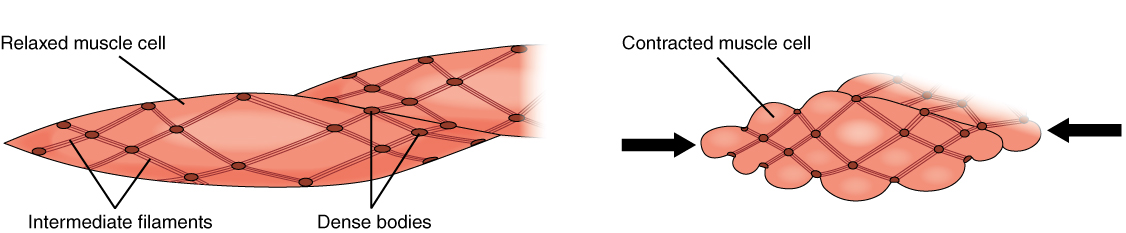
Sliding Filament Model of Contraction: Muscle fibers in relaxed and contracted positions. Muscle tone ensures that even when at rest the muscle is at least partially contracted.
Control of Muscle Tone
The main regulator of muscle tone is the muscle spindle, a small sensory unit that is closely associated with and lies parallel to a muscle. Connecting to the endomysium of a muscle fiber, muscle spindles are composed of nuclear bag fibers and nuclear chain fibers. Both are similar to muscle fibers in that they contain actin and myosin myofilaments that allow them to stretch with the muscle. However, unlike skeletal muscle fibers where the nuclei are spread out and located at the periphery of the cell, in nuclear bag and nuclear chain fibers the nuclei are located in a central region which is enlarged in nuclear bag fibers.
Both cells of the muscle spindle contain sensory neurons. When stretched, muscle spindles become activated, triggering impulses to the spinal cord that can generate an immediate reflex. Spindles can also trigger impulses to the cerebral cortex providing information about the degree of stretch within the muscle.
To maintain tone, spindles also operate a feedback loop by directly triggering motor neurons linked to their associated muscles. If tone decreases and the muscle stretches the spindle, an impulse results in a muscle contraction. With this contraction, the spindle is no longer stretched.
A similar system is found in the tendons attaching muscle to bone. Distinct stretch receptors called golgi tendon organs assess the level of stretch within the tendon. The sensitivity of the golgi tendon organ is significantly less than that of the spindle, so it is thought they exist to prevent damage rather than control muscle tone.
Smooth and Cardiac Muscles
Smooth and cardiac muscles do not have specialized muscle spindles. Tone is maintained through autonomous feedback from the muscle fibers, neurons, and associated tissues.
Types of Muscle Contractions: Isotonic and Isometric
Muscle contractions are defined by changes in the length of the muscle during contraction.
Key Points
Isotonic contractions generate force by changing the length of the muscle and can be concentric contractions or eccentric contractions.
A concentric contraction causes muscles to shorten, thereby generating force.
Eccentric contractions cause muscles to elongate in response to a greater opposing force.
Isometric contractions generate force without changing the length of the muscle.
Key Terms
Isometric: A muscular contraction in which the length of the muscle does not change.
isotonic: A muscular contraction in which the length of the muscle changes.
eccentric: An isotonic contraction where the muscle lengthens.
concentric: An isotonic contraction where the muscle shortens.
A muscle fiber generates tension through actin and myosin cross-bridge cycling. While under tension, the muscle may lengthen, shorten, or remain the same. Although the term contraction implies shortening, when referring to the muscular system, it means the generation of tension within a muscle fiber. Several types of muscle contractions occur and are defined by the changes in the length of the muscle during contraction.
Isotonic Contractions
Isotonic contractions maintain constant tension in the muscle as the muscle changes length. Isotonic muscle contractions can be either concentric or eccentric.
Concentric Contractions
A concentric contraction is a type of muscle contraction in which the muscles shorten while generating force, overcoming resistance. For example, when lifting a heavy weight, a concentric contraction of the biceps would cause the arm to bend at the elbow, lifting the weight towards the shoulder. Cross-bridge cycling occurs, shortening the sarcomere, muscle fiber, and muscle.
Eccentric Contractions
An eccentric contraction results in the elongation of a muscle while the muscle is still generating force; in effect, resistance is greater than force generated. Eccentric contractions can be both voluntary and involuntary. For example, a voluntary eccentric contraction would be the controlled lowering of the heavy weight raised during the above concentric contraction. An involuntary eccentric contraction may occur when a weight is too great for a muscle to bear and so it is slowly lowered while under tension. Cross-bridge cycling occurs even though the sarcomere, muscle fiber, and muscle are lengthening, controlling the extension of the muscle.

Types of Muscle Contraction: An isotonic concentric contraction results in the muscle shortening, an isotonic eccentric contraction results in the muscle lengthening. During an isometric contraction the muscle is under tension but neither shortens nor lengthens.
Isometric Contractions
In contrast to isotonic contractions, isometric contractions generate force without changing the length of the muscle, common in the muscles of the hand and forearm responsible for grip. Using the above example, the muscle contraction required to grip but not move a heavy object prior to lifting would be isometric. Isometric contractions are frequently used to maintain posture.
Isometric contractions are sometimes described as yielding or overcoming.
Yielding
A yielding contraction occurs when a muscle contraction is opposed by resistance. For example, when holding a heavy weight steady, neither raising nor lowering it.
Overcoming
An overcoming contraction occurs when a muscle contraction is opposed by an immovable object, such as the contraction generated in the muscles when pushing against a wall.
In both instances, cross-bridge cycling is maintaining tension in the muscle; the sarcomere, muscle fibers, and muscle are not changing length.
Skeletal muscle
.jpg)
Organization of skeletal muscle
Excluding reflexes, all skeletal muscles contractions occur as a result of conscious effort originating in the brain. The brain sends electrochemical signals through the nervous system to the motor neuron that innervates several muscle fibers.[rx] In the case of some reflexes, the signal to contract can originate in the spinal cord through a feedback loop with the grey matter. Other actions such as locomotion, breathing, and chewing have a reflex aspect to them: the contractions can be initiated both consciously or unconsciously.
Smooth Muscle Contraction
Classification of smooth muscle cells based on contraction features
Different contraction behaviors determine the classification of smooth muscles into single- and multi-unit smooth muscles. The single-unit smooth muscle is coupled by gap junctions and, therefore, works as one coherent functional unit, i.e. the muscles contract together. This muscle type is found predominantly along organ walls and blood vessels.
Multi-unit smooth muscles are capable of contracting independently of one another due to the predominantly autonomic innervation. Since there are relatively few gap junctions, the electric coupling occurs through a basal membrane-like layer. Furthermore, the neurotransmitters are distributed by varicosities. This type of smooth muscle can be found, among others, in the ciliary muscle of the iris and the arrector pili muscle.
Special features of smooth muscle contraction
- Spontaneous, autonomous contraction: Due to the innervation through the autonomic nervous system (involuntary processes), existing automatic processes can be adjusted according to the situation.
- The predominant share of the incoming Ca2+ ions comes from the extracellular space (vs. sarcoplasmic reticulum in skeletal muscle).

Smooth muscle contraction step by step:
- Ca2+ binds to calmodulin
- Activation of myosin-light-chain-kinase (MLCK) through the Ca2+-calmodulin complex
- Accretion of the complex to caldesmon → activation of a certain enzyme to facilitate phosphorylation
- Phosphorylation of the light chain of the myosin head through MLCK, using ATP
- Contraction via cross-bridge formation
- The separation of the remaining phosphate from the light chain of the myosin head through myosin-light-chain phosphatase (MLCP) → dissolution of the actin-myosin-bond
Cardiac Muscle Contraction
Cardiac muscle contraction is facilitated by specialized cardiac muscle cells of the electrical system of the heart. The electrical impulses are transmitted via gap junctions to the cardiac muscle cells and, later on, to the working myocardium.
The specialized cardiac muscle cells can be divided into the electrical impulse formation and conduction systems. The systems have the following components:
- Sinus and AV nodes
- Bundle of His
- Bundle branches also called Tawara branches
- Purkinje fibers
- Working myocardium: These are cells, which are connected via gap junctions and form a syncytium, where the conduction of electrical impulses can occur rapidly. Syncytia are isolated from each other through the annulus fibrosus (valve connective tissue). The working myocardium is responsible for mechanical work.
From stimulation to cardiac muscle contraction
The stimulation from the sinus node, i.e., the impulse formation system, is transmitted from the syncytium to the AV node. This serves simultaneously as a ‘backup impulse formation system’. From there, the impulse is passed on from the bundle of His to the bundle branches. The function of the bundle branches is to rapidly spread the impulse to the apex of the heart and papillary muscles of the heart valves.
Electrophysiology of cardiac muscle contraction
Since the actual process of contraction occurs in the working myocardium, we will discuss the process of the genesis and cessation of an action potential in this section. First, it is important to note that localized processes of the action potential are similar to others as they pass through partially similar stages.
The action potential comprises 3 phases:
- Notch phase: Caused by the activation of Na+ channels with a membrane potential of about -60 mV. Na+ influx is triggered, which leads to the depolarization of the sarcolemma and increases membrane potential to about +30 mV (overshoot). This leads to the deactivation of the Na+ channels.
- Plateau phase: The depolarization leads to a temporary off-set activation of the Ca2+ channels, which results in a decreased membrane potential of about -30 mV. This leads to Ca2+ influx from the extracellular space, which enables the electromechanical coupling. In this phase, the action potential of the chamber myocardium is different from that of the nerve and muscle cells since there is a temporary equilibrium between the depolarizing and repolarizing currents.
- Rapid repolarization phase: Through the activation of tension-dependent K+ channels, the conductivity of Na+ and Ca2+ decreases more rapidly. Furthermore, the K+ channels serve to stabilize the membrane potential at rest.
The action potential of the myocardium lasts about 300–450 ms, depending on the exact location of measurement and heart rate.
References