Hemodynamics is the dynamics of blood flow. The circulatory system is controlled by homeostatic mechanisms of autoregulation, just as hydraulic circuits are controlled by control systems. The hemodynamic response continuously monitors and adjusts to conditions in the body and its environment. Thus, hemodynamics explains the physical laws that govern the flow of blood in the blood vessels.
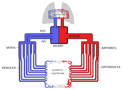
Blood flow through the body delivers oxygen, nutrients, hormones, cells, products of defense mechanisms for wound healing, and platelets. The heart pumps these products to the organs, while the vessels transport them to and from the organs. Arteries perfuse the organs and veins drain the organs of waste products. The lymphatic system helps in draining excess tissue fluid to the bloodstream. Two circulatory loops are most important to survival: pulmonary circulation and systemic circulation. The pulmonary circulation pumps blood from the right ventricle to the pulmonary artery. Blood exchanges carbon dioxide for oxygen while passing through the lung and the newly oxygenated blood drains into the left atrium from the pulmonary veins. The other circulatory loop is the systemic circulation, which pumps blood from the left ventricle to the aorta to the rest of the body. It transports nutrients to the intestines and hormones to the endocrine glands. Waste excretion then occurs via the kidneys, intestines, lungs, and skin. Blood returns to the right atrium from the superior and inferior vena cava.[rx]
Blood flow ensures the transportation of nutrients, hormones, metabolic waste products, O2, and CO2 throughout the body to maintain cell-level metabolism, the regulation of the pH, osmotic pressure, and temperature of the whole body, and the protection from microbial and mechanical harm.[rx]
Introduction to Blood Flow, Pressure, and Resistance
Blood flow
The heart is the driver of the circulatory system, pumping blood through rhythmic contraction and relaxation. The rate of blood flow out of the heart (often expressed in L/min) is known as cardiac output (CO).
Blood being pumped out of the heart first enters the aorta, the largest artery of the body. It then proceeds to divide into smaller and smaller arteries, then into arterioles, and eventually capillaries, where oxygen transfer occurs. The capillaries connect to venules, and the blood then travels back through the network of veins to the right heart. The micro-circulation — the arterioles, capillaries, and venules —constitutes most of the area of the vascular system and is the site of the transfer of O2, glucose, and enzyme substrates into the cells. The venous system returns the deoxygenated blood to the right heart where it is pumped into the lungs to become oxygenated and CO2 and other gaseous wastes exchanged and expelled during breathing. Blood then returns to the left side of the heart where it begins the process again.
In a normal circulatory system, the volume of blood returning to the heart each minute is approximately equal to the volume that is pumped out each minute (the cardiac output).[rx] Because of this, the velocity of blood flow across each level of the circulatory system is primarily determined by the total cross-sectional area of that level. This is mathematically expressed by the following equation:
- v = Q/A
where
- v = velocity (cm/s)
- Q = blood flow (ml/s)
- A = cross-sectional area (cm2)
Anatomical features
The circulatory system of species subjected to orthostatic blood pressure (such as arboreal snakes) has evolved with physiological and morphological features to overcome circulatory disturbance. For instance, in arboreal snakes, the heart is closer to the head, in comparison with aquatic snakes. This facilitates blood perfusion to the brain.[rx][rx]
Turbulence
Blood flow is also affected by the smoothness of the vessels, resulting in either turbulent (chaotic) or laminar (smooth) flow. Smoothness is reduced by the buildup of fatty deposits on the arterial walls.
The Reynolds number (denoted NR or Re) is a relationship that helps determine the behavior of a fluid in a tube, in this case, blood in the vessel.
The equation for this dimensionless relationship is written as:[rx]
- {\displaystyle NR={\frac {\rho vL}{\mu }}}
- ρ: density of the blood
- v: mean velocity of the blood
- L: the characteristic dimension of the vessel, in this case, diameter
- μ: viscosity of blood
The Reynolds number is directly proportional to the velocity and diameter of the tube. Note that NR is directly proportional to the mean velocity as well as the diameter. A Reynolds number of less than 2300 is laminar fluid flow, which is characterized by constant flow motion, whereas a value of over 4000, is represented as turbulent flow.[11] Due to its smaller radius and lowest velocity compared to other vessels, the Reynolds number at the capillaries is very low, resulting in laminar instead of turbulent flow.[rx]
Velocity
Often expressed in cm/s. This value is inversely related to the total cross-sectional area of the blood vessel and also differs per cross-section, because in normal conditions the blood flow has laminar characteristics. For this reason, the blood flow velocity is the fastest in the middle of the vessel and slowest at the vessel wall. In most cases, the mean velocity is used.[rx] There are many ways to measure blood flow velocities, like video capillary micro scoping with frame-to-frame analysis, or laser Doppler anemometry.[rx] Blood velocities in arteries are higher during systole than during diastole. One parameter to quantify this difference is the pulsatility index (PI), which is equal to the difference between the peak systolic velocity and the minimum diastolic velocity divided by the mean velocity during the cardiac cycle. This value decreases with distance from the heart.[rx]
- {\displaystyle PI={\frac {v_{systole}-v_{diastole}}{v_{mean}}}}
| Type of blood vessels | Total cross-section area | Blood velocity in cm/s |
|---|---|---|
| Aorta | 3–5 cm2 | 40 cm/s |
| Capillaries | 4500–6000 cm2 | 0.03 cm/s[rx] |
| Vena cavae inferior and superior | 14 cm2 | 15 cm/s |
The circulatory system is the continuous system of tubes that pumps blood to tissues and organs throughout the body.
Key Points
The pulmonary circulatory system circulates deoxygenated blood from the heart to the lungs via the pulmonary artery and returns it to the heart via the pulmonary vein.
The systemic circulatory system circulates oxygenated blood from the heart around the body into the tissues before it is returned to the heart.
The arteries divide into thin vessels called arterioles, which in turn divide into smaller capillaries that form a network between the cells of the body. The capillaries then join up again to make veins that return the blood to the heart.
The flow of blood along arteries, arterioles and capillaries is not constant but can be controlled depending upon the body’s requirements.
Vascular resistance generated by the blood vessels must be overcome by blood pressure generated in the heart to allow blood to flow through the circulatory system.
Key Terms
- vasodilation: The opening of a blood vessel.
- flow: The movement of blood around the body, closely controlled by alterations in resistance and pressure.
- vasoconstriction: The closing or tightening of a blood vessel.
- resistance: The resistance which must be overcome by pressure to maintain blood flow throughout the body.
- pressure: The force which overcomes resistance to maintain blood flow throughout the body.
The circulatory system is the continuous system of tubes through which the blood is pumped around the body. It supplies the tissues with their nutritional requirements and removes waste products. The pulmonary circulatory system circulates deoxygenated blood from the heart to the lungs via the pulmonary artery and returns it to the heart via the pulmonary vein. The systemic circulatory system circulates oxygenated blood from the heart around the body into the tissues before returning deoxygenated blood to the heart.

Pulmonary circulation: Pulmonary circulation is the half of the cardiovascular system that carries oxygen-depleted blood away from the heart to the lungs and returns oxygenated blood back to the heart.
Resistance, Pressure and Flow
Three key factors influence blood circulation.
Resistance
Resistance to flow must be overcome to push blood through the circulatory system. If resistance increases, either pressure must increase to maintain flow, or flow rate must reduce to maintain pressure. Numerous factors can alter resistance, but the three most important are vessel length, vessel radius, and blood viscosity. With increasing length, increasing viscosity, and decreasing radius, resistance is increased. The arterioles and capillary networks are the main regions of the circulatory system that generate resistance, due to the small caliber of their lumen. Arterioles in particular are able to rapidly alter resistance by altering their radius through vasodilation or vasoconstriction.
The resistance offered by peripheral circulation is known as systemic vascular resistance (SVR), while the resistance offered by the vasculature of the lungs is known as pulmonary vascular resistance (PVR).
Blood Pressure
Blood pressure is the pressure that blood exerts on the wall of the blood vessels. The pressure originates in the contraction of the heart, which forces blood out of the heart and into the blood vessels. If the flow is impaired through increased resistance then blood pressure must increase, so blood pressure is often used as a test for circulatory health. Blood pressure can be modulated through altering cardiac activity, vasoconstriction, or vasodilation.
Blood Flow
Flow is the movement of the blood around the circulatory system. A relatively constant flow is required by the body’s tissues, so pressure and resistance are altered to maintain this consistency. A too-high flow can damage blood vessels and tissue, while flow that’s too low means tissues served by the blood vessel may not receive sufficient oxygen to function.
Distribution of Blood Flow
Humans have a closed cardiovascular system, meaning that blood never leaves the network of arteries, veins, and capillaries.
Key Points
In humans, blood is pumped from the strong left ventricle of the heart through arteries to peripheral tissues and returns to the right atrium of the heart through veins.
After blood returns to the right atrium, it enters the right ventricle and is pumped through the pulmonary artery to the lungs, then returns to the left atrium through the pulmonary veins. Blood then enters the left ventricle to be circulated through the systemic circulation again.
The closing of blood vessels is termed vasoconstriction. Vasoconstriction occurs through contraction of the muscular walls of vessels and results in increased blood pressure.
Vasoconstriction is important for minimizing acute blood loss in the event of hemorrhage as well as retaining body heat and regulating mean arterial pressure.
Dilation, or opening of blood vessels, is termed vasodilation. Vasodilation occurs through relaxation of smooth muscle cells within vessel walls.
Vasodilation increases blood flow by reducing vascular resistance. Therefore, dilation of arterial blood vessels (mainly arterioles ) causes a decrease in blood pressure.
Key Terms
- vasoconstriction: The constriction of the blood vessels.
- vascular resistance: The resistance to flow that must be overcome to push blood through the circulatory system. The resistance offered by the peripheral circulation is known as systemic vascular resistance (SVR), while the resistance offered by the vasculature of the lungs is known as pulmonary vascular resistance (PVR).
- vasodilation: The dilation of the blood vessels.
- mean arterial pressure: The average arterial pressure during a single cardiac cycle.
Humans have a closed cardiovascular system, meaning that the blood never leaves the network of arteries, veins, and capillaries. Blood is circulated through blood vessels by the pumping action of the heart, pumped from the left ventricle through arteries to peripheral tissues and returning to the right atrium through veins. It then enters the right ventricle and is pumped through the pulmonary artery to the lungs and returns to the left atrium through the pulmonary veins. Blood then enters the left ventricle to be circulated again.
Pulmonary circuit: Diagram of pulmonary circulation. Oxygen-rich blood is shown in red; oxygen-depleted blood in blue.
The distribution of blood can be modulated by many factors, including increasing or decreasing heart rate and dilation or constriction of blood vessels.
Vasoconstriction

Blood distribution: Oxygenated arterial blood (red) and deoxygenated venous blood (blue) are distributed around the body.
Vasoconstriction is the narrowing of the blood vessels resulting from the contraction of the muscular wall of the vessels, particularly the large arteries and small arterioles. The process is the opposite of vasodilation, the widening of blood vessels. The process is particularly important in staunching hemorrhage and acute blood loss. When blood vessels constrict, the flow of blood is restricted or decreased, thus retaining body heat or increasing vascular resistance. This makes the skin turn paler because less blood reaches the surface, reducing the radiation of heat.
On a larger level, vasoconstriction is one mechanism by which the body regulates and maintains mean arterial pressure. Substances causing vasoconstriction are called vasoconstrictors or vasopressors. Generalized vasoconstriction usually results in an increase in systemic blood pressure, but it may also occur in specific tissues, causing a localized reduction in blood flow. The extent of vasoconstriction may be slight or severe depending on the substance or circumstance.
Vasodilation
Vasodilation refers to the widening of blood vessels resulting from the relaxation of smooth muscle cells within the vessel walls, particularly in the large veins, large arteries, and smaller arterioles. The process is essentially the opposite of vasoconstriction. When blood vessels dilate, the flow of blood is increased due to a decrease in vascular resistance. Therefore, dilation of arterial blood vessels (mainly the arterioles) causes a decrease in blood pressure. The response may be intrinsic (due to local processes in the surrounding tissue) or extrinsic (due to hormones or the nervous system). Additionally, the response may be localized to a specific organ (depending on the metabolic needs of a particular tissue, as during strenuous exercise), or it may be systemic (seen throughout the entire systemic circulation). Substances that cause vasodilation are termed vasodilators.
Blood Supply and Lymphatics Hemodynamics
Blood flow can either be laminar or turbulent. Laminar flow is linear flow, mainly found in the middle of the vessel. Turbulent flow is any disruption in the laminar flow. Reynold’s number predicts the chances of flow being turbulent. The higher the number, the increased likelihood of being turbulent and vice versa. Reynold’s number is proportional to density, velocity, and diameter and inversely proportional to viscosity.[rx] For example, high blood pressure causes increased velocity, which increases Reynold’s Number and increases the chances of turbulent flow. Anemia indicates low blood viscosity, which will also increase Reynold’s Number. Therefore, turbulence (which is identifiable on the physical exam via auscultation) could represent an underlying pathology. Shear forces can be a consequence of turbulent flow because velocity on the wall should be near zero. Disruption at the wall can damage the vessels and lead to atherosclerosis, thrombosis, and emboli.
Many organs, such as the heart, brain, and kidney, rely on autoregulatory mechanisms, or local control of blood flow, that affect perfusion. Other organs rely mostly on sympathetic stimulation or extrinsic control of blood flow. The coronary arteries are locally regulated by hypoxia and adenosine, which vasodilates the vessels to maintain oxygenation to the heart. When the heart increases in contractility, the oxygen demand of the coronary arteries increases. Therefore, vasodilation occurs to increase blood flow and oxygen to the arteries. The afferent arteries in the kidney are the main pressure-induced auto-regulators of renal blood flow and glomerular filtration rate via stretch and tubuloglomerular feedback. Carbon dioxide is the main autoregulator in the brain that stimulates cerebral vasodilation to maintain blood flow during ischemia.[rx] Astrocytes also play an important role in cerebral blood flow by mediating functional hyperemia, which states that blood flow is dependent on the amount of metabolic activity. Astrocytes release vasoactive substances depending on the oxygen state of the brain. For example, during normoxic conditions, astrocytes mediate vasodilation, and during hyperoxic states, they mediate vasoconstriction. These findings have shown that astrocyte disruption causes a lack of efficient cerebral blood flow in conditions such as Alzheimer’s disease and diabetic retinopathy.[rx] Autonomic receptors regulate blood flow to skeletal muscles at rest and metabolites during exercise. Lactate, potassium, and adenosine vasodilate the vessels during exercise.[rx] This vasodilation during exercise is essential for the proper delivery of oxygen skeletal muscle and the removal of waste products and heat.[rx] The skin has the highest amount of sympathetic innervation, mainly for temperature regulation. Vasoconstriction to maintain core body temperature during cold climates and vasodilation to dissipate the heat in hot climates.[rx]
Nerves
Baroreceptors located on the carotid sinus respond to the decreased pressure (low blood pressure), which signals to activate the sympathetic nerves and vasoconstrictors arteries and veins.[rx] The chemoreceptors in the carotid and aortic bodies are sensitive to oxygen pressures and respond with vasoconstriction if the partial pressure of oxygen is too low.[rx] Vasopressin or anti-diuretic hormone (ADH) is a vasoconstrictor released from the posterior pituitary in response to low blood volume. In contrast, atrial natriuretic peptide (ANP) is a vasodilator released from the atrium in response to fluid overload in the heart.
References


About the author