Category Archive Anatomy A – Z

ByRx Harun

Humoral Immune Response – Anatomy, Types, Functions

The humoral immune response is mediated by antibody molecules that are secreted by plasma cells. An antigen that binds to the B-cell antigen receptor signals B cells and is, at the same time, internalized and processed into peptides that activate armed helper. The extracellular spaces are protected by the humoral immune response, in which antibodies produced by B cells cause the destruction of extracellular microorganisms and prevent the spread of intracellular infections. The activation of B cells and their differentiation into antibody-secreting plasma cells is triggered by antigen and usually requires helper T cells. The term ‘helper T cell’ is often used to mean a cell from the TH2 class of CD4 T cells (see Chapter 8), but a subset of TH1 cells can also help in B-cell activation. In this case, we will therefore use the term helper T cell to mean any armed effector CD4 T cell that can activate a B cell. Helper T cells also control isotype switching and have a role in initiating somatic hypermutation of antibody variable V-region genes

Clonal Selection and B-Cell Differentiation

B cells mature in the bone marrow, where they undergo VDJ recombination to produce unique receptors that do not react to self-antigens.

Key Points

In the bone marrow, central tolerance of B cells is produced through negative selection. Immature B cells are tested for auto-reactivity before leaving the bone marrow. The immature B cells whose receptors (BCRs) bind too strongly to self-antigens will not be killed.

When the B cell receptor on the cell surface matches its cognate antigen in the body, the B cell proliferates and secretes a free form of those receptors ( antibodies ), with identical binding sites as on the original cell surface.

B cells that have not been exposed to an antigen, also known as naïve B cells, can be activated in a T cell-dependent or independent manner.

There are two types of T cell-independent activation: Type 1 T cell-independent (polyclonal) activation, and type 2 T cell-independent activation.

During clonal selection, random mutations during clonal expansion cause the production of B cells with an increased antibody-binding affinity for their antigens.

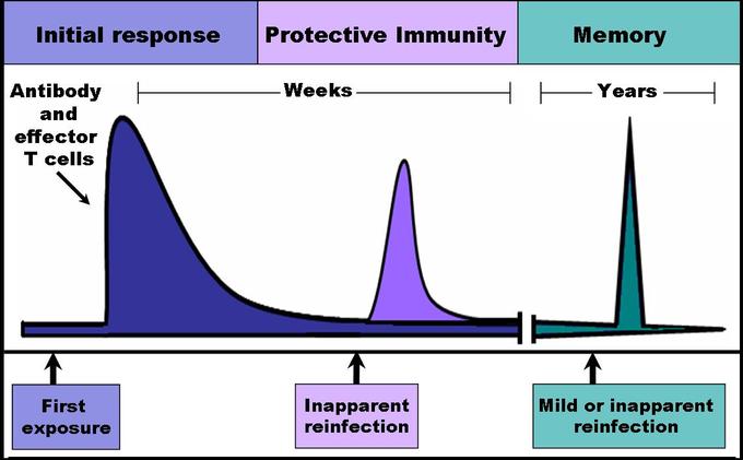
The clonal selection hypothesis may explain why secondary immune responses are so effective at preventing reinfection by the same pathogen.

Key Terms

  • Clonal selection: The theory that lymphocytes bear antigen receptors before activation and that random mutations during clonal expansion cause the development of lymphocytes with high binding affinities for their antigens.
  • humoral: Of or relating to the body fluids or humor.
  • memory B cell: A B cell subtype formed following primary infection in which the cell recognizes a specific epitope.

B cells are lymphocytes that play a large role in the humoral immune response (as opposed to the cell-mediated immune response, which is governed by T cells). B cells primarily function to make antibodies against antigens, act as antigen-presenting cells (APCs), and eventually develop into memory B cells to provide long-term immunity. B cells undergo clonal selection and develop similarly to T cells with some notable differences.

B Cell Development

Immature B cells are produced in the bone marrow of most mammals. Their development occurs through several stages, each representing a change in the genome content at the antibody loci. An antibody is composed of two identical light (L) and two identical heavy (H) chains and the genes specifying them are found in the V (variable) region and C (constant) region. The heavy-chain V region has three segments, V, D and J. These segments recombine randomly in a process called VDJ recombination to produce a unique variable domain in the immunoglobulin of each individual B cell.

Similar rearrangements occur for the light-chain V region but with only two segments involved: V and J. When the B cell fails in any step of the maturation process, it will die by apoptosis, here called a clonal deletion. This is a form of positive selection. B cells are also tested for autoreactivity through negative selection. If these B cells have a high affinity for binding to self-antigens, they will die by clonal deletion or another pathway such as energy.

B Cell Activation

B cell activation refers to the differentiation and clonal expansion of B cells. When the B cell receptor on the cell surface matches its cognate antigen in the body, the B cell proliferates and secretes a free form of those receptors (antibodies) in the body, with binding sites identical to those on the original cell surface. However, B cell recognition of antigens is not the only element necessary for B cell activation. B cells that have not been exposed to an antigen, also known as naïve B cells, can be activated in a T cell-dependent or independent manner.

  • T cell-dependent activation is the activation of B cells by type 2 helper T cells in the lymph nodes.
  • T cell-independent activation occurs when antigens directly bind to B cells themselves, usually through cross-linking the antigen to the B cell receptor or receiving the antigen with a toll-like receptor.
This diagram of T-dependent B cell activation indicates T helper cells, CD40, CD40L, TCR, MHC II peptide, ILR, B cell, BCR, and antigen. 

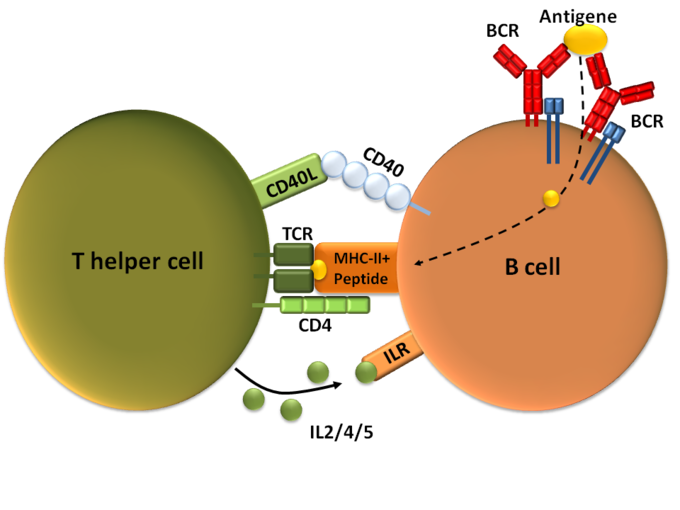
T-dependent B cell activation: T cell-dependent B cell activation, showing a TH2-cell (left), B cell (right), and several interaction molecules.

B Cell Differentiation and Clonal Expansion

After activation, the B cell undergoes differentiation and clonal expansion, which usually involves migration to germinal centers if the activation takes place in a lymph node. B cell differentiation is the process by which B cells change into different types, such as plasma cells and plasmablasts. Clonal expansion is the process by which daughter cells arise from a parent cell. During B cell clonal expansion, many copies of that B cell are produced that share affinity with and specificity of the same antigen.

Clonal Selection

Clonal selection is a theory stating that B cells express antigen-specific receptors before antigens are ever encountered in the body. After B cell activation, the B cells clone themselves through clonal expansion, but during each cellular division, random mutations occur that gradually increase the binding affinity for B cell-produced antibodies to antigens.

For example, memory B cells that differentiate after an adaptive immune response are thought to undergo clonal selection so that antibodies produced by newer memory B cells have considerably higher binding affinities to their antigens. This theory may explain why secondary immune responses from memory cells are so effective that repeated infections by the same pathogen are stopped before symptoms even develop. Following the initial infection, random mutations during clonal selection could produce memory B cells that can more easily bind to antigens than can the original B cells.

Structure and Function of Antibodies

An antibody is a Y-shaped protein produced by B cells to identify and neutralize antigens in the body.

Key Points

An antibody, also known as an immunoglobulin, is a large Y-shaped protein produced by B- cells and used by the immune system to identify and neutralize foreign objects such as bacteria and viruses.

Each tip of the “Y” of an antibody contains a paratope (a structure analogous to a lock) that is specific for one particular epitope (similarly analogous to a key) on an antigen, allowing these two structures to bind together with precision.

Though the general structure of antibodies is very similar, a small region at the tip of the protein is extremely variable, allowing millions of antibodies with different antigen-binding sites to exist. This region is known as the hypervariable region.

Five isotypes of antibodies are found in different locations and perform different specific functions.

The base of the Y plays a role in modulating immune cell activity. This region is called the Fc region, and phagocytes may bind to it to initiate phagocytosis.

Antibodies that bind to surface antigens on a bacterium attract the first component of the complement cascade with their Fc region and initiate activation of the classical complement system.

Key Terms

  • epitope: Part of a biomolecule (such as a protein) that is the target of an immune response.
  • paratope: Part of the molecule of an antibody that binds to an antigen.
  • isotype: A marker corresponding to an antigen found in all members of a subclass of a specific class of immunoglobulins.

An antibody (formally called immunoglobulin) is a large Y-shaped glycoprotein produced by B-cells and used by the immune system to identify and neutralize pathogens. Antibodies are produced by B cells, and are either secreted into circulation or remain expressed on the surface of the B cell.

Structure of Antibodies

The antibody recognizes a unique part of an antigen (foreign object). Each tip of the “Y” of an antibody contains a paratope (a structure analogous to a lock) that is specific for one particular epitope (similarly analogous to a key) on an antigen, allowing these two structures to bind together with precision. Using this binding mechanism, an antibody can neutralize its target directly or tag it for attack by other parts of the immune system.

image 

Antibody: Each antibody binds to a specific antigen, an interaction similar to a lock and key.

Antibodies are glycoproteins belonging to the immunoglobulin superfamily, typically made of basic structural units each with two large heavy chains and two small light chains. Most antibodies exist as a monomer, in which they have a single “Y” shaped sub-unit, but some antibodies can exist as dimers (two subunits) or pentamers (five subunits). The paratope is considered a hypervariable region and has the same specificity and antigen-binding affinity as the B cell receptor of the B cell that created the antibody. In some isotypes, the tail end of the antibody is called the constant region and faces away from the “Y-shaped” paratope ends, functioning as an Fc tail to which phagocytes can bind.

Isotypes

Five different isotypes of antibodies each perform different functions and are generally found in different parts of the body.

  • IgA: A dimer secreted into mucosal surfaces, such as the gut, respiratory tract, and urogenital tract, that prevents mucosal invasion into the body by pathogens. It is resistant to the proteolytic enzymes found in the gastrointestinal mucosae.
  • IgD: Functions mainly as an antigen receptor on B cells that have not been exposed to antigens. It has been shown to activate basophils and mast cells to produce antimicrobial factors.
  • IgE: Found in circulation and binds to allergens, triggering histamine release from mast cells and basophils. Also protects against parasitic worms.
  • IgG: Has four different forms and provides the majority of antibody-based immunity against invading pathogens as the best opsonin of any type of antibody. This is because it expresses a tail for Fc receptors on phagocytes to bind to, which activates phagocytosis. It is the only antibody capable of crossing the placenta to give passive immunity to fetus and can activate the classical complement system.
  • IgM: Expressed on the surface of B cells (monomer) and in a secreted pentamer with very high avidity. Eliminates pathogens in the early stages of B cell-mediated (humoral) immunity before there is sufficient IgG. Like IgG, it can also activate the classical complement system.

Function of Antibodies

Circulating antibodies are produced by clonal B cells that specifically respond to only one antigen. Antibodies contribute to immunity in three ways: preventing pathogens from entering or damaging cells by binding to them (neutralization); stimulating the removal of pathogens by macrophages and other cells by coating the pathogen (opsonization); and triggering the destruction of pathogens by stimulating other immune responses such as the complement pathway. The complement system starts a long cascade of protein productions that either opsonize a pathogen for phagocytosis or lyse it directly by forming a membrane attack complex. During opsonization, the antibody expresses the tail for an Fc receptor on a macrophage, neutrophil, or natural killer cell. The immune cell will then bind to the antibody’s Fc tail instead of the pathogen itself, which speeds up the process of finding pathogens to phagocytize. Additionally, because antibodies have two or more paratopes, they can sometimes link pathogens together, making phagocytosis more efficient.

Role of the Complement System in Immunity

The complement system is the ability of antibodies and phagocytic cells to remove pathogens from an organism.

Key Points

The complement system helps antibodies and phagocytic cells clear pathogens from an organism.

The complement system consists of a number of small proteins produced by the acute phase reaction in the liver during inflammation.

The complement system might play a role in diseases with an immune component and those of the central nervous system. Complement protein deficiency is a form of primary immunodeficiency.

The classical complement pathway starts with antibody binding, which causes a cascade reaction of complement proteins that gradually form a membrane attack complex.

The alternative complement pathway is usually stimulated by pathogen antigens or toxins rather than antibodies and cleaves C3 until there is enough to continue the steps of the classical complement pathway from the C5 convertase step.

The lectin pathway is homologous to the classical pathway, but with the opsonin, mannose-binding lectin (MBL), and ficolins, instead of C1 from the antibody. This pathway uses proteases on the MBL to form C3 convertase, which continues the steps of the classical complement pathway from the C3 convertase step.

The complement system is regulated by complement control proteins, such as the decay-accelerating pathway, which prevent complement proteins from forming MAC on the body’s cells.

Key Terms

  • C5a: A complementing protein that is an acute phase, inflammatory mediator, causing vasodilation and neutrophil chemotaxis.
  • membrane attack complex: The final complex of all complement system pathways that lyses the pathogen. It is composed of C5b, C6, C7, C8, and C9.
  • mannan-binding-lectin: A protein that binds to carbohydrates on pathogens to activate the lectin complement pathway.

The complement system or “complements” the ability of antibodies and phagocytic cells to clear pathogens from an organism. The complement system consists of a number of small proteins found in the blood, generally synthesized by the liver as a part of the acute phase reaction during systemic inflammation (from TNF-alpha release). When stimulated by one of several triggers, proteases in the system cleave specific proteins to release cytokines and initiate an amplifying cascade of further cleavages. The end result of this activation cascade is massive amplification of the response and activation of the cell-killing membrane attack complex (also called MAC). There are three different pathways by which the complement system may occur.

Classical Complement Pathway

The classical complement pathway is the main pathway by which the complement system occurs. It is comprised of a cascade of many steps with complement proteins cleaving one another in sequential order:

  • The antibody binds to an antigen on the surface of a pathogen, activating the C1 complement protein.
  • C1 acts as a protease and cleaves C2 and C4 to form C4b2b.
  • C42b converts C3 into C3a and C3b, which forms a C5 convertase.
  • C5 convertase cleaves C5 into C5a and C5b.
  • C5b forms a complex with C6, C7, and then C8, and C9, which becomes the membrane attack complex that lyses the pathogen.

Note that C5a has a number of other functions in the immune system, such as causing vasodilation during inflammation and stimulating neutrophil chemotaxis. Additionally, the body’s cells express a glycoprotein called decay accelerating factor, which decays C3 and C5 convertase on the body’s cells. This factor prevents membrane attack complexes from forming on the body’s cells under normal conditions.

This diagram outlines the classical and alternative pathways as described in the text. 

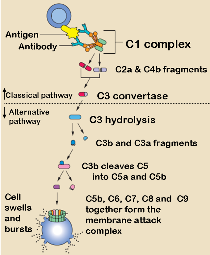
The Classical and Alternative Complement Pathways: The classical and alternative complement pathways start off differently, but end in the same cascade of complement proteins that combine to form a membrane attack complex.

The Alternative Complement Pathway

The alternative pathway may be a leftover evolutionary precursor to the classical pathway. Unlike the classical pathway, the alternative pathway is generally activated by microbial inflammatory mediators instead of antibodies. For example, lipopolysaccharide, the toxin of gram-negative bacteria, may activate this pathway. The steps for the alternative pathway are:

  • The pathogenic antigen (such as LPS) activates C3 so it creates a C3B complex
  • Factor D cleaves the C3B complex so that C3bBb is created.
  • C3bBb is a C3 convertase, which converts more C3 into C3a and C3b.
  • Similar to the classical pathway, C3b forms a C42b complex, and the rest of the steps are essentially the same as the classical pathway, ending with C5b forming a membrane attack complex with C6, C7, C8, and C9.

Lectin Pathway

The lectin pathway is not caused by antibody binding, but by a carbohydrate -binding-protein called mannan-binding-lectin (MBL). It is an acute-phase reactant produced in the liver and binds to the carbohydrates on the surfaces of many pathogens. The steps for the lectin pathway are:

  • MBL binds to the carbohydrates on a pathogen.
  • Proteases bound on the other side of the MBL cleaves C4 into C4a and C4b.
  • C4b creates C3 convertase, and the rest of the steps happen identically to the classical pathway from the C3 convertase step.

Problems with the Complement System

The complement system might play a role in diseases with an immune component, such as Barraquer-Simons Syndrome, asthma, lupus erythematosus, glomerulonephritis, various forms of arthritis, autoimmune heart disease, multiple sclerosis, inflammatory bowel disease, ischemia-reperfusion injuries, and rejection of transplanted organs. The complement system is also becoming increasingly implicated in diseases of the central nervous system such as Alzheimer’s disease and other neurodegenerative conditions such as spinal cord injuries. Additionally, deficiencies in complement proteins produced in the liver can lead to a form of primary (congenital) immunodeficiency, in which the body is more susceptible to disease, particularly autoimmune diseases and severe bacterial infections.

Immunological Memory

Immunological memory refers to the ability of B and T cells to produce long-lived memory cells that defend against specific pathogens.

Key Points

When B and T cells begin to replicate, some offspring will become long-lived memory cells.

Memory cells remember all specific pathogens encountered during the animal’s lifetime and can thus call forth a strong response if the pathogen ever invades the body again.

Passive immunity comes from IgG antibodies given through the mother during fetal development and through breast milk. This memory is short-term but protects the infant until its own adaptive immune system is functional.

During a secondary immune response, memory B and T cells work to rapidly eliminate the pathogen, preventing reinfection by the same pathogen.

During a vaccination, the antigen of a pathogen is introduced into the body through a weakened form of the pathogen that cannot cause an infection. This stimulates the immune system to develop specific immunity against that pathogen without actually causing the disease that the pathogen brings.

Vaccines do not exist for every pathogen due to frequent strain mutations and challenges in producing an immunization strong enough to work, but not strong enough to cause an infection.

Key Terms

  • secondary immune response: The act of exposure to the same pathogen after the initial immune response. Memory B and T cells work to rapidly eliminate the pathogen to prevent reinfection.
  • vaccination: Inoculation with the weakened form of a pathogen to protect against a particular disease or strain of disease by stimulating the development of immunological memory against that pathogen.

When B and T cells begin to replicate during an adaptive immune response, some offspring become long-lived memory cells. These memory cells remember all specific pathogens encountered during the animal’s lifetime and can thus call forth a stronger response, called the secondary immune response, if the pathogen ever invades the body again. The adaptive immune system is so-named because it is a result of an adaptation to an infection. Immunological memory can either exist in active long-term memory or passive short-term memory.

image 

Immune response: When B and T cells begin to replicate, some of the offspring that they produce will end up becoming long-lived memory cells. These memory cells will remember all specific pathogens encountered during the animal’s lifetime and can thus call forth a strong response if the pathogen ever invades the body again.

Passive Memory

Newborn infants are particularly vulnerable to infections since they have no prior exposure to pathogens. Thus, the mother protects the infant through several layers of passive protection. During pregnancy, IgG, a certain isotype of antibody, is transported to the baby from the mother through the placenta, so even babies have high levels of antibodies with similar antigen specificities as the mother. Even breast milk contains antibodies that are transferred to the infant’s gastrointestinal tract and protect against bacterial infections until the baby is capable of making its own antibodies. Since the fetus isn’t making any memory cells or antibodies, this is called passive immunity. Passive immunity is short-lived, ranging from a couple days to a couple months.

As the infant matures, their thymus and bone marrow work to raise a stock of mature lymphocytes that form the foundation for the infant’s personal adaptive immune system. Because the passive memory comes from antibodies instead of B cells themselves, infants do not inherit long-term immunological memory from the mother. Even if the infant receives antibodies specific to certain diseases from its mother, the infant wouldn’t be able to bolster a long-term memory that would direct antigen exposure and presentation.

Active Memory and Immunization

Following an infection, long-term active memory is acquired by activation of B and T cells. Memory cells derive from their parent B and T cells, and undergo clonal selection following infection, which increases antigen-binding affinity. Following reinfection, the secondary immune response typically eliminates the pathogen before symptoms of an infection can occur. During the secondary immune response, memory T cells rapidly proliferate into active helper and cytotoxic T cells specific to that antigen, while memory B cells rapidly produce antibodies to neutralize the pathogen. Long-term active memory consists of rapid response and form permanent immunological memory so long as those memory cells survive.

Vaccinations take advantage of memory lymphocyte development by artificially-generating active immunity, a process called immunization. During a vaccination, the antigen of a pathogen is introduced into the body and stimulates the immune system to develop a specific immunity against that pathogen. It doesn’t cause the disease that the pathogen brings because the vaccine uses an attenuated form of the pathogen that contains the same antigen but doesn’t have the capacity for replication. This deliberate introduction of the pathogen is successful since it exploits the immune system’s natural specificity and inducibility. Vaccination is an extremely effective manipulation of the immune system that helps fight diseases. Over the course of vaccine development, they have saved countless lives, and diseases like rubella and polio are not the widespread causes of disability they once were.

Despite the effectiveness of vaccines, methods do not yet exist to develop vaccines for every pathogen. Many pathogens undergo mutations that change the expression of their antigens, making immunization attempts fruitless for diseases like the common cold or norovirus. Many parasitic pathogens, such as the plasmodium protist that causes malaria, haven’t successfully been vaccinated against because it is challenging to develop a vaccine that is strong enough to stimulate an immune response (sufficient immunogenicity) without causing a live infection.

Major Histocompatibility Complex Antigens (Self-Antigens)humoral immune response

The major histocompatibility complex (MHC) is a cell surface molecule that regulates interactions between white blood cells and other cells.

Key Points

Major histocompatibility complex (MHC) is a cell-surface molecule that mediates interactions between white blood cells and other leukocytes or cells.

The MHC is divided into class I, class II, and class III subgroups, all encoded by the same gene.

MHC antigen presentation gains its diversity from the high degree of polymorphism of the MHC protein itself.

Differing HLA between members of the same species is the main reason why transplanted organs are rejected by the organ recipient.

Key Terms

  • human leukocyte antigen (HLA): The name of the major histocompatibility complex (MHC) in humans.

Major histocompatibility complex (MHC) is a cell-surface molecule encoded by a large gene family in all vertebrates. MHC molecules mediate the interactions of leukocytes with other leukocytes or body cells. MHC determines the compatibility of donors for organ transplant as well as one’s susceptibility to an autoimmune disease via cross-reacting immunization. In humans, MHC is also called human leukocyte antigen (HLA), because MHC can often act as an antigen for human leukocytes.

Types of MHC

MHC is a polymorphic protein attached to the surface of cells. Three different classes of MHC differ in structure and function in the immune system but are all encoded by the same set of gene sequences.

  • MHC I is presented on all cells of the body. It contains an epitope that forms the structural binding site for an antigen. MHC I interacts with natural killer (NK) cells and cytotoxic T cells to signal whether a cell is a self or non-self and whether it contains an antigen-specific to that T cell.
  • MHC II is presented mainly on macrophages, dendritic cells, and helper T cells, which are all involved in antigen presentation. It has a longer helical region than MHC I, which allows it to bind to CD4 (helper T cells) during antigen presentation.
  • MHC III is a secreted enzyme that is neither membrane-bound nor involved in antigen presentation like MHC I and II. It is merely included as an MHC protein because it is encoded by the same set of genes. It is involved in the production of complement proteins and inflammatory cytokines.

During antigen processing prior to presentation, protein peptides from pathogens and MHC (I or II) travel through the cytoplasm and then to the cell membrane surface. A complex series of vesicles enables MHC transport from the cell, and the endoplasmic reticulum and Golgi bodies facilitate this transport during antigen processing. After processing, the MHC can present the peptide antigen bound to it to naive T cells.

image 

MHC Class I processing: MHC class I pathway: proteins in the cytosol are degraded by the proteasome, liberating peptides internalized by the TAP channel in the endoplasmic reticulum, there associating with MHC-I molecules freshly synthesized. MHC-I/peptide complexes enter the Golgi apparatus, are glycosylated, enter secretory vesicles, fuse with the cell membrane, and externalize on the cell membrane interacting with T lymphocytes.

image 

MHC Class I: MHC class I protein molecule.

image 

MHC Class II: MHC class II protein molecule.

HLA and Organ Rejection

Organ transplantation is a complex procedure that can potentially cure many chronic diseases and acute injuries. However, surgically replaced organs are often rejected by the body’s immune system. MHC is also called human leukocyte antigen (HLA) and varies considerably among different members of the same species. If the T and B cells of the body recognize the HLA of the graft as foreign, they will attack the organ graft. The damage in organ rejection can be acute or chronic, cell-mediated or antibody-mediated, and often involves diffuse damage of the graft that causes necrosis and infarction ( tissue death from lack of oxygen) to the graft tissue by attacking its vascular components.

In nearly all cases, immunosuppressive chemotherapy is a requirement for successful organ transplantation. These drugs can stop acute organ rejection after the procedure, but will not stop chronic organ rejection, in which gradual vascular lesions and endothelial thickening slowly kill the graft. If an organ donor has HLA similar to that of the recipient, the risk of organ rejection is reduced. However, this isn’t feasible for heart, liver, or lung transplants because there generally isn’t enough time in these cases to find a matching organ donor.

References

ByRx Harun

Antigens – Anatomy, Types, Structure, Functions

Antigens are molecular structures on the surface of viruses that are recognized by the immune system and are capable of triggering an immune response (antibody production). On influenza viruses, the major antigens are found on the virus’ surface proteins.

When someone is exposed to an influenza virus (either through infection or vaccination) their immune system makes specific antibodies against the antigens (surface proteins) on that particular influenza virus. The term “antigenic properties” is used to describe the antibody or immune response triggered by the antigens on a particular virus. “Antigenic characterization” refers to the analysis of a virus’ antigenic properties to help assess how related it is to another virus.

antigen (Ag) is a molecule or molecular structure, such as may be present on the outside of a pathogen, that can be bound by an antigen-specific antibody or B-cell antigen receptor.[rx] The presence of antigens in the body normally triggers an immune response.[rx] The Ag abbreviation stands for an antibody generator.[rx]

Antigens and Antigen Receptors

Antigens are molecules that initiate the immune response and can be bound by antibodies.

Key Points

An antigen is a molecule that initiates the production of an antibody and causes an immune response.

Antigens are typically proteins, peptides, or polysaccharides. Lipids and nucleic acids can combine with those molecules to form more complex antigens, like lipopolysaccharide, a potent bacterial toxin.

An epitope is a molecular surface feature of an antigen that can be bound by an antibody. A paratope is the molecular surface feature of an antibody that binds to an epitope.

Antigens are classified as exogenous (entering from outside) endogenous (generated within cells ), an autoantigen, a tumor antigen, or a native antigen.

Antigenic specificity is the ability of host cells to recognize an antigen by its unique molecular structure, such as the relationship between antigen epitopes and antibody paratopes.

Key Terms

  • antigen: A substance that induces an immune response, usually foreign, but self antigens and internally produced antigens exist as well.
  • autoantigen: Any antigen that stimulates auto antibodies in the organism that produced it. These are “self” antigens that are involved in autoimmune disease pathogenesis.

EXAMPLES

Fluorescein, along with other haptens such as biotin, is used in various cell and molecular biological techniques. Fluorescein is often conjugated to a protein to allow scientists to examine its location using a fluorescent microscope.

In immunology, an antigen is a substance that evokes an immune response. Formally they are defined as a substance that causes the production of antibodies specific to that antigen, however they also cause T cell-mediated immune responses, and may lead to an inflammatory response. The substance may be from the external environment or formed within the body. The immune system will try to destroy or neutralize any antigen that is recognized as a foreign and potentially harmful invader.” Self” antigens are usually tolerated by the immune system; whereas “non-self” antigens can be identified as invaders and can be attacked by the immune system.

Molecular Structure of Antigens

At the molecular level, an antigen is characterized by its ability to be “bound” at the antigen-binding site of an antibody. Antibodies tend to discriminate between the specific molecular structures presented on the surface of the antigen. Antigens are usually either proteins, peptides, or polysaccharides. This includes parts (coats, capsules, cell walls, flagella, fimbriae, and toxins) of bacteria, viruses, and other microorganisms. Lipids and nucleic acids are antigenic only when combined with proteins and polysaccharides. For example, the combination of lipids and polysaccharides are lipopolysaccharides (LPS), which are the primary component of gram-negative bacterial endotoxin. LPS forms the cell wall of gram-negative bacteria and causes a powerful immune response when bound. Cells present their immunogenic-antigens to the immune system via a major histocompatibility (MHC) molecule. Depending on the antigen presented and the type of the histocompatibility molecule, several types of immune cells can become activated due to an antigen.

Antigens have several structural components of interaction that may be bound by different classes of antibodies. Each of these distinct structural components is considered to be an epitope, also called an antigenic determinant. Therefore, most antigens have the potential to be bound by several distinct antibodies, each of which is specific to a particular epitope. The antigen binding receptor on an antibody is called a paratope and is specific to the epitope of the antigen. Using the “lock and key” metaphor, the antigen itself can be seen as a string of keys – any epitope being a “key” – each of which can match a different lock.

Types of Antigens

Antigens are categorized into broad classes of antigens based on their origin. So many different molecules can function as an antigen in the body, and there is considerable diversity even within these categories.

These are the main classes of antigens that are involved in immune system activation. Their diversity is analogous to the immense diversity of the diseases that the immune system works to overcome.

Exogenous Antigens

Exogenous antigens are antigens that have entered the body from the outside, for example by inhalation, ingestion, or injection. Exogenous antigens are the most common kinds of antigens and include pollen or foods that may cause allergies, as well as the molecular components of bacteria and other pathogens that could cause an infection.

Endogenous Antigens

Endogenous antigens are that have been generated within previously normal cells as a result of normal cell metabolism or because of viral or intracellular bacterial infection (which both change cells from the inside in order to reproduce). The fragments are then presented on the surface of the infected cells in the complex with MHC class I molecules.

 Autoantigens

Autoantigens are normal “self” proteins or complex of proteins or nucleic acid that is attacked by the host’s immune system, causing an autoimmune disease. These antigens should, under normal conditions, not be the target of the immune system, but due to mainly genetic and environmental factors, the normal immunological tolerance for such an antigen has been lost.

Tumor Antigens (Neoantigens)

These antigens are presented by MHC I or MHC II molecules on the surface of tumor cells. These antigens result from a tumor-specific mutation during the malignant transformation of normal cells into cancer cells. Despite expressing this antigen, many tumors have developed ways to evade antigen recognition and immune system killing.

Native Antigens

A native antigen is an antigen that is not yet processed by an APC to smaller parts. T cells cannot bind native antigens, but require that they be digested and processed by APCs, whereas B cells can be activated by native ones without prior processing.

Neoantigens

Neoantigens are those that are entirely absent from the normal human genome. As compared with nonmutated self-proteins, neoantigens are of relevance to tumor control, as the quality of the T cell pool that is available for these antigens is not affected by central T cell tolerance. Technology to systematically analyze T cell reactivity against neoantigens became available only recently.[rx] Neoantigens can be directly detected and quantified through a method called MANA-SRM developed by a molecular diagnostics company, Complete Omics Inc., through collaborating with a team in Johns Hopkins University School of Medicine.[rx]

Viral antigens

For virus-associated tumors, such as cervical cancer and a subset of head and neck cancers, epitopes derived from viral open reading frames contribute to the pool of neoantigens.[rx]

Complete Antigens and Haptens

Haptens are molecules that create an immune response when attached to proteins.

Key Points

Haptens are incomplete antigens that do not cause an immune response upon binding because they cannot bind to MHC complexes.

Haptens may bind with a carrier protein to form an adduct, which is also a complete antigen.

While haptens don’t directly cause immune responses, they may sensitize the body towards hypersensitivity and autoimmune responses.

Haptens may inhibit antibody immune responses by binding with antibodies in place of the actual antigen until there aren’t enough antibodies left to bind to the complete antigen.

Key Terms

  • adduct: A complex molecule formed by the combination of two or more molecules, such as a complete antigen created by a hapten and a carrier.
  • hapten: Any small molecule that can elicit an immune response only when attached to a large carrier such as a protein.

Antigens are basic molecules that induce an immune response when detected by immune system cells. Antigens may be either complete or incomplete based on the nuances of their molecule structure.

Haptens

A hapten is essentially an incomplete antigen. These small molecules can elicit an immune response only when attached to a large carrier such as a protein; the carrier typically does not elicit an immune response by itself. Many hapten carriers are normal molecules that circulate through the body. When haptens and carriers combine, the resulting molecule is called an adduct, the combination of two or more molecules. Haptens cannot independently bind to MHC complexes, so they cannot be presented to T cells.

The first haptens used were aniline and its carboxyl derivatives (o-, m-, and p-aminobenzoic acid). One well-known hapten is urushiol, the toxin found in poison ivy and a common cause of cell-mediated contact dermatitis. When absorbed through the skin from a poison ivy plant, urushiol undergoes oxidation in the skin cells to generate the actual hapten, a reactive molecule called a quinone, which then reacts with skin proteins to form hapten adducts. Usually, the first exposure causes only sensitization, in which there is a proliferation of helper and cytotoxic T cells. After a second exposure, the proliferated T cells can become activated, generating an immune reaction and producing the characteristic blisters of poison ivy exposure.

image

Fluorescein Molecule: Fluorescein is an example of a hapten used in molecular biology.

Some haptens induce autoimmune disease. An example is a hydralazine, a blood pressure-lowering drug that occasionally causes lupus erythematosus (an autoimmune inflammatory disorder) in certain individuals with genetic predispositions to the disease. This also appears to be the mechanism by which the anesthetic gas halothane can cause life-threatening hepatitis and penicillin-class drugs cause autoimmune hemolytic anemia. Other haptens, such as fluorescein, detect proteins with which they form adducts. This makes them a common part of molecular biology lab techniques.

Complete Antigens

A complete antigen is essentially a hapten-carrier adduct. Once the body has generated antibodies to a hapten-carrier adduct, the small-molecule hapten may also be able to bind to the antibody, but will usually not initiate an immune response. In most cases, this can only be elicited by the only the hapten-carrier adduct. Sometimes the small-molecule hapten can block the immune response to the complete antigen by preventing the adduct from binding to the antibody, a process called hapten inhibition. In this case, the hapten acts as the epitope for the antigen, which binds to the antibodies without causing a response. If this happens with enough haptens, there will not be enough antibodies left to bind to the complete antigen, thus inhibiting the antibody response.

Antigenic Determinants and Processing Pathways of Antigens

Antigen epitopes make it possible for the immune system to recognize pathogens.

Key Points

An epitope (also known as an antigenic determinant) is part of an antigen that is recognized by the immune system, specifically by antibodies and B and T cells. Other immune cells like APCs cannot recognize epitopes (only PAMPS and DAMPS).

Antigenic determinants (epitopes) are divided into conformational epitopes and linear epitopes.

Antigen processing occurs within a cell and results in fragmentation of proteins, an association of the fragments with MHC molecules, and expression of the peptide -MHC molecules at the cell surface where they can be recognized by the T cell receptor on a T cell.

Antigen processing may be done through either the endogenous pathway (viral proteins from within an infected cell) or through the exogenous pathway (engulfing a pathogen and isolating its antigen from within the APC).

The endogenous pathway uses MHC class I and binds to cytotoxic T cells, while the exogenous pathway uses MHC class II and binds to helper T cells.

Some viruses can prevent antigen processing by disrupting the movement of MHC within the cell.

Key Terms

  • Linear epitopes: These consist of the primary amino acid structure of a protein that makes up the larger antigen.
  • The Exogenous Pathway: Phagocytized pathogens are broken down from within the cell and their broken-down antigens bind with MHC II, which then is expressed on the surface of the antigen-presenting cell.

An epitope, also known as an antigenic determinant, is the part of an antigen that is recognized by the immune system, specifically by antibodies, B cells, and T cells. The latter can use epitopes to distinguish between different antigens, and only binds to their specific antigen. In antibodies, the binding site for an epitope is called a paratope. Although epitopes are usually derived from non-self proteins, sequences derived from the host that can be recognized are also classified as epitopes. Epitopes determine how antigen binding and antigen presentation occur.

Types of Antigenic Determinants

The epitopes of protein antigens are divided into two categories based on their structures and interaction with the paratope.

  • A conformational epitope is composed of discontinuous sections of the antigen’s amino acid sequence. These epitopes interact with the paratope based on the 3-D surface features and tertiary structure (overall shape) of the antigen. Most epitopes are conformational.
  • Linear epitopes interact with the paratope based on their primary structure (shape of the protein’s components). A linear epitope is formed by a continuous sequence of amino acids from the antigen, which creates a “line” of sorts that builds the protein structure.

Antigenic determinants recognized by B cells and the antibodies secreted by B cells can be either conformational or linear epitopes. Antigenic determinants recognized by T cells are typically linear epitopes. T cells do not recognize polysaccharides or nucleic acid antigens. This is why polysaccharides are generally T-independent antigens and proteins are generally T-dependent antigens. The determinants need not be located on the exposed surface of the antigen in its original form, since recognition of the determinant by T cells requires that the antigen be first processed by antigen-presenting cells. Free peptides flowing through the body are not recognized by T cells, but the peptides associate with molecules coded for by the major histocompatibility complex (MHC). This combination of MHC molecules and peptides is recognized by T cells.

Antigen-Processing Pathways

image

Antigen-Binding Site of an Antibody: Antigen-binding sites can recognize different epitopes on an antigen.

In order for an antigen-presenting cell (APC) to present an antigen to a naive T cell, it must first be processed so it can be recognized by the T cell receptor. This occurs within an APC that phagocytizes an antigen and then digests it through fragmentation (proteolysis) of the antigen protein, an association of the fragments with MHC molecules, and expression of the peptide-MHC molecules at the cell surface. There, they are recognized by the T cell receptor on a T cell during antigen presentation. MHC molecules must move between the cell membrane and cytoplasm in order for antigen processing to occur properly. However, the pathway leading to the association of protein fragments with MHC molecules differs between class I and class II MHC, which are presented to cytotoxic or helper T cells respectively. There are two different pathways for antigen processing:

  • The endogenous pathway occurs when MHC class I molecules present antigens derived from intracellular (endogenous) proteins in the cytoplasm, such as the proteins produced within virus-infected cells. Generally, proteosomes are used to break up the viral proteins and combine them with MHC I.
  • The exogenous pathway occurs when MHC class II molecules present fragments derived from extracellular (exogenous) proteins that are located within the cell. First, pathogens are phagocytized, then endosomes within the cell break down antigens with proteases, which then combine with MHC II.

Some viral pathogens have developed ways to evade antigen processing. For example, cytomegalovirus and HIV-infected cells sometimes disrupt MHC movement through the cytoplasm, which may prevent them from binding to antigens or from moving back to the cell membrane after binding with an antigen.

References

ByRx Harun

Cytokines – Types and What About You Need To Know

Cytokines are small secreted proteins released by cells have a specific effect on the interactions and communications between cells. Cytokine is a general name; other names include lymphokine (cytokines made by lymphocytes), monokine (cytokines made by monocytes), chemokine (cytokines with chemotactic activities), and interleukin (cytokines made by one leukocyte and acting on other leukocytes). Cytokines may act on the cells that secrete them (autocrine action), on nearby cells (paracrine action), or in some instances on distant cells (endocrine action). There are both pro-inflammatory cytokines and anti-inflammatory cytokines.

Types of Cytokines Participating in Immune Response

Cytokines are small cell-signaling protein molecules secreted by numerous cells.

Nomenclature

Cytokines have been classed as lymphokines, interleukins, and chemokines, based on their presumed function, cell of secretion, or target of action. Because cytokines are characterized by considerable redundancy and pleiotropism, such distinctions, allowing for exceptions, are obsolete.

  • The term interleukin was initially used by researchers for those cytokines whose presumed targets are principally white blood cells (leukocytes). It is now used largely for the designation of newer cytokine molecules and bears little relation to their presumed function. The vast majority of these are produced by T-helper cells.
  • Lymphokines: produced by lymphocytes
  • Monokines: produced exclusively by monocytes
  • Interferons: involved in antiviral responses
  • Colony-stimulating factors: support the growth of cells in semisolid media
  • Chemokines: mediate chemoattraction (chemotaxis) between cells.

Structural

Structural homogeneity has been able to partially distinguish between cytokines that do not demonstrate a considerable degree of redundancy so that they can be classified into four types:

  • The four-α-helix bundle family (InterPro: IPR009079): member cytokines have three-dimensional structures with a bundle of four α-helices. This family, in turn, is divided into three sub-families:
    1. the IL-2 subfamily. This is the largest family. It contains several non-immunological cytokines including erythropoietin (EPO) and thrombopoietin (TPO).[rx] They can be grouped into long-chain and short-chain cytokines by topology.[rx] Some members share the common gamma chain as part of their receptor.[rx]
    2. the interferon (IFN) subfamily.
    3. the IL-10 subfamily.
  • The IL-1 family, which primarily includes IL-1 and IL-18.
  • The cysteine knot cytokines (IPR029034) include members of the transforming growth factor-beta superfamily, including TGF-β1, TGF-β2 and TGF-β3.
  • The IL-17 family, which has yet to be completely characterized, though member cytokines have a specific effect in promoting the proliferation of T-cells that cause cytotoxic effects.

Functional

A classification that proves more useful in clinical and experimental practice outside of structural biology divides immunological cytokines into those that enhance cellular immune responses, type 1 (TNFα, IFN-γ, etc.), and those that enhance antibody responses, type 2 (TGF-β, IL-4, IL-10, IL-13, etc.). A key focus of interest has been that cytokines in one of these two sub-sets tend to inhibit the effects of those in the other. Dysregulation of this tendency is under intensive study for its possible role in the pathogenesis of autoimmune disorders. Several inflammatory cytokines are induced by oxidative stress.[15][16] The fact that cytokines themselves trigger the release of other cytokines [17][18][19] and also lead to increased oxidative stress makes them important in chronic inflammation, as well as other immune responses, such as fever and acute-phase proteins of the liver (IL-1,6,12, IFN-a). Cytokines also play a role in anti-inflammatory pathways and are a possible therapeutic treatment for pathological pain from inflammation or peripheral nerve injury.[rx] There are both pro-inflammatory and anti-inflammatory cytokines that regulate this pathway.

Key Points

Cytokines are immune system regulatory agents that work at both systemic and local levels. Cytokines are also involved in several developmental processes during embryogenesis.

Each cytokine has a matching cell-surface receptor. Upon binding, intracellular signaling and gene expression regulations are altered, leading to the production of other cytokines, surface receptors, or feedback inhibition.

Interleukins principally target leukocytes. They include common inflammatory and anti-inflammatory mediators and lymphokines.

Chemokines mediate chemoattraction (chemotaxis) between cells.

Interferons have an antiviral function and can act as pyrogen.

Tumor necrosis factor causes long-lasting inflammatory effects and fever during systemic immune responses. It stimulates the acute phase reaction in the liver and is responsible for much of the immune system-caused damage in severe infections.

Key Terms

  • chemokine: Any of various cytokines produced during inflammation that organize the leukocytes by providing a stimulus for chemotaxis.
  • IL-10: Also known as human cytokine synthesis inhibitory factor (CSIF), an anti-inflammatory cytokine.
  • cytokine: Any of various small regulatory proteins or glycoproteins that regulate the cells of the immune system.

Cytokines are small cell-signaling protein molecules secreted by numerous cells and used extensively in intercellular communication. Cytokines can be classified as proteins, peptides, or glycoproteins. They provide the signaling pathways that orchestrate the complex immune responses of the human body. Cytokines are similar to hormones, which are also chemical messengers, but hormones have considerably more variation in molecular structure and are involved more in tissue signaling than cellular signaling.

Each cytokine has a matching cell-surface receptor. Subsequent cascades of intracellular signalling then alter cell functions. This may include the upregulation (increased expression) and/or downregulation (decreased expression) of several genes and their transcription factors resulting in the production of other cytokines, an increase in the number of surface receptors for other molecules, or the suppression of their own effects by feedback inhibition.

Interleukins

Interleukins are a class of cytokines primarily expressed by leukocytes. They are glycoproteins involved in the signaling of many types of immune system functions. There are 17 different families of interleukins. Some of the more important ones include inflammatory mediators such as IL-1, IL-4, and IL-6, the potent anti-inflammatory IL-10, and other interleukins involved with T and B cell signaling following antigen presentation. Many interleukins are also considered lymphokines, interleukins released by helper T cells to organize immune responses.

Interferons

Interferons are protein cytokines that have antiviral functions. They can activate macrophages and natural killer (NK) cells to attack and lyse virus-infected cells. One common interferon is IFN-gamma, a pyrogen involved in inflammatory response and macrophage and NK cell activation. IFN-gamma is produced by T cells (both CD4 and CD8) and NK cells.

Chemokines

Chemokines are protein cytokines that are mainly involved in facilitating chemotaxis (chemical-stimulated movement) in immune cells. Leukocytes travel along chemotactic gradients that guide them to sites of injury, infection, or inflammation. By definition, inflammatory mediators in other classes of cytokines are also considered chemokines. This category also includes cytokines that are only involved in leukocyte migration, such as CCL2 which causes monocyte chemotaxis and stimulates its differentiation into macrophages inside of tissues.

Tumor Necrosis Factor

Tumor necrosis factors (TNF) are cytokines that induce apoptosis in abnormal cells such as tumor cells. It is a protein released by NK cells, macrophages, and helper T cells, typically in systemic immune responses. TNF-alpha is the most notable example. This long-lasting inflammatory mediator and pyrogen can cause fever and inflammation for up to 24 hours. It also stimulates acute phase reaction in the liver, a component of systemic immune system activation where the liver makes proteins involved in immune system response such as complement proteins. TNF-alpha is released in very high amounts in response to lipopolysaccharide  (infection with gram-negative bacteria), which facilitates much of the self-destructive immune response in septic shock. In these cases, TNF-alpha can cause organ failure from tissue hypoperfusion, caused by damage and blood clotting from an overactive immune response.

Selected cytokines and their primary activities

Cytokines Principal Source Primary Activity
GM-CSF Th cells Growth and differentiation of monocytes and dendritic cells
IL-1α
IL-β
Macrophages and another antigen
presenting cells (APCs)
Costimulation of APCs and T cells, inflammation and fever, acute phase response,
hematopoiesis
IL-2 Activated Th1 cells, NK cells With the proliferation of B cells and activated T cells, NK functions
IL-3 Activated T cells Growth of hematopoietic progenitor cells
IL-4 Activated T cells B cell proliferation, eosinophil and mast cell growth and function, IgE and class II MHC
the expression on B cells, inhibition of monokine production
IL-5 Th2 and mast cells Eosinophil growth and function
IL-6 Activated Th2 cells, APCs, other
somatic cells
Acute-phase response, B cell proliferation, thrombopoiesis, synergistic with IL-1 and TNF on
T cells
IL-7 Thymic and marrow stromal cells T and B lymphopoiesis
IL-8 macrophages, somatic cells Chemoattractant for neutrophils and T cells
IL-9 T cells Hematopoietic and thymopoiesis effects
IL-10 Activated Th2 cells, CD8+ T and B
cells, macrophages
Inhibits cytokine production, promotes B cell proliferation and antibody production,
suppresses cellular immunity, mast cell growth
IL-11 Atromal cells Synergistic hematopoietic and thrombopoiesis effects
IL-12 B cells, macrophages The proliferation of NK cells, IFN production, promotes cell-mediated immune functions
IL-13 Th2 cells IL-4-like activities
IL-18 Macrophages potent inducer of interferon-+ by T cells and NK cells
IFN-α
IFN-β
Macrophages, neutrophils and
some somatic cells
Antiviral effects, induction of class I MHC on all somatic cells, activation of NK cells and
macrophages
IFN-γ Activated Th1 and NK cells Induces of class I MHC on all somatic cells, induces class II MHC on APCs and somatic
cells, activates macrophages, neutrophils, NK cells, promotes cell-mediated immunity,
antiviral effects
MIP-1α Macrophages Chemotaxis
MIP-1β Lymphocytes Chemotaxis
TGF-β T cells, monocytes Chemotaxis, IL-1 synthesis, IgA synthesis, inhibit proliferation
TNF-α macrophages, mast cells, NK
cells, sensory neurons
Cell death, inflammation, pain
TNF-β Th1 and Tc cells phagocytosis, NO production, cell death

Basic mechanisms of cell communication.

Intracrine actions: intracellular action by regulation of intracellular events within the cytoplasm and/or nucleus.

Autocrine: action produced within the cell through surface cell receptors.

Intercrine: communication between cells. This type of cell interaction can be classified into:

  • Paracrine: signaling produced by soluble mediators through neighboring cells.
  • Matricrine: cytokines are immobilized in the extracellular matrix (ECM) by its binding to proteoglycans, and they are then stored in an inactive form. These cytokines will be released by the action of proteases such as Metalloproteinases (MMPs) by a mechanism know as Protease-triggered matricrine (PTM). Glycocalyx, which is made of glycoprotein carbohydrate motifs with proteoglycan on its surface, could play the same role.
  • Cytokine secretion by exchange of membrane fragments between cells through mechanisms such as trogocytosis, formation of tunneling nanotubes (TNTs), and release, secretion, and transportation of microvesicles (MVs)/Exosomes.
  • Juxtacrine: neighboring adjacent cells send signals through membrane-anchored mediators. The classic example is the action of the endothelium on the smooth muscle of the tunica media of certain vessels. Some cytokines have the ability to bind to extracellular matrix soluble proteoglycans or to proteoglycan-cell surfaces (for example, CD44, Glypicans, Syndecans, Betaglycan/TGFBR3, inter alia), where this mechanism serves as a reservoir, or as an enabler of these mediators to act on specific receptors in a juxtracrine manner.
  • Endocrine: this refers to the distal or systemic action which depends on secreted cytokine and its transportation within the blood.

Note that the autocrine, paracrine, juxtacrine, and endocrine actions are exerted by the binding to transducing signals (second messenger cascade) through specific cell surface receptors. Cytokines have limited biological half-lives and they act locally for the most part. Furthermore, they have overlapping actions characterized by a very broad range of functions, e.g., hematopoiesis, cell growth, and differentiation, angiogenesis, tissue remodeling, wound healing, effector immune cell activity, and life/death decisions. Moreover, cytokines with endocrine action circulate in picomolar concentrations, but under the influence of strong immune activation circumstances, they can surge up to 1,000-fold (cytokine mix).

In immunological jargon, terms such as interleukins (IL’s), monokines, lymphokines, haematopoietins, lymphopoietins, myelopoietins, leucopoietins, basophilopoietins, chalones, leucokines, macrophage-activator factors (MAF), macrophage inhibitor factors (MIF), histamine-releasing factors (HRF), endogenous pyrogens, tumor necrosis factors, and interferons were originally used to identify the cellular source, the target cell and/or their action-type. However, at present, it is clearly understood that these substances are produced by a multiplicity of cell populations, depending on whether the cell is in a physiological resting state, activated state, or in the pathological context of a specific scenario. The evolution of protein domain families is evident in the genesis and in the great diversity of different cytokines and cytokine-receptor families.

Chemokines (Chemoattractant cytokines) are a particular class of heat immune system cell communication mediators. They are a family of low molecular mass (8–14 kDa) proteins, most basic and structurally related, which exhibit a wide variety of immunological activities such as cell trafficking ().

Fundamental classification

The nomenclature for genes and related diseases, which is used throughout this chapter is assigned by the Human Genome Organisation (HUGO), the Gene Nomenclature Committee (HGNC) by the National Human Genome Research Institute (NHGRI) (http://www.genenames.org/), and the Online Mendelian Inheritance in Man (OMIM) catalog, which is a registered trademark of Johns Hopkins. (http://omim.org/)

Archetypical cytokines signaling through classical-cytokine receptors

  1. Type I helical Cytokine families signaling through Class I cytokine receptors (CRF1 family or Hematopoietic family)
    1. IL-2 Family or Common gamma Chain Receptor Family: IL-2, IL-9, IL-15, IL-21, IL-4 subfamily (IL-4, IL-13), and IL7 subfamily (IL-7, TSLP)
    2. Common beta Chain Receptor Cytokine Family: IL-3, IL-5, and Colony Stimulating Factor 2/Granulocyte Monocyte-stimulating factor (CSF2/GMCSF)
    3. Prolactin family: PRL, GH Subfamily (GH1, GH2), Chorionic somatomammotropin Subfamily (CSH1, CSH2), Erythropoietin (EPO), Thrombopoietin (TPO), and Colony Stimulating Factor 3/Granulocyte-stimulating factor (CSF3/GCSF).
    4. IL-6 Family: IL-6, IL-11, IL-31, LIF, Ciliary Neurotrophic Factor (CNTF), Oncostatin M (OSM), and Cardiotrophin subfamily (CT1, CLC)
    5. IL-12 Family: IL-12, IL-23, IL-27/30, and IL-35
  2. Type II Cytokine families signaling through Class II cytokine receptors (CRF2 family or IL-10/IFN superfamily)
    1. L-10 Family: IL-10, IL-22, and IL-26
    2. IL-19 Family: IL-19, IL-20, and IL-24
    3. Type I IFN: IFN-α Family (IFN-α1, IFN-α2, IFN-α 4, IFN-α 5, IFN-α 6, IFN-α 7, IFN-α 8, IFN-α10, IFN-α13, IFN-α14, IFN-α16, IFN-α17, and IFN-α21)
    4. Type II IFN: IFN-γ, and IFN-λ Family (IFN-λ1/IL-29, IFN-λ2/IL-28A, IFNλ3/IL-28B, and IFN-λ4)
    5. IFN-β/ω: IFN-type I β/ω Family (IFN-β1 and IFN-ω1)
    6. Tissue Factor-VIIa system

Cytokine families signaling through immunoglobulin(Ig) superfamily cytokine receptors

  1. Receptor tyrosine kinase class III-ligands –RTKIII/PDGFR family: MCSF Family (CSF1/MCSF and IL-34), Flt3/Flk2, and Stem Cell Factor/KitL (SCF/KitL)
    1. CSF1/MCSF
    2. IL-34
    3. FLT3LG (Fms-like tyrosine kinase 3 ligands)
    4. Stem Cell Factor(SCF)/KitL
  2. non-Receptor tyrosine-kinase(RTK)
    1. IL-1 Family: IL-1s, IL-18, IL-33, IL-36, IL-37, and IL-38
    2. IL-16
    3. HMG1B (High Mobility Group 1B)

Cytokine TNF family signaling through TNF receptor family

  1. LTA/TNFSF1, TNF-α/TNFSF2, LTB/TNFSF3, OX40L/TNFSF4, CD40L/TNFSF5, FasL/TNFSF6, CD70/TNFSF7, CD30L/TNFSF-8, 4-1BBL/TNFSF-9, TRAIL/TNFSF10, RANKL/TNFSF11, TWEAK/TNFSF12, APRIL/TNFSF13, BLYS/TNFSF13B, LIGHT/TNFSF14, VEGI/TNFSF15, GITRL/TNFSF18, and EDA (Ectodysplasin).
  2. Non-TNF-ligand: Granulin/Epithelin (GRN) and Nerve Growth Factor (NGF).

Chemokine superfamily signaling through chemokine receptors (seven-transmembrane heptahelical (serpentine) receptors associated with G-protein trimeric system)

  1. Chemokine CC Motif Ligand Family (CCL): CCL1, CCL2, CCL3 Subfamily (CCL3, CCL3L1, and CCL3L3), CCL4 Subfamily (CCL4, CCL4L1, and CCL4L2), CCL5, CCL7, CCL8, CCL11, CCL13, CCL14, CCL15, CCL16, CCL17, CCL18, CCL19, CCL20, CCL21, CCL22, CCL23, CCL24, CCL25, CCL26, CCL27, and CCL28
  2. Chemokine CXC Motif Ligand Family (CXCL): CXCL1, CXCL2, CXCL3, CXCL4 Subfamily(CXCL4/PF4 and CXCL4L1/PF4V1), CXCL5, CXCL6, CXCL7/PPBP, CXCL8/IL8, CXCL9, CXCL10, CXCL11, CXCL12, CXCL13, CXCL14, CXCL16, and CXCL17
  3. Chemokine XC Motif Ligand Family (XCL): XCL1 and XCL2
  4. Chemokine CX3C Motif Ligand (CXC3L)
  5. Other non-Chemokine ligands signaling through Chemokine receptors: Chemerin

Orphan and other cytokine family members

  1. IL-17 Family: IL-17A, IL-17B, IL-17C, IL-17D, IL-17E/IL-25, and IL-17F.
  2. Other Cytokines: IL-14, IL-25/SF20, IL-32, MIF family (MIF1 and MIF2), Osteopontin (OPN), Thymic peptide family [Thymosin Family (Prothymosin-α, Thymosin-β4, Thymosin-β10, Thymosin-β15, and Parathymosin), and LEM Family (Thymopoietin)]

Growth factors and other glycoprotein hormones with immunological functions

  1. Insulin
  2. Cardiac Natriuretic Hormones/Natriuretic Peptides (CNH/NPs)
  3. TGF-β Superfamily members with immunological functions: TGF-β family (TGF-β1, TGFβ-2, and TGFβ-3), Activin family (Activin A, Activin B, and Activin AB), and GDF15/Mic1

Morphogen factors with immunological function

  1. Notch Receptor/Notch Receptor Ligand System: Notch Receptor Family (Notch1, Notch2, Notch3, and Notch4), Notch Receptor Ligand Jagged Subfamily (Jag1 and Jag2), Notch Receptor Ligand Delta-like Subfamily (DLL1, DLL3, and DLL4)
  2. Hedgehog (HH) system: SHH, IHH, DHH
  3. Wnt system

Adipokine functional family

Cytokines and cytokine-receptors general characteristics and properties

General Cytokine characteristics and properties are:

  • A functional network.
  • Usually, many cytokine families have evolved from common ancestral genes which, through mechanisms such as ancestral gene duplication, have created a wide variety of cytokines that often have the benefit of generating functional diversity.
  • Some cytokines are produced constitutively while others are inducible. Many of them are produced as pre-pro-cytokine or pro-cytokines, which require convertase-dependent processing for their activation.
  • Many have clearly inferred redundant actions; as a result, some cytokines carry out similar functions.
  • Many cytokines are immobilized in the extracellular matrix (ECM) by binding to proteoglycans (see Matricrine action example in the Introduction) ().
  • Many cytokines will have different effects depending on the concentration and on whether or not their action is during the acute or chronic immune response phase. For example, Prolactin (PRL) has a dual effect, so low PRL concentrations stimulate T-lymphocytes, whereas high concentrations are anti-inflammatory/immunosuppressors.
  • A number of them are pleiotropic; therefore, they act on a wide variety of cell types.
  • They act on hematoinmune system cells as well as on almost any cell type in the body. Since they also function as metabolic modulators and response coordinators against systemic stressors within all tissues, organs, and body systems, some cytokines exert systemic effects that go beyond their immunoregulator role.
  • At any given time of life, a cell may have a wide variety of receptors to various cytokines.
  • Certain cytokines are read through products. This means that they are proteins decoded from contiguous genes. For example, TNFSF12-TNFSF13 read through genes produces a cytokine called TWEAKPRIL. In other situations, the contiguous cytokine gene mRNA transcripts probably encode miscRNA, which is a term used for a series of miscellaneous small RNA. The miRNA have a wide variety of functions, e.g., enzyme-like catalysis and processing after RNA synthesis. An example of this is the CCL15-CCL14 read-through gene chemokines (non-protein coding).
  • Some cytokines are capable of forming hybrids with other cytokines within the same or different families, which increases the response diversity, and signaling system versatility. These cytokines are called Hybridokines. For example, chemokine CXCL5-CCL5, or HGF/SF-IL7 heteromerization produces pre-pro-B cell growth-stimulating factor -PPBSF.
  • Typically, cytokine production tends to be local (tissue) and limited in time (transient) unless there is a pathological background phenomenon.
  • Many cytokines may be synergistic or antagonistic depending on the situation.
  • Cytokines may regulate or modulate the production/ activity of other cytokines.
  • A cytokine can have a differential effect on the same cell depending on the cell basal state (standby or activation) as well as the cell type and variety of expressed receptors. Moreover, one different cytokine could modulate the effect of another cytokine.
  • Some cytokines are pro-inflammatory, others anti-inflammatory (IL-2, IL-10, TGF-β family, IL-27, IL-35, and IL-37) or both (for example, IL-6), and others are regulatory, thus allowing tolerance to self – components as well as pathogen control and destruction with minimum tissue damage. For example, promoting the induction of immunocyte regulation (e.g., Treg, and the Breg) ().
  • There is an association between the heat immune and skeletal systems–a phenomenon called Osteoimmunology. Numerous proinflammatory cytokines affect bone cells as in the case of IL-1, IL-6, TNF-α, IL-8, IL-11, IL-15, IL-17, and IL-32 which are osteoclastogenic cytokines. In contrast, IFN-γ, IFN-β, IFN-α, IL-4, IL-10, IL-13, IL-18, and IL-33 are anti-osteoclastogenic. There are other cytokines with dual roles such as IL-17, IL-12, and IL-23. Note that the RANK-RANKL system is a member of the Tumor Necrosis Factor Superfamily-Tumor Necrosis Factor Superfamily Receptor system (TNFSF-TNFRSF) ().
  • Cytokines are key factors in reproduction physiology through the regulation of specialized gonadal processes and during gestation ().
  • Many cytokines are endogenous molecules called alarmins. They are rapidly released after non-programmed cell death, but they are not released by apoptotic cells from injured tissues or as a result of stress. These cytokines promote the adaptive immune responses and restore homeostasis by promoting tissue reconstruction ().
  • Certain cytokines are rapidly inactivated by proteases such as the Neutral Serine Proteases (NSP) secreted by human polymorphonuclear neutrophils (PMNLs) and thus generate a negative feedback loop. So, for example, IL-2, IL6, and TNF-α are inactivated by human leukocyte elastase (HLE), proteinase 3 (PR3), or cathepsin G (Cat G). In other cases, proteases generate cytokine fragments that may function as antagonists of the active cytokines such as in the case of IL-2.
  • The hematopoietic cytokines have pro-and anti-angiogenic effects. pro-angiogenic cytokines are erythropoietin (EPO), Granulocyte-colony stimulating factor (GCSF), Granulocyte-macrophage colony-stimulating factor (GMCSF), IL-1, IL-3, IL-4, IL-6, IL-8, IL-10, IL-15, and IL-17. Some of the anti-angiogenic cytokines are IL-2, IL-4, IL-12, and IL-13 ().
  • The genome of certain viruses encodes cytokine-like molecules, which are referred to as “virokines.” They function as competitive inhibitors or competitive stimulators of host cytokine-receptors. So they subvert host immune responses and thereby favor the development of an infection-pathogenic scenario. Some examples are the vIL-6 (viral Interleukin 6) encoded by the Human Herpes Virus 8/Kaposi’s Sarcoma Virus genome (HHV8/KSV), vIL-8 encoded by Marek’s disease virus (MDV) genome, vIL-10/BCRF1 (Bam HI C fragment rightward reading frame 1) encoded by Human Herpes Virus 4/Epstein-Barr virus genome, vIL-10/UL111a (open reading frame) encoded by Human Herpes Virus 5/Citomegalovirus genome, and vIL-17 encoded by Herpesvirus saimiri genome, inter alia. The herpes virus and poxvirus family genome encode proteins that modulate chemokine activity, e.g., proteins with homology to chemokines or secreted chemokine-binding proteins (CKBPs). These CKBPs competitively interact with chemokines and prevent chemokine interactions with chemokine-receptors or the extracellular matrix. (See section chemokine superfamily signaling through chemokine receptors seven-transmembrane–heptahelical (serpentine) receptors associated with g-protein trimeric system) ().
  • Certain cytokines are found within biological fluids and are considered biomarkers for disease.
  • Cytokines have been used as contemporary therapeutic intervention targets. In some cases, they have been used to block or inhibit their own receptors or signalings pathways. In other cases, they have been used to stimulate the immune response (primary immunodeficiency, secondary immunodeficiency, severe infections, cancer, and vaccine adjuvant) or hematopoiesis (recombinant DNA technology) ().

Cytokines as well as other cell communication mediators are able to regulate various cell events within the cell, tissue, and system dynamics. Therefore, it is said that they can have many types of effects:

  • Geotropic: effect on gene expression regulation.
  • Metabotropic: metabolic process regulation.
  • Ionotropic: regulation of ion flow through cell membranes and related physiological processes.
  • Redoxtropic: oxidation-reduction potential regulation in the cell, which is also related to the physiological and pathological role of antioxidant neutralization and free radicals from reactive oxygen, nitrogen, and sulfur species (ROS/ROI, RNS/RNI, and RSS/RSI respectively).
  • Mitotropic, also know as mitogen cytokine: cell proliferative capacity regulation, but in addition to this function, they also regulate the entire cell cycle.
  • Morphogenic/morphogenetic, also know as morphogen cytokine: regulates ontogenetic development processes; commitment, proliferation, and differentiation during histogenesis; embryogenesis, ketogenesis, and organogenesis.
  • Motitropic/mitogenic, also called motogen cytokine: cytoskeletal activity regulation, cell motility capability, and cell contractility regulation. Some of these molecules are involved in the migration processes during embryogenesis and ketogenesis, e.g., operating through gradients as motomorphogens.
  • Trophotropic, or organotrophic factor, also known as morphogen cytokine: many cytokines such as growth factors favor cell and tissue tropism, which promotes their survival and proper function. Because of that, these cytokines are called morphogens.
  • Cytoprotection (against harmful and stressful agents) and reparation/regeneration factors.
  • Deathtropic: under certain circumstances, some cytokines are death cell signals (type I/apoptosis, type II/autophagy, type III/necrosis, or mixed/special states such as necroptosis, pyroptosis, and pyronecrosis, etc). The withdrawal of some cytokines also produces cell death, e.g., in the case of certain growth factors.

Throughout this section of the chapter, general cytokine characteristics and properties as well as the different effects they have on the cells or tissues have been discussed. Therefore, the general characteristics and properties of the Cytokine-receptors will be dealt with next (,):

  • The cytokine-receptors are (glyco) proteins signaling through different signal transduction mechanisms.
  • Usually, many cytokine-receptor families have evolved from common ancestral genes. Hence, a wide variety of cytokine-receptors have been created through mechanisms of ancestral gene duplication, often with the benefit of generating functional diversity.
  • In other cases, cytokine-receptors are obtained through secretion or exchange of membrane fragments between cells using mechanisms such as trogocytosis, formation of tunneling nanotubes (TNTs), and the release, secretion, and transportation of microvesicles (MVs)/Exosomes.
  • Cytokine-receptors satisfy the general properties of receptors including specificity, selectivity, induced fit (in previous scientific models, it is called lock-key model), desensitization/adaptation, signaling amplification, integration, saturability, and reversibility.
  • The receptors are usually located in specialized membrane microdomains known as lipid rafts, detergent-resistant membrane-DRM, and caveolae. These microdomains may be of various kinds depending on the variable presence of sphingolipids, glycosphingolipids, cholesterol, Glycosylphosphatidylinositol-proteins (GPI-linked), and certain specialized proteins such as caveolins (caveolin1, caveolin 2, and caveolin 3), Myelin and Lymphocyte protein (MAL) family members (MAL, MAL2, and MALL), and flotillins (FLOT1, and FLOT2). This is a way to centralize and coordinate the intracellular signaling platforms (receptor clustering). Moreover, note that there is even functional diversity when the receptor signals come either from inside of these domains or from outside of them (,).
  • Certain cytokine-receptors are numeric, and others are dimeric, oligomeric, or polymeric complexes. There is usually a cytokine-binding receptor and cytokine signaling-coreceptors in the dimeric, oligomeric or polymeric receptors.
  • Some receptors are part of polymeric complexes, in which several transmembrane proteins are recruited from different families including membrane proteoglycans (mPG) and CAMs (cell adhesion molecules) such as integrins. This is a key biological mechanism of receptor cross-talk (e.g., transphosphorylation) which generates a diversity of cell responses to a single ligand, even when there is an intracellular signaling modulation.
  • Some cytokine-receptors lack transmembrane and cytoplasmic domains. They function as scavengers, interceptors, and silent or decoy receptors and bind to ligands without inducing cell signaling. Many of these receptors are usually anchored to the cell surface via Glycosylphosphatidylinositol (GPI-linked). Some cytokine-receptors are kidnapper receptors acting via endocytosis, and some of them, in turn, are even in cytokine intracrine-pathways or cytokine-catabolisms.
  • Many receptors undergo the internalization and degradation that is dependent on the proteasome-ubiquitin route. Endocytic proteins that contain ubiquitin-interaction motifs (UIM) recognize the ubiquitylated receptors and direct them into clathrin-coated vesicles and, ultimately, into lysosomes. So, degradation is achieved through the action of the E3-ubiquitin-ligase Cbl member (Casitas B-lineage lymphoma proto-oncogene) family. In humans, four members of the Cbl-family are recognized: Cbl, CblB, CblC, and Cbll1 ().
  • Some cytokine-receptors, particularly receptor tyrosine kinase (RTK) type autocatalytic, are inhibited by the Sprouty (Spry) protein, which contains the SPRY/B30.2 homologous domain. Various human SPRY family members (SPRY1, SPRY2, SPRY3, SPRY4, SPRED1, SPRED2, and SPRED3) and ERBB receptor feedback inhibitor 1/Mitogen-inducible gene 6 protein (ERRFI1/Mig6) modulate the actions of RTKs and have inhibitory effects on signal transduction. Spry proteins in some cases are genetically induced by the same cytokines, and in other cases, they are phosphorylated as part of the signaling cascade. This promotes their inhibitory action, in both cases as a feedback negative ().
  • There are soluble versions of some receptors which are produced from mRNA splicing and/or a receptor ectodomain undergoing shedding Regulated Intramembrane Proteolysis (RIP). During RIP cleavage are involved enzymes called “Sheddases” e.g., the MMPs, Adams (A Disintegrin and Metalloproteinase), and the gamma-secretase/presenilin complex are involved. Other examples are IL-2R, TNFRII, CSF1/MCSF-R, CD44, NOTCH-Receptors, and IL-6R shedding by PMN-serine proteases. These soluble versions function in some cases as decoy-receptors. In other cases, they act as binding soluble receptors carrying cytokines towards membrane receptors, or as conveyors. Likewise, the cytosolic fraction receptor-free or intracellular domain (ICD), functions as a transcription factor ().
  • Some receptors are not directly activated by binding their ligands. Activation occurs upon binding another ligand to its specific receptor. This phenomenon is called “Receptor transactivation,” and it refers to the ability of a ligand to bind to its specific receptor and thus activate another ligand-receptor. The mechanism involved during this process is transphosphorylation.
  • Some receptors are anchored to the membrane and are recognized as entry routes for certain infectious pathogenic agents. An example of this is the HGF/SF-R (Hepatocyte growth factor/scatter factor receptor), which is involved in the recognition and internalization of Listeria monocytogenes. A surface protein of this bacteria – internalin B (InlB) – interacts with the HGF/SF-R (Hepatocyte growth factor/scatter factor-receptor) and thus favors the entry of the bacteria. The chemokine-receptor Duffy antigen receptor for chemokines (DARC) serves as the erythroid receptor for the human malaria parasite (Plasmodium vivax) and the monkey malaria parasite (Plasmodium knowlesi). Another example is the Human immunodeficiency virus (HIV)-coreceptors by chemokine family (See chemokine superfamily signaling through chemokine receptors (seven-transmembrane –heptahelical(serpentine) receptors associated with g-protein trimeric system).
  • The cytokine-receptors suffer negative regulation by specialized inhibitory receptors, which belong to the Paired-receptor Superfamily. Some inhibitory receptors of this family are: SIRPα/CD172a, CD200R, CEACAM1/CD66a, 5 Leukocyte immunoglobulin-like receptor (LILR) members (LIR1/ILT2/CD85j, LIR2/ILT4/CD85d, LIR3/ILT5/CD85a, LIR5/ILT3/CD85K, and LIR8/CD85c), NKG2A/CD159a, DCIR/CLEC4A, CMRF35H/IREM1, PILRA, 12 Sialic acid-binding immunoglobulin-type lectin (SIGLE) family members (SIGLEC1, SIGLEC2/CD22, SIGLEC3/CD33, SIGLEC4/MAG, SIGLEC5, SIGLEC6/CD327, SIGLEC7/CD328, SIGLEC8, SIGLEC9/CD329, SIGLEC10, SIGLEC11, and SIGLEC12), and 8 Killer cell immunoglobulin-like receptor (KIR) family members (KIR2DL1/CD158a, KIR2DL2/CD158B1, KIR2DL3/CD158B2, KIR2DL5A/CD158F, KIR2DL5B/CD158F2, KIR3DL1/CD158E, KIR3DL2/CD158K, and KIR3DL3/CD158Z). These inhibitory receptors have immunoreceptor tyrosine-based inhibitory–motifs (ITIM) in their cytoplasmic region that upon receptor triggering, recruit cytosolic tyrosine-phosphatases ().
  • The genome of certain viruses encodes cytokine-receptor-like molecules referred as “receptors,” which function as competitive receptors of host cytokine-receptors. For example, herpes virus and poxvirus family genomes encoded a large number of proteins that modulate chemokine activity such as proteins with homology chemokine-receptors (See section chemokine superfamily signaling through chemokine receptors seven-transmembrane –heptahelical (serpentine) receptors associated with g-protein trimeric system) ().
  • Some receptors are part of the blood group system. The main example is the chemokine-receptor DARC (Duffy antigen receptor complex)/CD234. Therefore, variations in DARC generate the basis of the Duffy minor blood group system.
  • Some receptors are expressed abnormally in target cells, tissues, and organs involved in some diseases. Therefore, they can be considered pathophysiological and diagnostic biomarkers. The same happens with some soluble receptors when they are detected in biological fluids.
  • Within the current biological therapy, the use of soluble receptors produced by recombinant DNA technology emerges. An example of this is the biopharmaceutical molecule Etanercept (Enbrel ®), which was produced by the fusion of the Tumor Necrosis Factor Receptor (TNFR) protein linked to the IgG1 Fc portion of an antibody ().

Cytokines, cytokine receptors, and their signaling pathways specific characteristics and properties

Archetypical cytokine families signaling through class I cytokine receptors (CRF1 or hematopoietic family) and class II cytokine receptors (CRF2 or IL-10/IFN superfamily)

Type I cytokines have limited homology between the sequences of their family members. However, they are characterized by four α-helical bundle structures with an ‘up-up-down-down’ configuration. They can be further divided into short-chain and long-chain four α-helical bundle cytokines because of their α-helices length as well as some other structural and topological characteristics. Short-chain type I cytokines are those cytokines sharing the γc (gamma common chain) receptor, the βc (beta common chain) receptor, and also MCSF and SCF/KitL as atypical examples. Within of long-chain type I cytokines, there are cytokines sharing the gp130 common chain receptor, GH, Leptin, EPO, TPO, IL-12, PRL, and CSF3/GCSF. Unlike the type I cytokines, the type II cytokines have different structures and correspond to the members of the IL-10/IFN superfamily ().

CRF1 family members are characterized by conserved extracellular domains of approximately 200 amino acids, known as cytokine receptor homology domain (CHD), hematopoietic receptor domain (HRD), or D200. The CHD consists of two tandem fibronectin type III (FBN/FNIII) folds. It also contains two pairs of conserved cysteines (four conserved cysteines – C4) linked via disulfide-bonds, and it is arranged in a CX-(9–10)-CXWX-(26–32)-CX-(10–15)-C motif within the first FBN/FNIII fold. The second FBN/FNIII fold (proximal to the transmembrane domain) has a highly conserved tryptophan-serine doublet [(WS)2]=Trp-Ser-X-Trp-Ser motif (WSXWS-motif) in its carboxyl extreme. The C4 (WS) 2 motif represents a common signature to define Class I cytokine receptors. Additionally, this family has other module domains including the extracellular Ig-like, a transmembrane (TM) domain, and conserved intracellular motifs such as Box1- and Box2-motifs. These last two motifs are associated with cytosolic tyrosine Janus Kinase (JAK)-docking. These conserved intracellular motifs are part of the “Intracellular Homology Region (IHR) sequences”.

Jean-Louis Boulay, John J. O’Shea, and William E. Paul did a comparative study on the molecular phylogeny of type I cytokines and proposed that they be classified into 5 groups based on certain characteristics:

  • Group 1: receptor chains have an extracellular domain consisting solely of a CHD. For example, erythropoietin receptor (EPOR), thrombopoietin receptor (TPOR), prolactin receptor (PRLR), and growth hormone receptor (GHR) chains. Each of them produces homodimers in the presence of their respective ligands.
  • Group 2: receptors with polypeptidic chains structurally related to the prototypical glycoprotein 130 (gp130), which has an N-terminal Ig-domain and FBN modules between their CHD and transmembrane domains.
  • Group 3: receptor chains generally possess an N-terminal Ig domain in addition to the CHD. They are soluble and have short intracellular regions.
  • Group 4: receptors chains consist solely of an extracellular CHD domain and long intracellular domains.
  • Group 5: receptor chains possess extracellular Ig-domains in addition to the CHD and have short intracellular regions.

Receptor chains from Groups 2 and 3 constitute the large IL-6R family receptor complexes, which variably share the gp130 as a common signal transducer. Group 4 and 5 receptor chains associate in order to generate the IL-2R and IL-3R family receptor complexes with IL-2Rγc and IL-3Rβc respectively. IL-2Rα and IL1-5Rα receptor chains are not members of the class I family receptors. Instead, they contain a distinctive ‘sushi domain’, also known as Complement control protein (CCP) modules or short consensus repeats (SCR) (,).

However, for academic purposes, we proceeded to classify receptors and cytokine systems of this type as follows:

  • Family or Common gamma Chain Receptor Family: IL-2, IL-5, IL-9, IL-15, IL-21, IL-4 subfamily (IL-4 and IL-13), and IL-7 subfamily (IL-7 and TSLP).
  • Common beta Chain Receptor Cytokine Family: IL3, IL5, and CSF2/GMCSF.
  • Prolactin family: PRL, GH Subfamily (GH-1, and GH-2), CSH Subfamily (CSH-1 and CSH-2), EPO, TPO, and CSF3/GCSF
  • IL-6 Family: IL-6, IL-11, IL-31, LIF, CNTF, OSM, and Cardiotrophin subfamily (CT1 and CLC)
  • IL-12 Family: IL-12, IL-23, IL-27/30, and IL-35

Type II cytokines have different structures in comparison to type I. However, they retain the Box1/2 regions and they are classified as follows:

  • IL-10 Family: IL-10, IL-22, and IL-26.
  • IL-19 Family: IL-19, IL-20, and IL-24.
  • Type III: IFN- λ Family (IFN-λ1/IL-29, IFN-λ2/IL-28A, IFNλ3/IL-28B, and IFN-λ4).
  • Type I: IFN-α–IFN Family (IFN-α1, IFN-α2, IFN-α 4, IFN-α 5, IFN-α 6, IFN-α 7, IFN-α 8, IFN-α10, IFN-α13, IFN-α14, IFN-α16, IFN-α17, and IFN-α21).
  • Type II – IFN: IFN-γ.
  • Type I IFN-β/ω: IFN β/ω Family (IFN-β1 and IFN-ω1).
  • Tissue Factor – VIIa system.

References

ByRx Harun

Interleukins – Anatomy, Structure, Functions

Interleukins (IL) are a type of cytokine first thought to be expressed by leukocytes alone but have later been found to be produced by many other body cells. They play essential roles in the activation and differentiation of immune cells, as well as proliferation, maturation, migration, and adhesion. They also have pro-inflammatory and anti-inflammatory properties. The primary function of interleukins is, therefore, to modulate growth, differentiation, and activation during inflammatory and immune responses. Interleukins consist of a large group of proteins that can elicit many reactions in cells and tissues by binding to high-affinity receptors in cell surfaces. They have both paracrine and autocrine functions. Interleukins are also used in animal studies to investigate aspects related to clinical medicine.

Interleukins (IL) are a type of cytokine first thought to be expressed by leukocytes alone but have later been found to be produced by many other body cells. They play essential roles in the activation and differentiation of immune cells, as well as proliferation, maturation, migration, and adhesion. They also have pro-inflammatory and anti-inflammatory properties. The primary function of interleukins is, therefore, to modulate growth, differentiation, and activation during inflammatory and immune responses. Interleukins consist of a large group of proteins that can elicit many reactions in cells and tissues by binding to high-affinity receptors on cell surfaces. They have both paracrine and autocrine functions. Interleukins are also used in animal studies to investigate aspects related to clinical medicine.

General Properties of Cytokines/Interleukins

  • Cytokines are proteins made in response to pathogens and other antigens that regulate and mediate inflammatory and immune responses.
  • Interleukin production is a self-limited process. The messenger RNA encoding most interleukins is unstable and causes a transient synthesis. These molecules are rapidly secreted once synthesized.
  • Cellular responses to interleukins include up- and down-regulatory mechanisms with the induction and participation of genes that encode inhibitors of the cytokine receptors.
  • Interleukins have redundant functions. For instance, IL-4, IL-5, and IL-13 are B-cell growth factors and stimulate B-cell differentiation.
  • Cytokines stimulate switching of antibody isotypes in B cells, differentiation of helper T cells into Th-1 and Th-2 subsets, and activation of microbicidal mechanisms in phagocytes.
  • Interleukins often influence other interleukin synthesis and actions. For instance, IL-1 promotes lymphocyte activation that leads to the release of IL-2.
  • Cellular responses to cytokines are stimulated and regulated by external signals or high-affinity receptors. For example, stimulation of B-cells by pathogens leads to increased expression of cytokine receptors.
  • Most cytokines act either on the same cell that secretes the cytokine, for instance, IL-2 produced by T cells operates on the same T cells that made it or on a nearby cell. Besides, cytokines may enter the circulation and act far from the site of production, for example, IL-1 is an endogenous pyrogen that works on the central nervous system (CNS) and causes fever.
  • Small quantities of a cytokine are needed to occupy receptors and elicit biological effects.

Function

  • Interleukin-1 (IL-1) – Macrophages, large granular lymphocytes, B cells, endothelium, fibroblasts, and astrocytes secrete IL-1. T cells, B cells, macrophages, endothelium and tissue cells are the principal targets. IL-1 causes lymphocyte activation, macrophage stimulation, increased leukocyte/endothelial adhesion, fever due to hypothalamus stimulation, and release of acute-phase proteins by the liver. It may also cause apoptosis in many cell types and cachexia.
  • Interleukin-2 (IL-2) – T cells produce IL-2. The principal targets are T cells. Its primary effects are T-cell proliferation and differentiation, increased cytokine synthesis, potentiating Fas-mediated apoptosis, and promoting regulatory T cell development. It causes proliferation and activation of NK cells and B-cell proliferation and antibody synthesis. Also, it stimulates the activation of cytotoxic lymphocytes and macrophages.
  • Interleukin-3 (IL-3) – T cells and stem cells make IL-3. It functions as a multilineage colony-stimulating factor.
  • Interleukin-4 (IL-4) – CD4+T cells (Th2) synthesize IL-4, and it acts on both B and T cells. It is a B-cell growth factor and causes IgE and IgG1 isotype selection. It causes Th2 differentiation and proliferation, and it inhibits IFN gamma-mediated activation on macrophages. It promotes mast cell proliferation in vivo.
  • Interleukin-5 (IL-5) – CD4+T cells (Th2) produce IL-5, and its principal targets are B cells. It causes B-cell growth factor and differentiation and IgA selection. Besides, causes eosinophil activation and increased production of these innate immune cells.
  • Interleukin-6 (IL-6) – T and B lymphocytes, fibroblasts, and macrophages make IL-6. B lymphocytes and hepatocytes are its principal targets. IL-6’s primary effects include B-cell differentiation and stimulation of acute-phase proteins.
  • Interleukin-7 (IL-7) – Bone marrow stromal cells produce IL-7 that acts on pre-B cells and T cells. It causes B-cell and T-cell proliferation.
  • Interleukin-8 (IL-8) – Monocytes and fibroblasts make IL-8. Its principal targets are neutrophils, basophils, mast cells, macrophages, and keratinocytes. It causes neutrophil chemotaxis, angiogenesis, superoxide release, and granule release.
  • Interleukin-9 (IL-9)  – Th9, Th2, Th17, mast cells, NKT cells, and regulatory T cells produce this cytokine. It enhances T-cell survival, mast cell activation and synergy with erythropoietin.
  • Interleukin-10 (IL-10) – Th2 cells produce IL-10. Its principal targets are Th1 cells. It causes inhibition of IL-2 and interferon-gamma. It decreases the antigen presentation, and MHC class II expression of dendritic cells, co-stimulatory molecules on macrophages and it also downregulates pathogenic Th17 cell responses. It inhibits IL-12 production by macrophages.
  • Interleukin-11 (IL-11) – Bone marrow stromal cells and fibroblasts produce IL-11. The IL-11 principal targets are hemopoietic progenitors and osteoclasts. The IL-11 primary effects include osteoclast formation, colony-stimulating factor, raised platelet count in vivo, and inhibition of pro-inflammatory cytokine production.
  • Interleukin-12 (IL-12) – Monocytes produce IL-12. Its principal targets are T cells. It causes induction of Th1 cells. Besides, it is a potent inducer of interferon-gamma production by T lymphocytes and NK cells.
  • Interleukin-13 (IL-13) – CD4+T cells (Th2), NKT cells, and mast cells synthesize IL-13. It acts on monocytes, fibroblasts, epithelial cells, and B cells. The IL-13 significant effects are B-cell growth and differentiation, stimulates isotype switching to IgE. It causes increased mucus production by epithelial cells, increased collagen synthesis by fibroblasts and inhibits pro-inflammatory cytokine production. Also, IL-13 works together with IL-4 in producing biological effects associated with allergic inflammation and in defense against parasites.
  • Interleukin-14 (IL-14) – T cells produce IL-14, and its principal effects are stimulation of activated B cell proliferation and inhibition of immunoglobulin secretion.
  • Interleukin-15 (IL-15) – Monocytes, epithelium, and muscles make IL-15. It acts on T cells and activated B cells. It causes the proliferation of both B and T cells. It causes NK cell memory and CD8+ T cell proliferation.
  • Interleukin-16 (IL-16) – Eosinophils and CD8+T cells synthesize IL-16. Its principal target is CD4+ T cells. It causes CD4+ T cell chemoattraction.
  • Interleukin-17 (IL-17) – This cytokine is produced by Th-17. It acts on epithelial and endothelial cells. IL-17 main effects are the release of IL-6 and other pro-inflammatory cytokines. It enhances the activities of antigen-presenting cells. It stimulates chemokine synthesis by endothelial cells.
  • Interleukin-18 (IL-18) – Macrophages mostly make IL-18, which can be produced by hepatocytes and keratinocytes. Its principal target is a co-factor in Th1 cell induction. It causes interferon-gamma production and enhances NK cell activity.
  • Interleukin-19 (IL-19) – Th2 lymphocytes synthesize IL-19 and acts on resident vascular cells in addition to immune cells. It is an anti-inflammatory molecule. It promotes immune responses mediated by regulatory lymphocytes and has substantial activity on microvascular.
  • Interleukin-20 (IL-20) – Immune cells and activated epithelial cells secrete IL-20. It acts on epithelial cells. It plays a vital role in the cellular communication between epithelial cells and the immune system under inflammatory conditions.
  • Interleukin-21 (IL-21) – NK cells and CD4+ T lymphocytes make IL-21. It acts on various immune cells of innate and adaptive immune systems. IL-21 promotes B and T lymphocyte proliferation and differentiation. It enhances NK cell activity.
  • Interleukin-22 (IL-22) – Different cells in both innate and acquired immunities produce IL-22, but the primary sources are T cells. Th22 cell is a new line of CD4+ T cells, which differentiated from naive T cells in the presence of various pro-inflammatory cytokines including IL-6. IL-22 inhibits IL-4 production. It also has essential functions in mucosal surface protection and tissue repair.
  • Interleukin-23 (IL-23) – Macrophages and dendritic cells mainly synthesize IL-23. It acts on T cells causing maintenance of IL-17 producing T cells.
  • Interleukin-24 (IL-24) – Monocytes, T and B cells mostly make IL-24. It causes cancer-specific cell death, causes wound healing and protects against bacterial infections and cardiovascular diseases.
  • Interleukin-25 (IL-25) – Dendritic cells produced predominantly IL-25. It acts on various types of cells, including Th2 cells. It stimulates the synthesis of Th2 cytokine profiles including IL-4 and IL-13.
  • Interleukin-26 (IL-26) – It is strongly associated with inflammatory activity with IL-26. Th17 cells produce this interleukin. It acts on epithelial cells and intestinal epithelial cells. It induces IL-10 expression, stimulates the production of IL-1-beta, IL-6, and IL-8 and causes Th17 cell generation.
  • Interleukin-27 (IL-27) – T cells make IL-27 which activates STAT-1 and STAT-3, which regulates immune responses. IL-27 stimulates IL-10 production. It is a pro-inflammatory molecule and upregulates type-2 interferon synthesis by natural killer cells.
  • Interleukin-28 (IL-28) – Regulatory T-cells synthesize IL-28, which acts on keratinocytes and melanocytes. It stimulates cell presentation of viral antigens to CD8+T lymphocytes. IL-28 also upregulates TLR-2 and TLR-3 expression. IL-28 enhances the keratinocyte capacity to recognize pathogens in healthy skin.
  • Interleukin-29 (IL-29) – IL-29 is type-3 interferon and produced by virus-infected cells, dendritic cells, and regulatory T-cells. It upregulates viral protective responses. Virus-infected cells may regulate the IL-29 genome.
  • Interleukin-30 (IL-30) – Monocytes mainly produce IL-30 in response to TLR agonists including bacterial LPS. It acts on monocytes, macrophages, dendritic cells, T and B lymphocytes, natural killer cells, mast cells, and endothelial cells.
  • Interleukin-31 (IL-31) – IL-31 is produced mainly by Th2 cells and dendritic cells. It is a proinflammatory cytokine and a chemotactic factor that direct polymorphonuclear cells, monocytes, and T cells to inflammatory lesions. IL-31 induces chemokines production and synthesis of IL-6, IL-16, and IL-32.
  • Interleukin-32 (IL-32) – IL-32 is a pro-inflammatory molecule. Natural killer cells and monocytes mainly produce it. IL-32 induces the synthesis of various cytokines including IL-6, and IL-1beta. It inhibits IL-15 production.
  • Interleukin-33 (IL-33) – Mast cells and Th2 lymphocytes express IL-33 that acts on various innate and immune cells including dendritic cells and T and B lymphocytes. It mediates Th2 responses and therefore participates in the protection against parasites and type-I hypersensitivity reaction.
  • Interleukin-34 (IL-34) – Various phagocytes and epithelial cells synthesize Interleukin-34 (IL-34). It enhances IL-6 production and participates in the differentiation and development of antigen-presenting cells including microglia.
  • Interleukin-35 (IL-35) – Regulatory B cells mainly secrete it. One of the primary functions of this interleukin is its involvement in lymphocyte differentiation. It exhibits an immune-suppressive effect.
  • Interleukin-36 (IL-36) – Phagocytes mainly make IL-36. It acts on T lymphocytes and NK cells regulating the IFN-γ synthesis. It stimulates the hematopoiesis and expression of both MHC class I and II molecules as well as intracellular adhesion molecules (ICAM)-1.
  • Interleukin-37 (IL-37) – IL-37 plays an essential role in the regulation of the innate immunity causing immunosuppression. Phagocytes and organs including the uterus, testis, and thymus express it. IL-37 upregulates immune responses and inflammation in autoimmune disorders.
  • Interleukin-38 (IL-38) – Il-38 acts on T cells and inhibits the synthesis of IL-17 and IL-22.  The placenta, tonsil’s B lymphocytes, spleen, skin, and thymus widely express IL-38.
  • Interleukin (IL-39) – B lymphocytes mainly produce IL-39. It acts on neutrophils inducing their differentiation or expansion.
  • Interleukin-40 (IL-40) – IL-40 is produced in the bone marrow, fetal liver, and by activated B cells. IL-40 plays a vital role in the development of humoral immune responses.

Clinical Significance

The clinical significance of some cytokines are listed below:

  • IL-1 acts on the hypothalamus to induce fever and is therefore called an endogenous pyrogen. It operates on hepatocytes to increase the synthesis of specific serum proteins, such as amyloid A protein and fibrinogen. It causes a fall in blood pressure or shock in large amounts. Corticosteroids inhibit the IL-1 effect.
  • Gene knockout mouse studies have provided evidence that the primary IL-2 function in vivo is the suppression of T responses. Mice lacking IL-2 or its receptor (CD25) develop lymphadenopathy and T cell-mediated autoimmunity.
  • Knockout mice lacking IL-10 develop inflammatory bowel disease, probably because of uncontrolled activation of macrophages reacting to enteric microbes.
  • IL-12 overproduction causes allergic disorders. Corticosteroids inhibit the effects of IL-12.
  • IL-19 may be used to induce angiogenesis in ischemic tissue.
  • The administration of IL-21 may be considered for use as a preventive and therapeutic approach when dealing with Th2-mediated allergic diseases.
  • IL-26 shows high expression in psoriatic skin lesions, colonic lesions from individuals with inflammatory bowel disease, and synovia of individuals with rheumatoid arthritis. It may constitute a promising target to treat chronic inflammatory disorders.
  • IL-27 was found to exerts anti-inflammatory effects in several experimental autoimmune models. IL-27 treatment suppressed autoimmune diabetes.
  • IL-28 may be a sufficient treatment of HCV patients.
  • IL-29 is a marker of osteoarthritis as joint inflammation implicates it.
  • IL-36 also seems to play a significant role in human psoriasis. In psoriatic lesion tissues, IL-36 levels were found to be elevated, and generalized pustular psoriasis was also discovered, which is rare and life-threatening.
  • In lupus patients were elevated IL-37 levels in comparison with healthy controls, and mucocutaneous and renal involvement was correlated with high disease activity.
  • Recent studies point to an association between IL-38 and autoimmune diseases. Its role in carcinogenesis or cancer growth is unclear.
  • IL-39 secreted by activated B cells may be a critical pro-inflammatory cytokine and a potential therapeutic target for the treatment of autoimmune diseases such as systemic lupus erythematosus.
  • IL-40 expression in several human B-cell lymphomas suggests that it may play a role in the pathogenesis of these diseases.

References

ByRx Harun

Immunoglobulins – Anatomy, Types, Functions

Immunoglobulins (Ig) or antibodies are glycoproteins that are produced by plasma cells. B cells are instructed by specific immunogens, for, example, bacterial proteins, to differentiate into plasma cells, which are protein-making cells that participate in humoral immune responses against bacteria, viruses, fungi, parasites, cellular antigens, chemicals, and synthetic substances. The immunogen or antigen reacts with a B-cell receptor (BCR) on the cell surface of B lymphocytes, and a signal is produced that directs the activation of transcription factors to stimulate the synthesis of antibodies, which are highly specific for the immunogen that stimulated the B cell. Furthermore, one clone of B cell makes an immunoglobulin (specificity). Besides, the immune system remembers the antigens that caused a previous reaction (memory) due to the development of memory B cells. These are intermediate, differentiated B cells with the capability to quickly become plasma cells. Circulating antibodies recognize antigen in tissue fluids and serum. This activity describes the physiology and pathophysiology of immunoglobulins.

Immunoglobulins (Ig) or antibodies are glycoproteins that are produced by plasma cells. B cells are instructed by specific immunogens, for, example, bacterial proteins, to differentiate into plasma cells, which are protein-making cells that participate in humoral immune responses against bacteria, viruses, fungi, parasites, cellular antigens, chemicals, and synthetic substances. Immunoglobulins constitute about 20% of the protein in plasma.

The immunogen or antigen reacts with a B-cell receptor (BCR) on the cell surface of B lymphocytes, and a signal is produced that directs the activation of transcription factors to stimulate the synthesis of antibodies, which are highly specific for the immunogen that stimulated the B cell. Furthermore, one clone of B cell makes an immunoglobulin (specificity). Besides, the immune system remembers the antigens that caused a previous reaction (memory) due to the development of memory B cells. These are intermediate, differentiated B cells with the capability to quickly become plasma cells. Circulating antibodies recognize antigen in tissue fluids and serum.

Types of Immunoglobulins

The following are 5 types of immunoglobulins in humans:

  1. IgM
  2. IgG
  3. IgA
  4. IgE
  5. IgD

Function

Basic immunoglobulin Structure and Function

Antibodies or immunoglobulins have two light chains and two heavy chains in a light-heavy-heavy-light structure arrangement. The heavy chains differ among classes. They have one Fc region that mediates biological functions (e.g., the binding capacity to cellular receptors) and a Fab region, where resides the antigen-binding sites. The chains are folded into regions called domains. There are 4 or 5 domains in the heavy chain, depending on their class, and two domains in the light chain. In the hypervariable regions (HRR) reside the antigen-binding sites. There are three HRR in the V domains of each light and heavy chain. These fold into regions that produce 2 antigen-binding sites at the tip of each monomer. All antibodies exhibit one or more functions (bifunctional) including activation of the complement system, opsonization of microbes to be easily phagocytosed, prevention of attachment of the microbes to mucosal surfaces, and neutralization of toxins and viruses.

  • Immunoglobulin M – IgM has a molecular weight of 970 Kd and an average serum concentration of 1.5 mg/ml. It is mainly produced in the primary immune response to infectious agents or antigens. It is a pentamer and activates the classical pathway of the complement system. IgM is regarded as a potent agglutinin (e.g., anti-A and anti-B isoagglutinin present in type B and type A blood respectively) and a monomer of IgM is used as a B cell receptor (BCR).
  • Immunoglobulin G – IgG is a monomer with an approximate molecular weight of 146 Kd and a serum concentration of 9.0 mg/mL. IgG is said to be divalent i-e it has two identical antigen-binding sites that comprise 2 L chains and 2 H chains joined by disulfide bonds. IgG is synthesized mostly in the secondary immune response to pathogens. IgG can activate the classical pathway of the complement system, and it also is highly protective. The four subclasses of IgG include IgG1, IgG2, IgG3, and IgG4. IgG1 is around 65% of the total IgG. IgG2 forms an important host defense against bacteria that are encapsulated. IgG is the only immunoglobulin that crosses the placentae as its Fc portion binds to the receptors present on the surface of the placenta, protecting the neonate from infectious diseases. IgG is thus the most abundant antibody present in newborns.
  • Immunoglobulin A – IgA appears in 2 different molecular structures: monomeric (serum) and dimeric structure (secretory). The serum IgA has a molecular weight of 160 Kd and a serum concentration of 3 mg/mL. Secretory IgA (sIgA) has a molecular weight of 385 Kd and a mean serum concentration of 0.05 mg/mL. Being the major antibody in secretions IgA is found in saliva, tears, colostrum, and intestinal, genital tract, and respiratory secretions. It appears in mucosa membranes as a dimer (with J chain when secreted) and protects the epithelial surfaces of the respiratory, digestive, and genitourinary systems. IgA possesses a secretory component that prevents its enzymatic digestion. It activates the alternative pathway of activation of the complement system.
  • Immunoglobulin E – IgE is a monomer. It has a molecular weight of 188 Kd and a serum concentration of 0.00005 mg/mL. It protects against parasites and also binds to high-affinity receptors on mast cells and basophils causing allergic reactions. IgE is regarded as the most important host defense against different parasitic infections which include Strongyloides stercoralis, Trichinella spiralis, Ascaris lumbricoides, and the hookworms Necator americanus and Ancylostoma duodenale.
  • Immunoglobulin D – IgD is a monomer with a molecular weight of 184 Kd. IgD is present in a meager amount in the serum (0.03 mg/mL) and has an unknown function against pathogens. It is regarded as a BCR. IgD may play an essential role in antigen-triggered lymphocyte differentiation.

Receptors for Immunoglobulins

For immunoglobulins to fulfill various biological functions, they should interact with receptors that are mainly expressed on mononuclear cells, mast cells, neutrophils, natural killer cells, and eosinophils. Again, binding to these receptors is essential for immunoglobulin functions. It promotes several activities including phagocytosis of bacteria (opsonization); mast cell degranulation (as seen in type I hypersensitivity or allergic response); killing of tumors; and activation of antigen-presenting cells including macrophages and dendritic cells, which present antigens to T lymphocytes for the generation of cellular and humoral immune responses.

The following are immunoglobulin receptors:

  • Fc gamma RI (CD64) binds to monomeric IgG is expressed on phagocytes and is involved in the phagocytosis of immune complexes.
  • Fc gamma RII (CD32) attaches to B-cells, monocyte/macrophages (phagocytes), and granulocytes. On B cells regulates cell activation in the presence of a high titer of antibodies.
  • Fc gamma RIII (CD16) has 2 types. Fc gamma RIIIa is expressed on macrophages, NK cells, and some T cells. Fc gamma RIIIb is expressed on granulocytes and has a low affinity for IgG.
  • Fc epsilon RI is a high-affinity receptor for IgE that is shown on mast cells and basophils. It involves an allergic response.
  • Fc epsilon RII  is expressed on leukocytes and lymphocytes and has homology with mannose-binding lectin.

Genetics of Immunoglobulins

The immune system can respond to many antigens by generating a vast diversity in immunoglobulins produced by plasma cells. V and J gene segments encode immunoglobulin light chains. The above genes, in addition to D gene segments, encode the heavy chains. The mechanisms that contribute to this great diversity of immunoglobulin specificities include somatic mutation (immunoglobulin heavy and light chain genes undergo structural modifications after antigen stimulation) and the presence of multiple V-region genes in the germline (antibody diversity also arises when numerous V genes are recombining with J and D segments). Gene conversion, recombinational inaccuracies, nucleotide addition, and assorted heavy and light chains also contribute to the diversity of immunoglobulin molecules.

Clinical Significance

Immunoglobulins or antibodies play an essential role in the protection against bacteria, viruses, and fungi. When there is a deficiency of these glycoproteins, recurrent infectious diseases occur as seen in the following antibody deficiency disorders:

  • X-linked agammaglobulinemia
  • Transient hypogammaglobulinemia of infancy
  • IgA deficiency
  • IgG subclass deficiency
  • Immunodeficiency with increased IgM
  • Common variable immunodeficiency

The most common immunodeficiency is Selective IgA deficiency, characterized by recurrent infections that affect the respiratory, digestive, and genitourinary systems. Recurrent pneumonia, Giardia lamblia infestation, and urinary sepsis are prevalent. The majority of patients can, however, be asymptomatic. They are at higher risks for autoimmune diseases, atopy, and anaphylaxis to IgA-containing products.

Another common problem is the transient hypogammaglobulinemia of infancy. During the first 3 to 5 months the child is healthy, but he becomes sick because of a physiological deficit of immunoglobulins. This disease is characterized by recurrent bacterial infections including pneumonia, meningitis, otitis, arthritis, osteomyelitis, among others. This problem heals once the child starts producing immunoglobulins.

X-linked agammaglobulinemia is also called Bruton agammaglobulinemia. It occurs due to a defect in Bruton Tyrosine Kinase (BTK) gene that prevents B-cell maturation. This condition is X-linked recessive and seen mostly in males. They present with recurrent bacterial and enteroviral infections after 6 months, once the maternal IgG is low. No B cells are seen in peripheral blood and immunoglobulins of all classes are absent. Patients also have absent or scanty lymph nodes and tonsils. Live vaccines are contraindicated.

In common variable immunodeficiency (CVID), individuals acquire the immunodeficiency in the second or third decade of life or later. Both males and females can develop this problem. CVID may follow a viral infection, such as infectious mononucleosis. Giardia lamblia infestation and recurrent pyogenic infections characterize CVID. It may be due to a defect in B-cell differentiation. The patients have a risk of autoimmune disease, bronchiectasis, lymphoma, and sinopulmonary infections.

Laboratory Assessment of Immunoglobulins

The quantification of immunoglobulins and the study of their functions are vital for the immunodiagnosis of immunodeficiencies, autoimmunity, hypersensitivity reactions, and inflammatory disorders. The following examinations are routinely performed for the study of the behavior of antibodies

Quantitative serum immunoglobulins (classes and subclasses)

  • IgG
  • IgM
  • IgA
  • IgE

This assay is used to test for the presence of immunodeficiency disorders such as those in X-linked agammaglobulinemia. There are insufficient amounts of all classes of immunoglobulins, or they are absent. The presence of low IgA may be associated with recurrent diarrhea and lung and sinus infections. Low IgG is associated with pyogenic infections, and a high IgE may be found in parasitic infections.

IgG antibodies (post-immunization)

  • Tetanus toxoid
  • Diphtheria toxoid
  • Pneumococcal polysaccharide
  • Polio

This assay evaluates the quality of the immune response after vaccination. In healthy individuals, there is at least a 1:16 titer of antibody.

IgG antibodies (post-exposure)

  • Measles
  • Varicella-Zoster

This test is to evaluate the production of antibodies against antigens after the infectious disease has occurred.

Detection of isohemagglutinins (IgM)

  • Anti-type A blood
  • Anti-type B blood

Isohemagglutinins are IgM antibodies produced by the immune system in response to bacterial antigens present in the digestive system. It has been shown that their titers may be below 1:4 in antibody deficiency disorders.

Other assays

  • Test for heterophile antibody to measure the presence of antibodies against Epstein-Barr virus
  • Serum protein electrophoresis evaluates the level of antibodies qualitatively. For example, in multiple myeloma, it shows a monoclonal peak in the gamma region of the electrophoresis that is consistent with a monoclonal antibody.

Clinical use of immunoglobulins

Immunoglobulins or antibodies can be used as a form of immunotherapy. Like drugs, they are prepared from a pool of blood donated at blood collection centers and processed through fractionation to separate the protein fraction from the cellular component. The purified immunoglobulin can be used in the treatment of many immunological problems, including antibody deficiencies, severe combined immunodeficiency disorders (SCID), multiple sclerosis, myasthenia gravis, Kawasaki disease, systemic lupus erythematosus (SLE), organ transplantations, and many others.

References

ByRx Harun

Immune Response – Anatomy, Structure, Functions

The Immune response is the body’s ability to stay safe by affording protection against harmful agents and involves lines of defense against most microbes as well as a specialized and highly specific response to a particular offender. This immune response classifies as either innate which is non-specific and adaptive acquired which is highly specific. The innate response, often our first line of defense against anything foreign,  defends the body against a pathogen in a similar fashion at all times. These natural mechanisms include the skin barrier, saliva, tears, various cytokines, complement proteins, lysozyme, bacterial flora, and numerous cells including neutrophils, basophils, eosinophils, monocytes, macrophages, reticuloendothelial system, natural killer cells (NK cells), epithelial cells, endothelial cells, red blood cells, and platelets.

The adaptive acquired immune response will utilize the ability of specific lymphocytes and their products (immunoglobulins, and cytokines) to generate a response against the  invading microbes and its typical features are:

  • Specificity – as the triggering mechanism is a particular pathogen, immunogen or antigen.
  • Heterogeneity – signifies the production of millions of different effectors of the immune response (antibodies) against millions of intruders.
  • Memory – The immune system has the ability not only to recognize the pathogen on its second contact but to generate a faster and stronger response.

The inflammatory immune response is an example of innate immunity as it blocks the entry of invading pathogens through the skin, respiratory or gastrointestinal tract. If pathogens can breach the epithelial surfaces, they encounter macrophages in the subepithelial tissues that will not only attempt to engulf them but also produce cytokines to amplify the inflammatory response.

Active immunity results from the immune system’s response to an antigen and therefore is acquired. Immunity resulting from the transfer of immune cells or antibodies from an immunized individual is passive immunity.

The immune system has evolved for the maintenance of homeostasis, as it can discriminate between foreign antigens and self; however, when this specificity is affected an autoimmune reaction or disease develops.

Issues of Concern

While the immune system is meant to protect the individual against threats, at times an exaggerated immune response generates a reaction against self-antigens leading to autoimmunity. Also, the immune system is not able to defend against all threats at all times.

  • Transplantation rejections are immune-mediated responses, represent a hindrance to transplantation
  • The etiology of many autoimmune disorders is obscure – the reality is that the prevalence of these disorders increases and manifests more aggressively
  • Type-I hypersensitivity disorders are immune-mediated and include allergic bronchial asthma, food allergy, and anaphylactic shock
  • Immunodeficiency disorders are rare, but they affect some children

Vaccination is required to induce an adequate active immune response to specific pathogens

  • Live attenuated vaccines: Induce both humoral and cellular response. Contraindicated in pregnancy and immunocompromised states. Examples include adenovirus, Polio (Sabin), Varicella, Smallpox, BCG, Yellow fever, Influenza (intranasal), MMR, Rotavirus, etc
  • Killed or inactivated vaccines: Induce only humoral response. Examples include rabies, influenza (injection), Polio (Salk), Hepatitis A, etc
  • Subunit vaccines: Examples include HBV, HPV (types 6,11,16 and 18), acellular pertussis, Neisseria meningitides, Streptococcus pneumoniae, Hemophilus influenza type b, etc
  • Toxoid vaccine: Examples include Clostridium tetani, Corynebacterium diphtheria, etc.

Cellular

Cells of the innate immunity are:

  • Phagocytes (monocytes, macrophages, neutrophils, and dendritic cells)
  • Natural killer (NK) cells

Cells of the adaptive response are:

  • T Lymphocytes classified as  CD4+T cells and CD8+T cells
  • B Lymphocytes differentiate into plasma cells, which produce specific antibodies

Organ Systems Involved

The organ systems involved in the immune response are primarily lymphoid organs which include, spleen, thymus, bone marrow, lymph nodes, tonsils, and liver. The lymphoid organ system classifies according to the following:

  • Primary lymphoid organs (thymus and bone marrow), where T and B cells first express antigen receptors and become mature functionally.
  • Secondary lymphoid organs like the spleen, tonsils, lymph nodes, the cutaneous and mucosal immune system; this is where B and T lymphocytes recognize foreign antigens and develop appropriate immune responses.

T lymphocytes mature in the thymus, where these cells reach a stage of functional competence while B lymphocytes mature in the bone marrow the site of generation of all circulating blood cells. Excessive release of cytokines stimulated by these organisms can cause tissue damage, such as endotoxin shock syndrome.

Function

The immune system responds variedly to different microorganisms often determined by the features of the microorganism. These are some different ways in which the immune system acts

Immune Response to Bacteria

The response often depends on the pathogenicity of the bacteria:

  • Neutralizing antibodies are synthesized if the bacterial pathogenicity is due to a toxin
  • Opsonizing antibodies – produced as they are essential in destroying extracellular bacteria
  • The complement system is activated especially by gram-negative bacterial lipid layers
  • Phagocytes kill most bacteria utilizing positive chemotaxis, attachment, uptake and finally engulfing the bacteria
  •  CD8+ T cells can kill cells infected by bacteria

Immune Response to Fungi 

  • The innate immunity to fungi includes defensins and phagocytes
  •  CD4+ T helper cells are responsible for the adaptive immune response against fungi
  • Dendritic cells secrete IL-12 after ingesting fungi, and IL-12 activates the synthesis of gamma interferon which activates the cell-mediated immunity

Immune Response to Viruses 

  • Interferon, NK cells, and phagocytes prevent the spread of viruses in the early stage
  • Specific antibodies and complement proteins participate in viral neutralization and can limit spread and reinfection
  • Adaptive immunity is of foremost importance in the protection against viruses – these include CD8+ T cells that kill them and CD4+ T cells as the dominant effector cell population in response to many virus infections

Immune response to parasites

  • Parasitic infection stimulates various mechanisms of immunity due to their complex life cycle
  • Both CD4+ and CD8+ Cells protect against parasites
  • Macrophages, eosinophils, neutrophils, and platelets can kill protozoa and worms by releasing reactive oxygen radicals and nitric oxide
  • Increased eosinophil number and the stimulation of IgE by Th-2 CD4+ T cells are necessary for the killing of intestinal worms
  • Inflammatory responses also combat parasitic infections

Despite Immune response(s) generated by intact and functional Immune systems we still fall sick, and this is often due to evasive mechanisms employed by these microbes. Here are some of those.

Strategies of Viruses to Evade the Immune System

Antigenic variation: It is a mutation in proteins that are typically recognized by antibodies and lymphocytes. HIV continually mutates, thus making it difficult for either the immune system to protect against it and also hinders the development of a vaccine. By disrupting 2′,5′-oligoadenylate synthetase activity or by the production of soluble interferon receptors viruses disrupt the Interferon response.

By several mechanisms, Viruses affects the expression of MHC molecules.   

A virus can infect immune cells: Normal T and B cells are also sites of virus persistence. HIV hides in CD4+T cells and EBV in B cells.

Strategies of Bacteria to Evade the Immune System

Intracellular pathogens may hide in cells: Bacteria can live inside metabolically damaged host leukocytes, and escaping from phagolysosomes (Shigella spp).

Other mechanisms: 

  • Production of toxins that inhibit the phagocytosis
  • They are preventing killing by encapsulation
  • The release of catalase inactivates hydrogen peroxide
  • They infect cells and then cause impaired antigenic presentation
  • The organism may kill the phagocyte by apoptosis or necrosis

Strategies of Fungi to Evade the Immune System

  • Fungi produce a polysaccharide capsule, which inhibits the process of phagocytosis and overcoming opsonization, complement, and antibodies
  • Some fungi inhibit the activities of host T cells from delaying cell-mediated killing
  • Other organisms (e.g., Histoplasma capsulatum) evade macrophage killing by entering the cells via CR3 and their escape from phagosome formation

Strategies of Parasites to Evade the Immune System 

  • Parasites can resist destruction by complement
  • Intracellular parasites can avoid being killed by lysosomal enzymes and oxygen metabolites
  • Parasites disguise themselves as a protection mechanism
  • Antigenic variation (e.g., African trypanosome) is an essential mechanism to evade the immune system
  • Parasites release molecules that interfere with the immune system normal function

Mechanism

The most important mechanisms of the immune system by which it generates immune response include:

Macrophages produce lysosomal enzymes and reactive oxygen species to eliminate the ingested pathogens. These cells produce cytokines that attract other leukocytes to the site of infection to protect the body. The innate response to viruses includes the synthesis and release of interferons and activation of natural killer cells that recognize and destroys the virus-infected cells. The innate immunity against bacterial consist of the activation of neutrophils that ingest pathogens and the movement of monocytes to the inflamed tissue where it becomes in macrophages. They can engulf, and process the antigen and then present it to a group of specialized cells of the acquired immune response. Eosinophils protect against parasitic infections by releasing the content of their granules.

  • Antibody-dependent cell-mediated cytotoxicity (ADCC): A cytotoxic reaction in which Fc-receptor expressing killer cells recognize target cells via specific antibodies.
  • Affinity maturation: The increase in average antibody affinity mostly seen during a secondary immune response.
  • Complement system: It is a molecular cascade of serum proteins involved in the control of inflammation, lytic attack on cell membranes, and activation of phagocytes. The system can undergo activation by interaction with IgG or IgM (classical pathway) or by involving factors B, D, H, P, I, and C3, which interact closely with an activator surface to generate an alternative pathway C3 convertase.
  • Energy: It is the failure to induce an immune response following stimulation with a potential immunogen.
  • Antigen processing: Conversion of an antigen into a form that can be recognized by lymphocytes. It is the initial stimulus for the generation of an immune response.
  • Antigen presentation: It is a process in which specific cells of the immune system express antigenic peptides in their cell membrane along with alleles of the major histocompatibility complex (MHC) which is recognizable by lymphocytes.
  • Apoptosis: Programmed cell death involving nuclear fragmentation and the formation of apoptotic bodies.
  • Chemotaxis: Migration of cells in response to concentration gradients of chemotactic factors.
    Hypersensitivity reaction: A robust immune response that causes tissue damage more considerable than the one caused by an antigen or pathogen that induced the response. For instance, allergic bronchial asthma and systemic lupus erythematosus are an example of type I and type III hypersensitivity reactions respectively.
  • Inflammation: Certain reactions that attract cells and molecules of the immune system to the site of infection or damage. It featured increased blood supply, vascular permeability and increased transendothelial migration of blood cells (leukocytes).
  • Opsonization: A process of facilitated phagocytosis by deposition of opsonins (IgG and C3b) on the antigen.
  • Phagocytosis: The process by which cells (e.g., macrophages and dendritic cells) take up or engulf an antigenic material or microbe and enclose it within a phagosome in the cytoplasm.
  • Immunological tolerance: A state of specific immunological unresponsiveness.

Hypersensitivity Reactions

They are overreactive immune responses to antigens that would not normally cause an immune reaction.

Type 1 hypersensitivity reactions: Initial exposure to the antigen causes stimulates Th2 cells. They release IL-4 leading the B cells to switch their production of IgM to IgE antibodies which are antigen-specific. The IgE antibodies bind to mast cells and basophils, sensitizing them to the antigen.

When the body is exposed to the allergen again, it cross-links the IgE bound to the sensitized mast cells and basophils, causing the degranulation and release of preformed mediators including histamine, leukotrienes, and prostaglandins. This causes systemic vasodilation, bronchoconstriction, and increased permeability of vascular endothelium.

The reaction can be divided into two stages –

  • 1) Immediate, in which release of preformed mediators cause the immune response, and
  • 2) Late-phase response 8-12 hours later, in which the cytokines released in the immediate stage stimulate basophils, eosinophils, and neutrophils even though the allergen is removed.

Type 2 hypersensitivity reactions (Antibody dependant cytotoxic hypersensitivity): Immune response against the antigens present on the cell surface. Antibodies binding to the cell surface, activate the complement system and cause the degranulation of neutrophils and destruction of the cell. Such reactions can be targeted at self or non-self antigens. ABO blood group incompatibility leading to acute hemolytic transfusion reactions is an example of Type 2 hypersensitivity.

Type 3 hypersensitivity reactions – are also mediated by circulating antigen-antibody complex that may be deposited in and damage tissues. Antigens in type 3 relations are soluble as opposed to cell-bound antigens in type 2.

Type 4 hypersensitivity reactions (delayed-type hypersensitivity reactions): They are mediated by antigen-specific activated T-cells. When the antigen enters the body, it is processed by antigen-presenting cells and presented together with the MHC II to a Th1 cell. If the T-helper cell has already been sensitized to that particular antigen, it will be stimulated to release chemokines to recruit macrophages and cytokines such as interferon-γ to activate them. This causes local tissue damage. The reaction takes longer than all other types, around 24 to 72 hours. 

Transplant Rejection

  • Xenografts are grafts between members of different species, trigger the maximal immune response. Rapid rejection.
  • Allografts are grafts between members of the same species.
  • Autografts are grafts from one part of the body to another. No rejection.
  • Isografts are grafts between genetically identical individuals. No rejection.

Hyperacute Rejection: In hyperacute rejection, the transplanted tissue is rejected within minutes to hours because vascularization is rapidly destroyed. Hyperacute rejection is antibody-mediated and occurs because the recipient has preexisting antibodies against the graft, which can be due to prior blood transfusions, multiple pregnancies, prior transplantation, or xenografts. Activation of the complement system leads to thrombosis in the vessels preventing the vascularization of the graft.

Acute Rejection: Develops within weeks to months. Involves the activation of T lymphocytes against donor MHCs. May also involve humoral immune response, which antibodies developing after transplant. It manifests as vasculitis of graft vessels with dense interstitial lymphocytic infiltrate.

Chronic Rejection: Chronic rejection develops months to years after acute rejection episodes have subsided. Chronic rejections are both antibody- and cell-mediated. The use of immunosuppressive drugs and tissue-typing methods has increased the survival of allografts in the first year, but chronic rejection is not prevented in most cases. It generally presents as fibrosis and scarring. In heart transplants, chronic rejection manifests as accelerated atherosclerosis. In transplanted lungs, it manifests as bronchiolitis obliterans. In liver transplants, it manifests as vanishing bile duct syndrome. In kidney recipients, it manifests as fibrosis and glomerulopathy.

Graft-versus-host Disease: The onset of the disorder varies. Grafted immunocompetent T cells proliferate in the immunocompromised host and reject host cells which they consider ‘nonself’ leading to severe organ dysfunction. It is a type 4 hypersensitivity reaction and manifests as maculopapular rash, jaundice, diarrhea, hepatosplenomegaly. Usually occurs in the bone marrow and liver transplants, which are rich in lymphocytes.

Related Testing

The immunological investigations for the study of innate and adaptive immunity are listed below and include the assessment of immunoglobulins, B and T-lymphocyte counts, lymphocyte stimulation assays, quantification of components of the complement system and phagocytic activity.

Quantitative Serum Immunoglobulins

  • IgG
  • IgM
  • IgA
  • IgE

IgG Sub-Classes

  • IgG1
  • IgG2
  • IgG3
  • IgG4

Antibody Activity 

IgG antibodies (post-immunization)

  • Tetanus toxoid
  • Diphtheria toxoid
  • Pneumococcal polysaccharide
  • Polio

IgG antibodies (post-exposure)

  • Rubella
  • Measles
  • Varicella Zoster

Detection of isohemagglutinins (IgM)

  • Anti-type A blood
  • Anti-type B blood

Other assays

  • Test for heterophile antibody
  • Anti-streptolysin O titer
  • Immunodiagnosis of infectious diseases (HIV, hepatitis B, and C, HTLV and dengue)
  • Serum protein electrophoresis

Blood Lymphocyte Subpopulations

  • Total lymphocyte count
  • T lymphocytes (CD3, CD4, and CD8)
  • B lymphocytes (CD19 and CD20)
  • CD4/CD8 ratio

Lymphocyte Stimulation Assays

  • Phorbol ester and ionophore
  • Phytohemagglutinin
  • Antiserum to CD3

Phagocytic Function

Nitroblue tetrazolium (NBT) test (before and after stimulation with endotoxin)

  • Unstimulated
  • Stimulated

Neutrophil mobility

  • In medium alone
  • In the presence of chemoattractant

Complement System Evaluation

Measurement of individuals components by immunoprecipitation tests, ELISA, or Western blotting

  • C3 serum levels
  • C4 serum levels
  • Factor B serum levels
  • C1 inhibitor serum levels

Hemolytic assays

  • CH50
  • CH100
  • AH50

Complement system functional studies

  • Classical pathway assay (using IgM on a microtiter plate)
  • Alternative pathway assay (using LPS on a microtiter plate)
  • Mannose pathway assay (using mannose on a microtiter plate)

Measurement of complement-activating agents

  • Circulating immune complexes
  • Cold agglutinins

Assays for complement-binding

  • C1q autoantibody ELISA
  • C1 inhibitor autoantibody ELISA

Others complement assays

  • LPS activation assay
  • Specific properdin test
  • C1 inhibitor activity test

Autoimmunity Studies

  • Anti-nuclear antibodies (ANA)
  • Detection of specific auto-immune antibodies for systemic disorders (anti-ds DNA, rheumatoid factor, anti-histones, anti-Smith, anti-(SS-A) and anti-(SS-B)
  • Detection of anti-RBC, antiplatelet, and anti-neutrophil
  • Testing for organ-specific auto-immune antibodies

Microbiological Studies

  • Blood (bacterial culture, HIV by PCR, HTLV testing)
  • Urine (testing for cytomegalovirus, sepsis, and proteinuria)
  • Nasopharyngeal swab (testing for Rhinovirus)
  • Stool (testing for viral, bacterial or parasitic infection)
  • Sputum (bacterial culture and pneumocystis PCR)
  • Cerebrospinal fluid (culture, chemistry, and histopathology)

Coagulation Tests 

  • Factor V assay
  • Fibrinogen level
  • Prothrombin time
  • Thrombin time
  • Bleeding time

Other Investigations 

  • Complete blood cell count
  • Tuberculin test
  • Bone marrow biopsy
  • Histopathological studies
  • Liver function test
  • Blood chemistry
  • Tumoral markers
  • Serum levels of cytokines
  • Chest x-ray
  • Diagnostic ultrasound
  • CT scan
  • Fluorescent in situ hybridization (FISH)
  • DNA testing (for most congenital disorders)

Pathophysiology

The immune system protects the body against many diseases including recurrent infections, allergy, tumor, and autoimmunity. The consequences of an altered immunity will manifest in the development of many immunological disorders some of which are listed below:

  • X- linked agammaglobulinemia (Bruton disease)
  • Selective IgA Deficiency
  • Selective IgG deficiency
  • Congenital thymic aplasia (DiGeorge Syndrome)
  • Chronic mucocutaneous candidiasis
  • Hyper-IgM syndrome
  • Interleukin-12 receptor deficiency
  • Severe combined immunodeficiency disease (SCID)
  • ZAP-70 deficiency
  • Janus kinase 3 deficiency
  • RAG1 and RAG2 deficiency
  • Wiskott-Aldrich syndrome
  • Immunodeficiency with ataxia-telangiectasia
  • MHC deficiency (bare leukocyte syndrome)
  • Complement system deficiencies
  • Hereditary angioedema
  • Chronic granulomatous disease (CGD)
  • Leukocyte adhesion deficiency syndrome
  • Job syndrome
  • Chediak Higashi syndrome
  • Acquired immunodeficiency syndrome
  • Anaphylaxis
  • Allergic bronchial asthma
  • Allergic rhinitis
  • Allergic conjunctivitis
  • Food allergy
  • Atopic eczema
  • Drug allergy
  • Immune thrombocytopenia
  • Autoimmune hemolytic anemia
  • Autoimmune neutropenia
  • Systemic lupus erythematosus
  • Rheumatoid arthritis
  • Autoimmune hepatitis
  • Hemolytic disease of the fetus and the newborn (erythroblastosis fetalis)
  • Myasthenia gravis
  • Goodpasture syndrome
  • Pemphigus
  • Tuberculosis
  • Contact dermatitis
  • Leprosy
  • Insulin-dependent diabetes mellitus
  • Schistosomiasis
  • Sarcoidosis
  • Crohn disease
  • Autoimmune lymphoproliferative syndrome
  • X-linked lymphoproliferative disorder
  • Common variable immunodeficiency
  • B-cell chronic lymphocytic leukemia
  • B-cell prolymphocytic leukemia
  • Non-Hodgkin lymphoma (including mantle cell lymphoma) in leukemic phase
  • Hairy cell leukemia
  • Multiple myeloma
  • Splenic lymphoma with villous lymphocytes
  • Sezary syndrome
  • T-cell prolymphocytic leukemia
  • Adult T-cell leukemia-lymphoma
  • Large granulated lymphocyte leukemia
  • Leukocyte adhesion deficiency syndrome
  • Chronic active hepatitis
  • Coccidioidomycosis
  • Behcet disease
  • Aphthous stomatitis
  • Familial keratoacanthoma
  • Autoimmune polyendocrinopathy candidiasis ectodermal dystrophy
  • Idiopathic CD4+ lymphocytopenia
  • Complement system deficiencies
  • ADA-SCID
  • Artemis SCID
  • Newly diagnosed non-germinal center B-cell subtype of diffuse large B-cell lymphoma
  • Melanoma
  • Chagas disease

Functions

The immune system is organized into discrete compartments to provide the milieu for the development and maintenance of effective immunity. Those two overlapping compartments: the lymphoid and reticuloendothelial systems (RES) house the principal immunologic cells, the leukocytes. Leukocytes derived from pluripotent stem cells in the bone marrow during postnatal life include neutrophils, eosinophils, basophils, monocytes and macrophages, natural killer (NK) cells, and T and B lymphocytes. Hematopoietic and lymphoid precursor cells are derived from pluripotent stem cells. Cells that are specifically committed to each type of leukocyte (colony-forming units) are consequently produced with the assistance of special stimulating factors (e.g. cytokines).

Cells of the immune system intercommunicate by ligand-receptor interactions between cells and/or via secreted molecules called cytokines. Cytokines produced by lymphocytes are termed lymphokines (i.e., interleukins and interferon-γ) and those produced by monocytes and macrophages are termed monokines.

Lymphoid System

Cells of the lymphoid system provide highly specific protection against foreign agents and also orchestrate the functions of other parts of the immune system by producing immunoregulatory cytokines. The lymphoid system is divided into 1) central lymphoid organs, the thymus and bone marrow, and 2) peripheral lymphoid organs, lymph nodes, the spleen, and mucosal and submucosal tissues of the alimentary and respiratory tracts. The thymus instructs certain lymphocytes to differentiate into thymus-dependent (T) lymphocytes and selects most of them to die in the thymus (negative selection) and others to exit into the circulation (positive selection). T lymphocytes circulate through the blood, regulate antibody and cellular immunity, and help defend against many types of infections. The other classes of lymphocytes, B cells (antibody-forming cells) and natural killer (NK) cells are thymic-independent and remain principally in peripheral lymphoid organs.

Reticuloendothelial System

Cells of the RES provide natural immunity against microorganisms by

  • 1) a coupled process of phagocytosis and intracellular killing,
  • 2) recruiting other inflammatory cells through the production of cytokines, and
  • 3) presenting peptide antigens to lymphocytes for the production of antigen-specific immunity.

The RES consists of 1) circulating monocytes; 2) resident macrophages in the liver, spleen, lymph nodes, thymus, submucosal tissues of the respiratory and alimentary tracts, bone marrow, and connective tissues; and 3) macrophage-like cells including dendritic cells in lymph nodes, Langerhans cells in the skin, and glial cells in the central nervous system.

Leukocytes

Leukocytes, the main cells in the immune system, provide either innate or specific adaptive immunity. These cells are derived from myeloid or lymphoid lineage. Myeloid cells include highly phagocytic, motile neutrophils, monocytes, and macrophages that provide the first line of defense against most pathogens. The other myeloid cells, including eosinophils, basophils, and their tissue counterparts, mast cells, are involved in defense against parasites and in the genesis of allergic reactions. In contrast, lymphocytes regulate the action of other leukocytes and generate specific immune responses that prevent chronic or recurrent infections.

Myeloid Cells

Neutrophils: These are one of the major types of cells that are recruited to ingest, kill, and digest pathogens. Neutrophils are the most highly adherent, motile, phagocytic leukocytes and are the first cells recruited to acute inflammatory sites. Each of their functions is dependent upon special proteins, such as the adherence molecule CD11b/CD18, or biochemical pathways, such as the respiratory burst associated with cytochrome b558.

Eosinophils: Eosinophils defend against many types of parasites and participate in common hypersensitivity reactions via cytotoxicity. That cytotoxicity is mediated by large cytoplasmic granules, which contain the eosinophilic basic and cationic proteins.

Basophils: These cells and their tissue counterparts, mast cells, produce cytokines that help defend against parasites and engender allergic inflammation. These cells display high-affinity surface membrane receptors for IgE antibodies and have many large cytoplasmic granules, which contain heparin and histamine. When cell-bound IgE antibodies are cross-linked by antigens, the cells degranulate and produce low-molecular-weight vasoactive mediators (e.g. histamine) through which they exert their biological effects.

Monocytes/Macrophages: Monocytes and macrophages are involved in phagocytosis and intracellular killing of microorganisms. Macrophages process protein antigens and present peptides to T cells. These monocytes/macrophages are highly adherent, motile, and phagocytic; they marshal and regulate other cells of the immune system, such as T lymphocytes; serve as antigen processing-presenting cells, and act as cytotoxic cells when armed with specific IgG antibodies.

Macrophages are differentiated monocytes, which are one of the principal cells found to reside for long periods in the RES. Macrophages may also be recruited to inflammatory sites, and be further activated by exposure to certain cytokines to become more effective in their biologic functions.

Lymphoid Cells

These cells provide efficient, specific, and long-lasting immunity against microbes and are responsible for acquired immunity. Lymphocytes differentiate into three separate lines: thymic-dependent cells or T lymphocytes that operate in cellular and humoral immunity, B lymphocytes that differentiate into plasma cells to secrete antibodies, and natural killer (NK) cells. T and B lymphocytes are the only lymphoid cells that produce and express specific receptors for antigens.

T Lymphocytes: These cells are involved in the regulation of the immune response and in cell-mediated immunity and help B cells to produce antibodies (humoral immunity). Mature T cells express antigen-specific T cell receptors (TcR) that are clonally segregated (i.e., one cell lineage-one receptor specificity). Every mature T cell also expresses the CD3 molecule, which is associated with the TCR. In addition, mature T cells display one of two accessory molecules, CD4 or CD8. The TCR/CD3 complex recognizes antigens associated with the major histocompatibility complex (MHC) molecules on target cells (e.g. virus-infected cells). The TCR is a transmembrane heterodimer composed of two polypeptide chains (usually, α and β chains). Each chain consists of a constant (C) and a variable (V) region, and is formed by a gene-sorting mechanism similar to that found in antibody formation. The repertoire is generated by the combinatorial joining of variable (V), joining (J), and diversity (D) genes, and by N region (nucleotides inserted by the enzyme deoxynucleotidyl-transferase) diversification. Unlike immunoglobulin genes, genes encoding TcR do not undergo somatic mutation. Thus there is no change in the affinity of the TcR during activation, differentiation, and expansion.

T Helper Cells: These cells are the primary regulators of T cell- and B cell-mediated responses. They 1) aid antigen-stimulated subsets of B lymphocytes to proliferate and differentiate toward antibody-producing cells; 2) express the CD4 molecule; 3) recognize foreign antigen complexed with MHC class II molecules on B cells, macrophages or other antigen-presenting cells; and 4) aid effector T lymphocytes in cell-mediated immunity. Currently, it is believed that there are two functional subsets of T helper (Th) cells. Th1 cells aid in the regulation of cellular immunity, and Th2 cells aid B cells to produce certain classes of antibodies (e.g., IgA and IgE). The functions of these subsets of Th cells depend upon the specific types of cytokines that are generated, for example interleukin-2 (IL-2) and interferon-γ (IFN-γ) by Th1 cells and IL-4 and IL-10 by Th2 cells.

Cell-mediated immunity (delayed hypersensitivity) plays an important role in defense against many intracellular infections such as Mycobacterium tuberculosis. This inflammatory reaction is initiated by the recognition of specific antigens by Th1 cells. Consequently, lymphokines are generated which recruit activated macrophages to eliminate foreign antigens or altered host cells.

T Cytotoxic Cells: These cells are cytotoxic against tumor cells and host cells infected with intracellular pathogens. These cells 1) usually express CD8, 2) destroy infected cells in an antigen-specific manner that is dependent upon the expression of MHC class I molecules.

T Suppressor Cells: These cells suppress the T and B cell responses and express CD8 molecules.

Natural Killer Cells: NK cells are large granular lymphocytes that nonspecifically kill certain types of tumor cells and virus-infected cells. Killing by NK cells is enhanced by cytokines such as IL-2 and IFN-γ. NK cells are also activated by microorganisms to produce a number of cytokines [(IL-2, IFN-γ, IFN-α, and tumor necrosis factor-α (TNF-α)]. These circulating large granular lymphocytes do not express CD3, TCR or immunoglobulin, but display surface receptors (CD16) for the Fc fragment of IgG antibodies.

B Lymphocytes: These cells differentiate into plasma cells to secrete antibodies and are involved in processing proteins and presenting the resultant peptide antigen fragments in the context of MHC molecule to T cells. The genesis of μ and δ chain-positive, mature B cells from pre-B cells is antigen-independent. Pre-B cells in the bone marrow undergo gene rearrangement for IgM heavy (H) chains and consequently express those proteins in the cytoplasm (the μ chain), but no immunoglobulin light (L) chains. B cell development is characterized by recombinations of immunoglobulin H and L chain genes and the expression of specific surface monomeric IgM molecules. At this stage of development, B cells are highly susceptible to the induction of tolerance. Once these cells acquire IgD molecules on their surface, they become mature B cells that are able to differentiate after exposure to antigen into antibody-producing plasma cells.

The activation of B cells into antibody-producing/secreting cells (plasma cells) is antigen-dependent. Once a specific antigen binds to surface Ig molecule, the B cells differentiate into plasma cells that produce and secrete antibodies of the same antigen-binding specificity. If B cells also interact with Th cells, they proliferate and switch the isotype (class) of immunoglobulin that is produced, while retaining the same antigen-binding specificity. This occurs as a result of recombination of the same Ig VDJ genes (the variable region of the Ig) with a different constant (C) region gene such as IgG. In the case of protein antigens, Th2 cells are thought to be required for switching from IgM to IgG, IgA, or IgE isotypes.

IgM is therefore the principal antibody produced during a primary immunization. This primary antibody response is manifested by serum IgM antibodies as early as 3–5 days after the first exposure to an immunogen (immunizing antigen), peaks in 10 days, and persists for some weeks. Secondary or anamnestic antibody responses following repeated exposures to the same antigen appear more rapidly, are of longer duration, have higher affinity, and principally are IgG molecules.

When antibodies bind to antigens, they may 1) neutralize pathogenic features of antigens such as their toxins, 2) facilitate their ingestion by phagocytic cells (opsonization), 3) fix to and activate complement molecules to produce opsonins and chemoattractants (vide infra), or 4) participate in antibody-dependent cellular cytotoxicity (ADCC).

In addition to antibody formation, B cells also process and present protein antigens. After the antigen is internalized it is digested into fragments, some of which are complexed with MHC class II molecules and then presented on the cell surface to CD4+ T cells.

Immunoglobulin Supergene Family

Immunoglobulins (Ig)/Antibodies

Immunoglobulins (antibodies) are globular glycoproteins found in body fluids or on B cells where they act as antigen receptors. These molecules are either expressed on the surface of B cells or are secreted by terminally differentiated cells from this lineage (plasma cells) into the circulation or external secretions. An immunoglobulin molecule is an asymmetrical multi-chain peptide consisting of two identical H chains and two identical L chains. Each chain is divided into a V region that is responsible for specific antigen binding and a C region that carries out other functions such as the binding of IgG to complement or leukocytes. These antibody molecules are formed as a result of the assembly of separate germ-line genes for the V, J, and C regions of the H and L chains of the final immunoglobulin molecule. This combinatorial mechanism is responsible for the great diversity of antibody molecules.

There are five major isotypes (classes) of immunoglobulins (IgG, IgA, IgM, IgD, and IgE). These isotypes are distinguished by differences in the C regions of H chains of each immunoglobulin isotype (γ, α, μ, δ, and ε, respectively). These differences are responsible for the particular functions of immunoglobulin classes.

T Cell Receptor

The specific receptor for antigen on T lymphocytes, the TCR, is a heterodimeric protein with motifs that are similar to immunoglobulin molecules, but whose structure is encoded by a different set of V, J, D, and C genes. Moreover, T cells consist of two subsets carrying different receptors, that have been designated α/β and γ/δ. The T cell receptors act as specific antigen recognition molecules. Unlike antibody molecules, the TcR molecules cannot recognize soluble antigens. In contrast, they recognize protein antigens that have been processed and presented as peptides on the surface of antigen-presenting cells in the context of MHC class I or MHC class II molecules (vide infra).

Major Histocompatibility Complex (MHC)

These genes encode for cell surface molecules that are involved in the genesis and regulation of specific immune responses to T-cell-dependent antigens and in tissue transplantation. They principally encode cell surface protein molecules that bind antigenic peptides, which are recognized by T cells.

The MHC is a cluster of ~ 40–50 genes located on chromosome 6. These genes belong to the super-immunoglobulin gene family. There are three classes of these molecules. MHC class I molecules are found on all nucleated somatic cells and aid in presenting endogenously synthesized antigens, whereas MHC class II molecules are found principally on antigen processing/presenting cells (i.e., macrophages, B cells) and are involved in presenting processed exogenous protein antigens. The MHC class III region contains a heterogeneous group of genes that encode for some components of the complement system, heat shock proteins, tumor necrosis factor-α, and tumor necrosis factor-β.

T Cell Activation

The presentation of antigen in the context of MHC molecules is essential for T cell recognition of peptide antigens. However, interactions between the MHC-bound peptide and TcR and the MHC class I or class II molecules with CD8 or CD4, respectively, are not sufficient to activate T cells. Other ligands on antigen-presenting cells and their receptors (co-receptors/co-stimulators) on T cells are required to complete the process of T cell activation.

Tolerance-Autoimmunity

Immunologic tolerance (unresponsiveness) normally prevents reactions against self-antigens; if immunologic tolerance is broken, autoimmune reactions may occur. Much of the development of tolerance occurs in the thymus by the elimination (clonal deletion) or inactivation (clonal anergy) of self-reactive clones of T cells. Other mechanisms of tolerance occur extrathymically and include activation of antigen-specific T suppressor cells and clonal deletion, which results in the elimination of self-reactive B cells or T cells, and clonal anergy.

Tolerance may be broken because of a genetic predisposition to immune dysregulation, altered self-antigens, exposure to microbial antigens that cross-react with self-antigens, or exposure to a self-antigen that is normally not revealed to the immune system (e.g., an antigen in the eye). When tolerance against self-antigens is broken, autoimmunity is produced, which could result in autoimmune disease.

The Complement System

The complement system consists of inactive circulating glycoproteins that can be sequentially activated by antigen-antibody (IgG or IgM) complexes or bacterial products to enhance inflammation or to attack cellular membranes. The system consists of the classical and alternative pathways that converge to activate the membrane attack complex. After activation, opsonic, chemoattractant, or cytotoxic fragments are produced.

Defenses against Infections

Natural (innate) and acquired defenses are marshaled to combat infecting agents. The first line of defense includes the skin, mucous membranes, protective inhibitors, and IgA antibodies produced at mucosal sites. The second line of defense consists of local factors and cells that are activated or recruited to the site of microbial invasion. These include:

  • 1) the coagulation system,
  • 2) the fibrinolytic system,
  • 3) vasoactive peptides,
  • 4) the complement system,
  • 5) resident macrophages,
  • 6) recruited inflammatory leukocytes, and
  • 7) cytokines.

The third line of defense includes the expansion of populations of antigen-specific B cells and T cells, the production of systemic antibodies, and the activation of T cells. Successful defense is followed by a clearance of opsonized pathogens by the RES and tissue repair.

Immune Responses to Microorganisms Lead to Disease

Excessive or otherwise inappropriate immune responses to infecting agents may lead to disease. Examples of such excessive immunologic responses that can be protective or cause disease include: 1) circulating antigen-antibody (immune) complexes of microbial antigens bound to IgM or IgG antibodies, 2) antibodies to microorganisms that cross-react with self-antigens, 3) vasoactive compounds from the complement system and from the metabolism of arachidonic acid, 4) excessive production of proinflammatory cytokines, 5) delayed hypersensitivity reactions, and 6) cytotoxic T cells directed against the infected host cells.

Ontogeny of the Immune Response

The immune system undergoes an orderly development during the prenatal and postnatal periods. Mature T and B cells appear in the fetus, but are not activated until the infant is exposed to immunogens. Memory T cells are not present during early infancy and the antibody repertoire is not fully established for many months. IgM is the first type of antibody produced postnatally. IgG antibodies to protein antigens are formed in early infancy, but IgG antibodies to polysaccharides do not appear until 2–2.5 years of age. There are also developmental delays in the production of several cytokines such as the interferons.

Maternal Immunologic Contributions to the Infant

Maternal immune factors are transmitted to the fetus via the placenta and to the young infant by mammary gland secretions. These transferred maternal factors compensate for developmental delays in the production of those immune factors by the recipient fetus/infant. Developmental delay in the production of IgG is overcome by the transfer of maternal antibodies of that same isotype via the placenta. Other immune factors (whose production is developmentally delayed), such as secretory IgA, lactoferrin, and lysozyme; leukocytes; anti-inflammatory agents; and immunomodulating agents are provided by mammary gland secretions via human milk. These factors are not as well represented in non-human milk. Therefore, the breast-fed infant is less at risk for gastrointestinal and respiratory infections and for inflammatory disorders including common allergic diseases.

Immunologic Deficiency

Immune deficiencies are genetic or acquired and result in increased susceptibility to certain infections, the types of which depend upon the exact nature of the defect.

Genetic Defects: X-linked agammaglobulinemia is a genetic defect in a B cell progenitor kinase that is essential for B cell development. Consequently, few B cells and only low levels of antibodies are produced. This leads mainly to an increased susceptibility to highly virulent, encapsulated respiratory bacterial infections.

T cell deficiency is the primary problem in severe combined immunodeficiency (deficiencies of B and T cells). Most cases are due to an X-linked recessive defect in the formation of the γ-chain common to a number of cytokine receptors. Some autosomal recessive types are due to deficiencies in enzymes such as adenosine deaminases in the purine salvage pathway. Patients with these diseases display few T cells, decreased T cell functions, poor antibody formation, and increased susceptibility to opportunistic infections such as Pneumocystis carinii.

Hereditary defects also occur in neutrophils. For example, a decrease in leukocyte adherence is due to an autosomal recessive defect in the formation of the common CD18-subunit of leukocyte adherence glycoproteins, whereas deficiency in intracellular killing (chronic granulomatous disease) is due to a deficiency in the production of subunits of cytochrome b558 or ancillary proteins necessary for their stabilization. Consequently, reactive oxygen compounds required for intracellular killing are not produced.

Acquired Defects: Protein-energy malnutrition is the leading cause of the immunologic deficiency. A second, but important cause of acquired immunodeficiency is the human immunodeficiency virus (HIV) that attacks CD4+ T cells and macrophages. Also, certain other infections depress or destroy parts of the immune system by different mechanisms.

Evolution of the Immune System

The human defense system consists of factors that provide innate and acquired immunity against microorganisms. The system evolved from primitive but effective defenses found in more ancient animal species. The innate defenses include 1) structural barriers, 2) acids, bases, and other chemical agents produced at various sites, such as mucosal surfaces, and 3) highly phagocytic, motile scavenger cells that have well-developed killing and digestive powers. As a result of the evolutionary process, the mammalian immune system has become more specific, efficient, regulated, and complex. The development of specialized innate and acquired recognition/regulatory proteins (antibodies, cell receptors, and cytokines) expanded the repertoire, and control the magnitude of the protective responses. One of the most important consequences of this evolution is the ability of the immune system to discriminate between self and non-self antigens and maintain a memory of previous encounters with antigens, including those from microorganisms.

The evolutionary changes allowed the development of B and T cells which express antigen-specific receptors on their cell surface. These changes permit humans to survive in an environment laden with microbial pathogens and environmental toxins. The pathogenic features of those microorganisms include the ability to

  • 1) enter the body through portals such as the skin, respiratory system, and alimentary tract;
  • 2) utilize nutrients from those sites;
  • 3) adhere to epithelium;
  • 4) produce virulence factors and toxins;
  • 5) commandeer the replicative machinery of the host’s cells;
  • 6) evade the immunologic system;
  • 7) cripple the defenses of the host; and
  • 8) cause autoimmune responses by acting as cross-reactive antigens.

References

ByRx Harun

Adaptive Immunity – Anatomy, Types, Structure, Functions

Adaptive immunity has evolved to provide a broader and more finely tuned repertoire of recognition for both self-and nonself-antigens. Adaptive immunity involves a tightly regulated interplay between antigen-presenting cells and T and B lymphocytes, which facilitate pathogen-specific immunologic effector pathways, generation of immunologic memory, and regulation of host immune homeostasis. Lymphocytes develop and are activated within a series of lymphoid organs comprising the lymphatic system. During development, sets of gene segments are rearranged and assembled to create genes encoding the specific antigen receptors of T and B lymphocytes. The rearrangement mechanism generates a tremendously diverse repertoire of receptor specificities capable of recognizing components of all potential pathogens. In addition to specificity, another principal feature of adaptive immunity is the generation of immunologic memory. During the first encounter with an antigen (pathogen), sets of long-lived memory T and B cells are established. In subsequent encounters with the same pathogen, the memory cells are quickly activated to yield a more rapid and robust protective response.

Overview of Adaptive Immunity

The adaptive immune system is composed of highly specialized systemic cells and processes that eliminate or prevent pathogenic growth.

Key Points

The adaptive immune response provides the vertebrate immune system with the ability to recognize and remember specific pathogens to generate immunity, and mount stronger attacks each time the pathogen is encountered.

The cells of the adaptive immune system are a type of leukocyte called a lymphocyte. B cells and T cells are the major types of lymphocytes involved in adaptive immunity.

B and T cells can create memory cells to defend against future attacks by the same pathogen by mounting a stronger and faster adaptive immune response against that pathogen before it can even cause symptoms of infection.

Antigen-presenting cells present captured antigens to immature lymphocytes, which then mature to be specific to that antigen and work to destroy pathogens that express that antigen.

Hypersensitivity disorders (allergies) may occur when an adaptive immune response forms against antigens that aren’t associated with pathogens, such as pollen. More complex hypersensitivity disorders may involve cytotoxic T cells and cause chronic inflammation and damage to the body’s own tissues.

Key Terms

  • antigen: A substance that induces an immune response, usually a molecule found on a pathogen such as a toxin or a molecule expressed by the pathogen or pathogen-infected cells.
  • hypersensitivity: A disorder in which an adaptive immune response forms memory cells specific to antigens that aren’t associated with pathogens.
  • antigen-presenting cell: A cell that presents captured antigens to immature T-cells. Dendritic cells and macrophages are the best examples, but several other cells can present antigens as well.

The adaptive immune system, also known as the specific immune system, is composed of highly specialized systemic cells and processes that eliminate or prevent pathogenic growth. The adaptive immune system works to protect and heal the body when the innate immune system fails. It provides the body with the ability to recognize and remember specific pathogens through their antigens. This mechanism allows the immune system to mount stronger attacks each time the pathogen is encountered, thus preparing itself for future challenges and preventing reinfection by the same pathogen.

Functions of the Adaptive Immune System

The adaptive immune system starts to work after the innate immune system is activated. If the infection progresses despite the inflammation, fever, natural killer (NK) cell, and phagocyte activity of the innate immune system, a more coordinated response is required in order to destroy the pathogen. The adaptive immune response occurs a few days after the innate immune response is initiated. The major functions of the adaptive immune system include:

  • The recognition of specific “non-self” antigens in the presence of “self” during the process of antigen presentation
  • The generation of responses that are tailored to maximally eliminate specific pathogens or pathogen-infected cells
  • The development of immunological memory in which each pathogen is “remembered” by a signature antibody, which can then be called upon to quickly eliminate a pathogen should subsequent infections occur.

Formation of Adaptive Immunity

Adaptive immunity is triggered when a pathogen evades the innate immune system for long enough to generate a threshold level of an antigen. An antigen is any molecule that induces an immune response, such as a toxin or molecular component of a pathogen cell membrane, and is unique to each species of pathogen. A typical adaptive immune response includes several steps:

  • The antigen for the pathogen is taken up by an antigen-presenting cell (APC), such as a dendritic cell or macrophage, through phagocytosis.
  • The APC travels to a part of the body that contains immature T and B cells, such as a lymph node.
  • The antigen is processed by the APC and bound to MHC class II receptors and MHC class I receptors on the cell membrane of the APC.
  • The antigen is presented to immature helper T cells and cytotoxic T cells through binding the MHC II (helper T) or MHC I (cytotoxic T) to T-cell receptors.
  • These T lymphocytes mature and proliferate. Helper T cells activate B cells, which proliferate and produce antibodies specific to the antigen, while cytotoxic T cells destroy pathogens that bear the antigen that was presented to them by the APCs.
  • Memory B and T cells are formed after the infection ends.

image

Antigen Presentation: Antigen presentation stimulates T cells to become either “cytotoxic” CD8+ cells or “helper” CD4+ cells. Cytotoxic cells directly attack cells carrying certain foreign or abnormal molecules on their surfaces. Helper T cells, or Th cells, coordinate immune responses by communicating with other cells. In most cases, T cells only recognize an antigen if it is carried on the surface of a cell by one of the body’s own MHC, or major histocompatibility complex, molecules.

Immunological Memory

When B cells and T cells are activated, some become memory cells. Throughout the lifetime of an animal, these memory cells form a database of effective B and T lymphocytes. Upon interaction with a previously encountered antigen, the appropriate memory cells are selected and activated. In this manner, the second and subsequent exposures to an antigen produce a stronger and faster immune response. This is “adaptive” because the body’s immune system prepares itself for future challenges, which can stop an infection by the same pathogen before it can even cause symptoms.

image

Antibody: An antibody is made up of two heavy chains and two light chains. The unique variable region allows an antibody to recognize its matching antigen

Immunological memory can either be in the form of passive short-term memory or active long-term memory. Passive memory is usually short-term, lasting between a few days and several months, and is particularly important for newborn infants, who are given passive memory from maternal antibodies and immune cells before birth. Active immunity is generally long-term and can be acquired by infection followed by B cells and T cells activation, or artificially acquired by vaccines in a process called immunization.

The memory system does have a few flaws. Pathogens that undergo mutation often have different antigens than those known by memory B and T cells, meaning that different strains of the same pathogen can avoid the memory-enhanced immune response. Additionally, the memory cell function enables the development of hypersensitivity disorders, such as allergies and many chronic diseases (like multiple sclerosis or myasthenia gravis). In these cases, memory cells form for an antigen that elicits an immune response without actually being caused by a pathogen, which leads to immune system-mediated damage to the body from mast cell, antibody, or T-cell mediated activities and inflammation.

Types of Adaptive Immunity

The adaptive immune response is mediated by B and T cells and creates immunity memory.

Key Points

B cells and T cells, the major types of lymphocytes, are very important in the adaptive immune system.

B cells, type 2 helper T cells, antibodies, mast cells, and eosinophils are involved in the humoral immune response.

Type 1 helper T cells and cytotoxic T-cells are involved in the cell-mediated immune response.

Cytotoxic T cells kill pathogens through the release of perforin, granzymes, and proteases, which cause the target cell to undergo apoptosis.

Antibodies bind to pathogens to opsonize them, neutralize pathogen toxins, and activate the complement complex system. IgE also alerts circulating mast cells and eosinophils of known antigens, which causes a rapid inflammatory response.

Key Terms

  • cell-mediated immunity: Adaptive immunity that is not controlled by antibodies and is instead mediated directly by immune cells themselves, most notably type 1 helper T cells and cytotoxic T-cells.
  • humoral immunity: Adaptive immunity refers to antigen-specific components flowing through the plasma, such as antibodies, their function, and the cells that produce them.

The adaptive immune system mounts a stronger, antigen-specific immune response after the innate immune response fails to prevent a pathogen from causing an infection. There are two subdivisions of the adaptive immune system: cell-mediated immunity and humoral immunity.

Cell-Mediated Immunity

Cell-mediated immunity is controlled by type 1 helper T cells (Th1) and cytotoxic T cells. These cells are activated by antigen-presenting cells, which causes them to rapidly mature into forms specific to that antigen. T cells then circulate through the body to destroy pathogens in several ways. Helper T cells facilitate the immune response by guiding cytotoxic T cells to pathogens or pathogen-infected cells, which they will then destroy.

Cytotoxic T cells kill pathogens in several ways, including the release of granules that contain the cytotoxins perforin and granzyme, which lyse small pores in the membrane of a pathogen. Then T-cell produced proteases enter the pathogen and induce an apoptosis response within the cell. Helper T cells secrete cytokines such as interferon-gamma, which can activate cytotoxic T cells and macrophages.

Humoral Immunity

Humoral immunity refers to the component of the adaptive immune response that is caused by B cells, antibodies, and type 2 helper T cells (Th2), as well as circulating mast cells and eosinophils to a lesser extent. Its name comes from the idea that blood is one of the humors of the body since antibodies provide passive or active immunity through circulation in the bloodstream.

Type 2 helper T cells are included in the humoral immune system because they present antigens to immature B-cells, which undergo proliferation to become specific to the presented antigen. The B cells then rapidly produce a large number of antibodies that circulate through the body’s plasma.

Antibodies provide a number of functions in humoral immunity. Six different classes of antibodies provide distinct functions and interact with different cells in the immune system. All antibodies bind to pathogens to opsonize them, which makes it easier for phagocytic cells to bind to and destroy the pathogen. They also neutralize the toxins produced by certain pathogens and provide complement pathway activation, in which circulating proteins are combined in a complex cascade that forms a membrane attack complex on a pathogen cell membrane, which lyses the cell.

Mast cells and eosinophils are considered part of the humoral immune system because they can be sensitized towards certain antigens through circulating immunoglobin E (IgE), a specific type of antibody produced by B cells. IgE binds to the mast cells and eosinophils when an antigen is detected, using a type of Fc receptor on the mast cell or eosinophil that has a high binding affinity with IgE. This binding will cause degranulation and release of inflammatory mediators that start an immune response against the antigen. This process is the reason why memory B cells can cause hypersensitivity (allergy) formation, as circulating IgE from those memory cells will activate a rapid inflammatory and immune response.

image

Types of Adaptive Immunity: This diagram of adaptive immunity indicates the flow from antigen to APC, MHC2, CD4+, T helper cells, B cells, antibodies, macrophages, and killer T cells.

Maturation of T Cells

T cells originate from hematopoietic stem cells in the bone marrow and undergo positive and negative selection in the thymus to mature.

Key Points

Hematopoietic progenitors derived from hematopoietic stem cells populate the thymus and expand by cell division to generate a large population of immature thymocytes. After the thymus becomes inactive later in life, existing immature T cells will proliferate through clonal expansion.

About 98% of thymocytes die during the development processes in the thymus by failing either positive selection or negative selection, while the other 2% survive and leave the thymus to become mature immunocompetent T cells.

During positive selection, only T cells that can bind to MHC are kept alive. The rest are killed by an apoptotic signal so that non-functional T cells don’t get into the body and crowd out functional T cells.

During negative selection, most T cells that bind too easily to self-antigens are killed. Some are kept alive and differentiate into T reg cells, which help prevent overactive cell-mediated immune responses.

Autoimmune diseases may be caused either by antibodies or T cells that can bind to self-antigens, causing damage to self organs and tissues. They may be caused by failed negative selection and often have a genetic component.

Key Terms

  • thymus: A ductless gland consisting mainly of primary lymphatic tissue that is the site of lymphocyte maturation and selection.
  • negative selection: The process by which T cells are screened so that those with a high affinity for binding to self-antigens (and potentially causing autoimmunity) are destroyed.
  • Positive selection: The process by which T cells are screened so that only those capable of binding to MHC are kept alive.

T cells belong to a group of white blood cells known as lymphocytes and play a central role in the cell-mediated branch of the adaptive immune system. They are distinguished from other lymphocytes, such as B cells and natural killer cells (NK cells), by the presence of a T cell receptor (TCR) on the cell surface. T cells are produced in the bone marrow but travel to the thymus to mature. T cells can be either helper T cells or cytotoxic T cells based on whether they express CD4 (helper) or CD8 ( cytotoxic ) glycoprotein.

Maturation of T Cells

All T cells originate from hematopoietic stem cells in the bone marrow, which are capable of differentiating into any type of white blood cell. Immature T cells that migrate to the thymus are called thymocytes. The earliest thymocytes express neither CD4 nor CD8 and are therefore classed as double-negative (CD4-CD8-) cells. As they progress through their development they become double-positive thymocytes (CD4+CD8+) and finally mature to single-positive (CD4+CD8- or CD4-CD8+) thymocytes that are released from the thymus to peripheral tissues. Typically, these mature thymocytes are still referred to as either “immature” or “naive” because they have not been presented with an antigen. They travel to sites that contain secondary lymphoid tissue, such as the lymph nodes and tonsils, where antigen presentation. This facilitates the development of antigen-specific adaptive immunity.

The thymus contributes fewer cells as a person ages. As its functional mass shrinks by about 3% a year throughout middle age, there is a corresponding fall in the thymic production of naive T cells, leaving clonal expansion of immature T cells to play a greater role in protecting older subjects. The thymus is thus thought to be important in building a large stock of naive T cells soon after birth that can later function without thymus support.

Positive Selection of T Cells

During thymocyte maturation, 98% of T cells are discarded by the selection, which is a mechanism designed to ensure that T cells function without major problems. Positive selection designates T cells capable of interacting with MHC. Double-positive thymocytes (CD4+/CD8+) move deep into the thymic cortex tissue where they are presented with self-antigens. These are expressed by thymic cortical epithelial cells that express both MHC I and MHC II molecules on the surface of cortical cells. Only those thymocytes that interact with MHC I or MHC II will receive a vital “survival signal.” Those that can’t interact will undergo apoptosis (cell death). This ensures T cell functionality since T cells with non-functional receptors cannot receive antigens and are thus useless to the immune system. If non-functional T cells were allowed into circulation, they would crowd out functional T cells and slow down the rate at which adaptive immune responses are formed. The vast majority of thymocytes die during this process.

A thymocyte’s differentiation into either a helper or cytotoxic version is also determined during positive selection. Double-positive cells (CD4+/CD8+) that are positively selected on MHC class II molecules will eventually become CD4+ helper T cells, while cells positively selected on MHC class I molecules mature into CD8+ cytotoxic T cells. A T cell is then signaled by the thymus to become a CD4+ cell by reducing the expression of its CD8 cell surface receptors. If the cell does not lose its signal, it will continue reducing CD8 and become a CD4+, single positive cell. But if there is a signal interruption, it will instead reduce CD4 molecules, eventually becoming a CD8+, single positive cell. This process does not remove thymocytes that may become sensitized against self-antigens, which causes autoimmunity. The potentially autoimmune cells are removed by the process of negative selection.

Negative Selection of T Cells

Negative selection removes thymocytes that are capable of strongly binding with self-antigens presented by MHC. Thymocytes that survive positive selection migrate towards the boundary of the thymic cortex and thymic medulla (the part of the thymus where T cells enter circulation). While in the medulla, they are again presented with self-antigen in complex with MHC molecules on thymic epithelial cells. Thymocytes that interact too strongly with the antigen receive an apoptotic signal that leads to cell death.

image

Blood cells: Scanning electron micrograph of T lymphocyte (right), a platelet (center), and a red blood cell (left).

However, some cells are selected to become T-reg cells, which retain their ability to bind to self-antigens in order to suppress overactive immune responses. These cells may be protective against autoimmunity. The remaining cells exit the thymus as mature naive T cells. This process is an important component of central tolerance, a process that prevents the formation of self-reactive T cells that are capable of inducing autoimmune diseases in the host. Autoimmune diseases reflect a loss of central tolerance in which the body’s own B and T cells become sensitized towards self-antigens. Many autoimmune disorders are primarily antibody-mediated, but some are T cell-mediated. One example of the latter is Crohn’s disease, in which T cells attack the colon. These autoimmune disorders may be caused by problems in negative selection and tend to have genetic components.

Lymphocytes

A lymphocyte is a type of white blood cell in the vertebrate immune system.

Key Points

The main types of T cells are helper T cells, cytotoxic T cells, memory T cells, and regulatory T cells.

The main types of B cells are plasma cells, plasmablasts, memory B cells, and regulatory B cells.

T cells are involved in cell-mediated immunity, whereas B cells are primarily responsible for humoral (antibody-related) immunity.

If an antigen is detected again after the initial adaptive immune response, memory T cells create new helper and cytotoxic T cells, while memory B cells create new antibodies.

Regulatory T and B cells suppress immune responses at the end of infection and suppress T and B cells involved in autoimmunity.

Key Terms

  • Memory T cells: A type of T cell that rapidly differentiates into helper and cytotoxic T cells if its associated antigen is detected.
  • Plasma Cell: A type of B cell that produces most of the antibodies during the development of adaptive immune response.

A lymphocyte is a type of white blood cell in the immune system, including both the B and T cells of the adaptive immune system and natural killer (NK) cells of the innate immune system.

B and T cells and their various subdivisions perform many adaptive immune functions.

T Cells

T cells mature in the thymus and contain T cell receptors (TCRs) that allow them to bind to antigens on MHC complexes. T cells are a major component in cell-mediated adaptive immunity because they provide a pathway for the direct killing of pathogens. There are two main types of T cells that express either CD4 or CD8 depending on signals that occur during T cell maturation, as well as less common types:

  • Helper T cells (CD4s) facilitate the organization of immune responses and can bind to MHC class II. Subtype 2 helper T cells present antigens to B cells. Subtype 1 helper T cells produce cytokines that guide cytotoxic T cells to pathogens and activate macrophages.
  • Cytotoxic T cells (CD8s) destroy pathogens associated with an
    antigen. They function similarly to NK cells by binding to
    MHC class I and releasing perforin, granzymes, and proteases to induce apoptosis in a pathogen. They are different from NK cells because they only bind to
    cells that express their specific antigen, and are not large or granular like NK cells.
  • Suppressor T cells (T-reg cells) retain some of their ability to bind to self-cells. They have an immunosuppressive effect that inhibits cell-mediated immunity at the end of the response and destroys autoimmune T cells that aren’t filtered out by negative selection in the thymus.
  • Memory T cells are created after an adaptive immune response subsides, retaining the presented antigen. They rapidly proliferate and differentiate into helper and cytotoxic T cells that are specific to that antigen should it be detected in the body again.

While these are the main categories of T lymphocytes, there are other subtypes within these categories as well as additional categories that are not fully understood.

B Cells

image

Lymphocyte: A scanning electron microscope (SEM) image of a single human lymphocyte.

B cells are involved in humoral adaptive immunity, producing the antibodies that circulate through the plasma. They are produced and mature in bone marrow tissues and contain B cell receptors (BCRs) that bind to antigens. While in the bone marrow, B cells are sorted through positive and negative selection in a manner somewhat similar to T cell maturation in the thymus, with the same process of killing B cells that are nonreactive to antigens or reactive to self-antigens. Instead of apoptosis, though, defective B cells are killed through other mechanisms such as clonal deletion. Mature B cells leave the thymus and travel to secondary lymphoid tissue such as the lymph nodes.

During antigen presentation, antigen-presenting cells first present antigens to T cells. Then mature helper T cells bind their antigen to naive B cells through BCRs. After antigen presentation, the naive B cells migrate together to germinal centers within the lymphoid tissue, where they undergo extensive proliferation and differentiation into different types of mature B cells. Some of the major categories of B cells that arise include:

  • Plasma cell and long-lived B cells that are the main source of antibodies. They do not have the ability to proliferate and are considered terminally differentiated.
  • Plasmablasts are short-lived B cells produced early in an infection. Their antibodies have a weaker binding affinity than those of plasma cells.
  • Regulatory B cells (B reg cells) are immunosuppressive B cells that secrete anti-inflammatory cytokines (such as IL-10) to inhibit autoimmune lymphocytes.
  • Memory B cells are dormant B cells with the same BCR as the B cell from which they differentiated. They are specific to the antigen presented to that BCR and rapidly secrete large amounts of antigen-specific antibodies to prevent reinfection if that antigen is detected again.

Besides antibody production, B cells may also function in antigen presentation, though not to the degree of macrophages or dendritic cells. B cells are important to adaptive immune function but can cause problems as well. Autoreactive B cells may cause autoimmune disease that involves antibody-induced damage and inflammation. Certain B cells may undergo malignant transformation into cancer cells such as lymphoma, in which they continually divide and form solid tumors.

Antigen-Presenting Cells

Antigen presentation is a process by which immune cells capture antigens and then enable their recognition by T cells.

Key Points

The host’s cells express “self” antigens that identify them as such. These antigens are different from those in bacteria (“non-self” antigens) and in virus-infected host cells (“missing-self”).

Antigen presentation consists of pathogen recognition, phagocytosis of the pathogen or its molecular components, processing of the antigen, and then the presentation of the antigen to naive T cells.

The T cell receptor is restricted to recognizing antigenic peptides only when bound to appropriate molecules of the major histocompatibility complex (MHC), also known in humans as human leukocyte antigen (HLA).

Helper T cells receive antigens from MHC II on an APC, while cytotoxic T cells receive antigens from MHC I. Helper T cells present their antigen to B cells as well. Dendritic cells, B cells, and macrophages play a major role in the innate response and are the primary antigen-presenting cells (APC).

APCs use toll-like receptors to identify PAMPS and DAMPs, which are signs of an infection and may be processed into antigen peptides if phagocytized. Most APCs cannot tell the difference between different types of antigens like B and T cells can.

Key Terms

  • damage-associated molecular pattern: Protein or nucleic acid-based signs of pathogen-induced damage. Protein DAMPs may be phagocytized and processed for antigen presentation.
  • cytotoxic: A population of T cells specialized for inducing the deaths of other cells.

Antigen presentation is a process in the body’s immune system by which macrophages, dendritic cells and other cell types capture antigens, then present them to naive T-cells. The basis of adaptive immunity lies in the capacity of immune cells to distinguish between the body’s own cells and infectious pathogens. The host’s cells express “self” antigens that identify them as belonging to the self. These antigens are different from those in bacteria (“non-self” antigens) or in virally infected host cells (“missing-self”). Antigen presentation broadly consists of pathogen recognition, phagocytosis of the pathogen or its molecular components, processing of the antigen, and then the presentation of the antigen to naive (mature but not yet activated) T cells. The ability of the adaptive immune system to fight off pathogens and end an infection depends on antigen presentation.

Antigen-Presenting Cells

Antigen Presenting Cells (APCs) are cells that capture antigens from within the body and present them to naive T-cells. Many immune system cells can present antigens, but the most common types are macrophages and dendritic cells, which are two types of terminally differentiated leukocytes that arise from monocytes. Both of these APCs perform many immune functions that are important for both innate and adaptive immunity, such as removing leftover pathogens and dead neutrophils after an inflammatory response. Dendritic cells (DCs) are generally found in tissues that have contact with the external environment (such as the skin or respiratory epithelium) while macrophages are found in almost all tissues. Some types of B cells may also present antigens as well, though it is not their primary function.

APCs phagocytize exogenous pathogens such as bacteria, parasites, and toxins in the tissues and then migrate, via chemokine signals, to lymph nodes that contain naive T cells. During migration, APCs undergo a process of maturation in which they digest phagocytized pathogens and begin to express the antigen in the form of a peptide on their MHC complexes, which enables them to present the antigen to naive T cells. The antigen digestion phase is also called “antigen processing,” because it prepares the antigens for presentation. This MHC antigen complex is then recognized by T cells passing through the lymph node. Exogenous antigens are usually displayed on MHC Class II molecules, which interact with CD4+ helper T cells.

This maturation process is dependent on signaling from another pathogen-associated molecular pattern (PAMP) molecules (such as a toxin or component of a cell membrane from a pathogen) through pattern recognition receptors (PRRs), which are received by Toll-like receptors on the DC’s body. They may also recognize damage-associated molecular pattern (DAMP) molecules, which include degraded proteins or nucleic acids released from cells that undergo necrosis. PAMPs and DAMPS are not technically considered antigens themselves, but instead are signs of pathogen presence that alert APCs through Toll-like receptor binding. However, if a DC phagocytes a PAMP or DAMP, it could be used as an antigen during antigen presentation. APCs are unable to distinguish between different types of antigens themselves, but B and T cells can due to their specificity.

Antigen Presentation

T cells must be presented with antigens in order to perform immune system functions. The T cell receptor is restricted to recognizing antigenic peptides only when bound to appropriate molecules of the MHC complexes on APCs, also known in humans as Human leukocyte antigen (HLA).

Several different types of T cells can be activated by APCs, and each type of T cell is specially equipped to deal with different pathogens, whether the pathogen is bacterial, viral, or a toxin. The type of T cell activated, and therefore the type of response generated, depends on which MHC complex the processed antigen-peptide binds to.

MHC Class I molecules present antigen to CD8+ cytotoxic T cells, while MHC class II molecules present antigen to CD4+ helper T cells. With the exception of some cell types (such as erythrocytes), Class I MHC is expressed by almost all host cells. Cytotoxic T cells (also known as TC, killer T cell, or cytotoxic T-lymphocyte (CTL)) are a population of T cells that are specialized for inducing the death of other cells. Recognition of antigenic peptides through Class I by CTLs leads to the killing of the target cell, which is infected by a virus, intracytoplasmic bacterium, or are otherwise damaged or dysfunctional. Additionally, some helper T cells will present their antigen to B cells, which will activate their proliferation response.

image

Antigen presentation: In the upper pathway; foreign protein or antigen

  • (1) is taken up by an antigen-presenting cell
  • (2). The antigen is processed and displayed on an MHC II molecule
  • (3), which interacts with a T helper cell
  • (4). In the lower pathway; whole foreign proteins are bound by membrane antibodies
  • (5) and presented to B lymphocytes
  • (6), which process
  • (7) and present antigen on MHC II
  • (8) to a previously activated T helper cell
  • (10), spurring the production of antigen-specific antibodies.

References

ByRx Harun

Innate Immunity – Anatomy, Mechanism, Functions

Innate Immunity/ The innate immune system is essentially made up of barriers that aim to keep viruses, bacteria, parasites, and other foreign particles out of your body or limit their ability to spread and move throughout the body.

The innate immune response is the first mechanism for host defense found in all multicellular organisms. The innate immune system is more ancient than the acquired or adaptive immune response, and it has developed and evolved to protect the host from the surrounding environment in which a variety of toxins and infectious agents including bacteria, fungi, viruses and parasites are found ().

The system of defenses termed innate immunity involves immediate, nonspecific actions, including physical barriers such as the skin and mucous membranes of the gastrointestinal, respiratory, and urogenital tracts that prevent infections or penetration of the host body. Bacteria, fungi, and parasites that manage to penetrate these barriers are quickly removed by neutrophils

Summary of non-specific host-defense mechanisms for barriers of innate immunity []

Barrier Mechanism
Anatomic
 Skin • Mechanical barrier retards entry of microbes
• Acidic environment (pH 3–5) retards growth of microbes
 Mucous membrane • Normal flora compete with microbes for attachment sites
• Mucous entraps foreign microbes
• Cilia propel microbes out of body
Physiologic
 Temperature • Body temperature/fever response inhibits growth of some pathogens
 Low pH • Acidic pH of stomach kills most undigested microbes
 Chemical mediators • Lysozyme cleaves bacterial cell wall
• Interferon induces antiviral defenses in uninfected cells
• Complement lyses microbes or facilitates phagocytosis
Phagocytic/endocytic barriers
• Various cells internalize (endocytosis) and break down foreign macromolecules
• Specialized cells (blood monocytes, neutrophils, tissue macrophages) internalize (phagocytose), kill and digest whole organisms
Inflammatory barriers
• Tissue damage and infection induce leakage of vascular fluid containing serum protein with antibacterial activity, leading to influx of phagocytic cells into the affected area

Skin and Mucosae (Surface Barriers)

In mammals, the skin and mucosae constitute complex protective barriers that guard against infection and injury.

Key Points

The skin consists of the epidermis, dermis, and basement membrane.

The epidermis is the outermost layer of skin and forms a protective barrier over the body’s surface. Its layers continually grow outward as older layers shed away.

The dermis is below the epidermis and is made of connective tissue that cushions the body from stress and strain. It contains mechanoreceptors, blood vessels, and sweat glands.

The mucosae are linings covered in the epithelium and involved in absorption and secretion. They line cavities that are exposed to the external environment and internal organs.

The barrier immune system is part of the innate immune system and consists of anything that the skin, mucosae, and chemical secretions of the body do to prevent pathogens from invading.

The barrier system can fail when the skin breaks or when pathogens invade the mucosal epithelium, so other innate and adaptive immune system functions exist to destroy pathogens in these cases.

Key Terms

  • mucous membranes: Specialized epithelium for internal and semi-internal structures that usually secretes mucus and provides some barrier immune system function.
  • barrier immune system: A component of the innate immune system that refers to the physical and chemical barriers that prevent pathogens from entering and infecting the body.

The skin is one of the most important body parts because it interfaces with the environment and is the first line of defense from external factors. While it performs a wide range of functions, including sensation, heat regulation, control of evaporation, storage, synthesis, absorption, and water resistance, but its innate immune system functions as the barrier immune system are the most critical.

In humans, the outer covering of the body consists of the skin and mucosae, which together make up the barrier immune system.

Components of the Skin

The skin is made up of several layers that together protect the body, regulate temperature, keep water inside the body, and have a sensory function. The skin is the largest organ in the body, and consists of three components that differ greatly in structure and function:

  • The epidermis comprises the outermost layers of the skin. It forms a protective barrier over the body’s surface, responsible for keeping water in the body and preventing pathogens from entering. It is made of stratified squamous epithelium tissues, composed of proliferating basal and differentiated suprabasal keratinocytes that form an extracellular matrix that continually divides as the older outer layers of the epidermis shed. The epidermis also helps the skin regulate body temperature through sweat pores that connect to underlying sweat glands in the dermis.
  • The basement membrane is  a thin sheet of fibers called the basement membrane that separates the dermis and epidermis. It controls the traffic of cells and molecules between the dermis and epidermis and the release of cytokines and growth factors during wound healing.
  • The dermis is the layer of skin beneath the epidermis that consists of connective tissue and cushions the body from stress and strain. The dermis provides strength and elasticity to the skin through an extracellular matrix composed of collagen fibrils, microfibrils, and elastic fibers, embedded in proteoglycans. It harbors many mechanoreceptors (sensory nerve endings) that provide the sensations of touch and heat. It also contains the hair follicles, sweat glands, sebaceous glands, apocrine glands, lymphatic vessels, and blood vessels. The blood vessels in the dermis provide nourishment and waste removal for their own cells and for the epidermis.

Mucous Membranes

The mucous membranes (or mucosae; singular mucosa) are linings of mostly endodermal origin, covered in various types of epithelium, that are involved in absorption and secretion. They line cavities that are exposed to the external environment and internal organs. They attached to skin at the nostrils, mouth, lips, eyelids, and genital area, but are also located within the body cavities, such as in the stomach, anus, trachea, and ears.

Most mucous membranes secrete a sticky, thick fluid called mucus, which facilitates several barrier immune system functions and provides a moist environment for internal and semi-internal structures. The mucosae are highly specialized in each organ to deal with different conditions. The most variation is seen in the epithelium. In the esophagus and oropharynx, the epithelium is stratified, squamous and non-keratinizing, to protect these areas from harsh or acidic foods. In the stomach, it is columnar and organized into gastric pits and glands to secrete acids and pepsin. The small intestine epithelium is specialized for absorption, organized into simple columnar epithelium on protruding villi with narrow crypts that have a high surface area. The mucosal epithelium in the nasopharynx is pseudostratified and ciliated, which helps accumulate and remove mucus.

The Barrier Immune System

Together, the skin and mucosae from the barrier immune system technically considered a component of the innate immune system. These structures form physical barriers to infection that prevent pathogens from entering the body through a variety of methods. While the skin simply prevents pathogen entry, more specialized structures like the mucociliary escalator in the trachea trap pathogens in mucus secretions and use cilia to push them out of the trachea to prevent entry into the lungs.

The barrier system also includes chemical barriers that prevent pathogen entry. Notable examples include stomach acidity which kills most microbes, antimicrobial peptides on mucosal epithelial tissue, and even the flow of urine that flushes pathogens out of the urethra.

The barrier system is the first line of defense against pathogen invasion, though it is not perfect. The skin can be broken through cuts or abrasions that expose the bloodstream to the environment. Not every pathogen is caught nor inhibited in mucus, and some may infect the mucosal epithelium directly. Smoking and alcohol abuse damages the mucociliary escalator and make it easier for pathogens to invade the lungs. Fortunately, other mechanisms of the innate and adaptive immune systems defend the body when the barrier system fails.

image 

Dermis: A diagrammatic view of a skin section.

Phagocytes

Phagocytes are the white blood cells that protect the body by ingesting harmful foreign particles and help initiate an immune response.

Key Points

Many white blood cells and other cells in the body use phagocytosis to engulf and kill cells.

Phagocytosis occurs over several steps, which include binding to an opsonized pathogen with a receptor and killing it using an oxidative burst.

Monocytes are phagocytes that can differentiate into macrophages and dendritic cells based on conditions within the body.

Macrophages are the cleanup crew for the innate immune system. They remove debris, pathogens, and dead neutrophils after an inflammatory response.

Neutrophils are polymorphonuclear (PMN) granulocytes are the first responders to an inflammatory response. They kill pathogens through phagocytosis and degranulation but die as a result.

Mast cells are circulation PMN granulocytes that trigger an immune response by releasing an inflammatory mediator when they detect an antigen with their toll-like receptors.

Key Terms

  • oxidative burst: A chemical reaction that occurs in phagocytes in which an engulfed pathogen is destroyed by exposure to oxidative stress from reactive oxygen species.
  • PMN granulocyte: A type of phagocyte that contains PMN granules, most notably neutrophils and mast cells, but also basophils and eosinophils.

Any cell that undergoes phagocytosis, a process in which pathogens and other foreign particles and debris are engulfed by a cell to be destroyed, is considered a phagocyte. Most phagocytes are types of white blood cells that use phagocytosis to perform basic innate immune system function within the body.

The Mechanism of Phagocytosis

Phagocytosis is the process by which a phagocyte engulfs a pathogen or debris. It can occur in almost any tissue, most often in the bloodstream and interstitial space but also the alveoli of the lungs and the parenchyma of most other major organs in the body. Typical phagocytosis occurs over the course of a few steps:

  • A receptor on the phagocyte’s cell membrane binds to a foreign particle, such as a pathogenic microbe or a toxin. The Fc receptor is typically the receptor of use, which binds to antibodies that have opsonized (marked) a pathogen or toxin.
  • The cytoplasm surrounds and engulfs the bound pathogen through endocytosis.
  • The engulfed pathogen is kept in a vacuole called a phagosome, which then binds to the lysosomes inside the cell.
  • A series of chemical reactions called an oxidative burst occurs, which uses reactive oxygen species and NADPH oxidase to damage and kill the pathogen through oxidative stress. Oxidative stress can kill a cell through DNA, cell membrane, or mitochondrial damage.
  • The remains of the pathogen are expelled by exocytosis.

These are the general mechanisms used by phagocytosis to engulf and kill pathogens, but some variations can occur. For instance, other receptors may be used to engulf pathogens, and other non-oxidative methods (such as lysozyme) exist to kill the phagocytized pathogen.

Types of Phagocytes

There are many classes of phagocytes within the body, each with different specialized functions involving phagocytosis. Most phagocytes are derived from stem cells in the bone marrow. The main types of phagocytes are monocytes, macrophages, neutrophils, tissue dendritic cells, and mast cells. Other cells, such as epithelial cells and fibroblasts, may also engage in phagocytosis, but lack receptors to detect opsonized pathogens and are not primarily immune system cells.

Monocytes

Monocytes develop in the bone marrow and reach maturity in the blood. Mature monocytes have large, smooth, lobed nuclei and an abundant cytoplasm that contains granules, but are not technically considered granulocytes. Monocytes ingest foreign or dangerous substances and present antigens to other cells of the immune system. Monocytes form two groups: a circulating group and a marginal group that remains in other tissues (approximately 70% are in the marginal group). Most monocytes leave the bloodstream after 20–40 hours to travel to tissues and organs; during this process, they differentiate into macrophages or dendritic cells depending on the signals they receive.

Macrophages

Mature macrophages are derived from monocytes, granulocyte stem cells, or the cell division of pre-existing macrophages. Macrophages do not have granules but contain many lysosomes. They are found throughout the body in almost all tissues and organs but are rarely found in the bloodstream. Macrophages cause inflammation through the production of interleukin-1, interleukin-6, and TNF-alpha. Macrophages are activated in a number of ways, including by T cells, cytokines such as IFN-gamma, or pathogen-derived compounds such as LPS toxins from bacteria. During inflammation, they enter about 72 hours after the initial response to clean up debris and dead neutrophils.

Dendritic Cells


Dendritic cells are specialized antigen-presenting cells that have long outgrowths called dendrites, which help to engulf microbes and other invaders. They express MHC class II molecules, which makes them the ideal antigen-presenting cell. Dendritic cells are present in the tissues that are in contact with the external environment, mainly the skin, the inner lining of the nose, the lungs, the stomach, and the intestines.

Once activated, they mature and migrate to the lymphoid tissues, where they present antigens to T and B cells to initiate the adaptive immune response. This involves deriving T and B cells that are specific towards a single antigen from naive lymphocytes.

Neutrophils

Neutrophils are a type of PMN granulocyte normally found in the bloodstream. They are the most abundant type of phagocyte and the first responder during inflammation. Once they have received the appropriate chemokine signals, neutrophils leave the bloodstream and reach the site of an infection through adhering to the vascular endothelium to squeeze into the tissues. There, they rapidly engulf invaders coated with antibodies, damaged cells, or cellular debris. They also degranulate to release perforin, granzyme, proteases, and other chemicals to cause cytotoxic damage to pathogens (and occasionally normal bodily tissues as well). Neutrophils die after phagocytosis, becoming pus that is later cleaned up by macrophages.

image 

Extraversion of Neutrophils: Neutrophils move through the blood to the site of infection by rolling onto the vascular endothelium and adhering to it to slip through small gaps into the tissues during an inflammatory response.

Mast Cells

Mast cells are PMN granulocytes with toll-like receptors that tend to trigger inflammatory responses. Mast cells express MHC class II molecules and can participate in antigen presentation; however, the mast cell’s role in antigen presentation is not well-understood. Mast cells can consume, kill, and process their antigens. In addition to these functions, mast cells produce cytokines kept in their granules, such as histamine,  that induce an inflammatory response when a pathogen is detected. Because of this function, allergic inflammatory responses occur when a mast cell is sensitized to an antigen that it normally wouldn’t react to.

This diagram of leukocyte differentiation indicates the self-renewing stem cell, B lymphocyte, T lymphocyte, NK cell, bi-potential cell, phagocytes, dendritic cell, macrophage, neutrophils, monocytes, and mast cells. 

Leukocyte Differentiation: Phagocytes derive from stem cells in the bone marrow. Monocytes differentiate into dendritic cells and macrophages, while mast cells and neutrophils are in a separate group of PMN granulocytes as well.

Overview of the defining features of innate and adaptive immunity []

Innate immune system Adaptive immune system
Cells Hematopoietic cells:
• Macrophages
• Dendritic cells
• Mast cells
• Neutrophils
• Basophils
• Eosinophils
• NK cells
• T cells
Non-hematopoietic cells
• Epithelial cells (skin, airways, gastrointestinal tract)
Hematopoietic cells:
• T cells
• B cells
Molecules • Cytokines
• Complement
• Proteins and glycoprotein
• Antibodies (Ig)
• Cytokines
Response time • Immediate • Delayed by hours to days
Immunologic memory • None: responses are the same with each exposure • Responsiveness enhanced by repeated antigen exposure

Self vs. Non-self: How does the body know?

In order to be effective, the immune system needs to be able to identify which particles are foreign, and which are a part of your body. Let’s define some terms before we jump in to how this works:
  • Self refers to particles, such as proteins and other molecules, that are a part of, or made by, your body. They can be found circulating in your blood or attached to different tissues. Something that is self should not be targeted and destroyed by the immune system. The non-reactivity of the immune system to self particles is called tolerance.
  • Non-self refers to particles that are not made by your body, and are recognized as potentially harmful. These are sometimes called foreign bodies. Non-self particles or bodies can be bacteria, viruses, parasites, pollen, dust, and toxic chemicals. The non-self particles and foreign bodies that are infectious or pathogenic, like bacteria, viruses, and parasites, make proteins called antigens that allow the human body to know that they intend to cause damage.
  • Antigens are anything that causes an immune response. Antigens can be entire pathogens, like bacteria, viruses, fungi, and parasites, or smaller proteins that pathogens express. Antigens are like a name tag for each pathogen that announce the pathogens’ presence to your immune system. Some pathogens are general, whereas others are very specific. A general antigen would announce “I’m dangerous”, whereas a specific antigen would announce “I’m a bacteria that will cause an infection in your gastrointestinal tract” or “I’m the influenza virus”.
  • Cytokines are molecules that are used for cell signaling, or cell-to-cell communication. Cytokines are similar to chemokines, wherein they can be used to communicate with neighboring or distant cells about initiating an immune response. Cytokines are also used to trigger cell trafficking, or movement, to a specific area of the body.
  • Chemokines are a type of cytokines that are released by infected cells. Infected host cells release chemokines in order to initiate an immune response, and to warn neighboring cells of the threat.

Innate Immune System

The innate immune system is made of defenses against infection that can be activated immediately once a pathogen attacks. The innate immune system is essentially made up of barriers that aim to keep viruses, bacteria, parasites, and other foreign particles out of your body or limit their ability to spread and move throughout the body. The innate immune system includes:
  • Physical Barriers

    • such as skin, the gastrointestinal tract, the respiratory tract, the nasopharynx, cilia, eyelashes and other body hair.
  • Defense Mechanisms

    • such as secretions, mucous, bile, gastric acid, saliva, tears, and sweat.
  • General Immune Responses

    • such as inflammation, complement, and non-specific cellular responses. The inflammatory response actively brings immune cells to the site of an infection by increasing blood flow to the area. Complement is an immune response that marks pathogens for destruction and makes holes in the cell membrane of the pathogen.
The innate immune system is always general, or nonspecific, meaning anything that is identified as foreign or non-self is a target for the innate immune response. The innate immune system is activated by the presence of antigens and their chemical properties.

Cells of the Innate Immune System

There are many types of white blood cells, or leukocytes, that work to defend and protect the human body. In order to patrol the entire body, leukocytes travel by way of the circulatory system.
The following cells are leukocytes of the innate immune system:
  • Phagocytes, or Phagocytic cells: Phagocyte means “eating cell”, which describes what role phagocytes play in the immune response. Phagocytes circulate throughout the body, looking for potential threats, like bacteria and viruses, to engulf and destroy. You can think of phagocytes as security guards on patrol.

Phagocytosis diagram
  • Macrophages: Macrophages, commonly abbreviated as “Mφ”, are efficient phagocytic cells that can leave the circulatory system by moving across the walls of capillary vessels. The ability to roam outside of the circulatory system is important, because it allows macrophages to hunt pathogens with less limits. Macrophages can also release cytokines in order to signal and recruit other cells to an area with pathogens.

Macrophage and cytokines diagram
  • Mast cells: Mast cells are found in mucous membranes and connective tissues, and are important for wound healing and defense against pathogens via the inflammatory response. When mast cells are activated, they release cytokines and granules that contain chemical molecules to create an inflammatory cascade. Mediators, such as histamine, cause blood vessels to dilate, increasing blood flow and cell trafficking to the area of infection. The cytokines released during this process act as a messenger service, alerting other immune cells, like neutrophils and macrophages, to make their way to the area of infection, or to be on alert for circulating threats.

Mast cell and histamine diagram
  • Neutrophils: Neutrophils are phagocytic cells that are also classified as granulocytes because they contain granules in their cytoplasm. These granules are very toxic to bacteria and fungi, and cause them to stop proliferating or die on contact.

Neutrophil and granules diagram
The bone marrow of an average healthy adult makes approximately 100 billion new neutrophils per day. Neutrophils are typically the first cells to arrive at the site of an infection because there are so many of them in circulation at any given time.
  • Eosinophils: Eosinophils are granulocytes target multicellular parasites. Eosinophils secrete a range of highly toxic proteins and free radicals that kill bacteria and parasites. The use of toxic proteins and free radicals also causes tissue damage during allergic reactions, so activation and toxin release by eosinophils is highly regulated to prevent any unnecessary tissue damage.
    While eosinophils only make up 1-6% of the white blood cells, they are found in many locations, including the thymus, lower gastrointestinal tract, ovaries, uterus, spleen, and lymph nodes.

Eosinophil and granules diagram
  • Basophils: Basophils are also granulocytes that attack multicellular parasites. Basophils release histamine, much like mast cells. The use of histamine makes basophils and mast cells key players in mounting an allergic response.
  • Natural Killer cells: Natural Killer cells (NK cells), do not attack pathogens directly. Instead, natural killer cells destroy infected host cells in order to stop the spread of an infection. Infected or compromised host cells can signal natural kill cells for destruction through the expression of specific receptors and antigen presentation.
  • Dendritic cells: Dendritic cells are antigen-presenting cells that are located in tissues, and can contact external environments through the skin, the inner mucosal lining of the nose, lungs, stomach, and intestines. Since dendritic cells are located in tissues that are common points for initial infection, they can identify threats and act as messengers for the rest of the immune system by antigen presentation. Dendritic cells also act as bridge between the innate immune system and the adaptive immune system.

Dendritic cell diagram

The Complement System

The complement system (also called the complement cascade) is a mechanism that complements other aspects of the immune response. Typically, the complement system acts as a part of the innate immune system, but it can work with the adaptive immune system if necessary.
The complement system is made of a variety of proteins that, when inactive, circulate in the blood. When activated, these proteins come together to initiate the complement cascade, which starts the following steps:
  1. Opsonization: Opsonization is a process in which foreign particles are marked for phagocytosis. All of the pathways require an antigen to signal that there is a threat present. Opsonization tags infected cells and identifies circulating pathogens expressing the same antigens.
  2. Chemotaxis: Chemotaxis is the attraction and movement of macrophages to a chemical signal. Chemotaxis uses cytokines and chemokines to attract macrophages and neutrophils to the site of infection, ensuring that pathogens in the area will be destroyed. By bringing immune cells to an area with identified pathogens, it improves the likelihood that the threats will be destroyed and the infection will be treated.
  3. Cell Lysis: Lysis is the breaking down or destruction of the membrane of a cell. The proteins of the complement system puncture the membranes of foreign cells, destroying the integrity of the pathogen. Destroying the membrane of foreign cells or pathogens weakens their ability to proliferate, and helps to stop the spread of infection.
  4. Agglutination: Agglutination uses antibodies to cluster and bind pathogens together, much like a cowboy rounds up his cattle. By bringing as many pathogens together in the same area, the cells of the immune system can mount an attack and weaken the infection. Other innate immune system cells continue to circulate throughout the body in order to track down any other pathogens that have not been clustered and bound for destruction.

Complement cascade diagram
The steps of the complement cascade facilitate the search for and removal of antigens by placing them in large clumps, making it easier for other aspects of the immune system to do their jobs. Remember that the complement system is a supplemental cascade of proteins that assists, or “complements” the other aspects of the innate immune system.
The innate immune system works to fight off pathogens before they can start an active infection. For some cases, the innate immune response is not enough, or the pathogen is able to exploit the innate immune response for a way into the host cells. In such situations, the innate immune system works with the adaptive immune system to reduce the severity of infection, and to fight off any additional invaders while the adaptive immune system is busy destroying the initial infection.

Natural Killer Cells

Natural killer (NK) cells are cytotoxic lymphocytes critical for the innate immune system.

Key Points

NK cells differentiate from lymphocyte progenitor cells and are a critical part of the innate immune system.

NK cells recognize abnormal or infected cells with activating receptors and inhibitory receptors.

All normal cells in the body express MHC I to signal that those cells are part of the body.

Inhibitory receptors recognize MHC class I alleles, which inhibits the NK killing response and explains why NK cells will kill cells possessing few or no MHC class I molecules.

Activating receptors recognize antigens, antibodies, or other opsonins on a cell’s surface and activate a killing response.

NK cells are cytotoxic; small granules in their cytoplasm contain proteins such as perforin and proteases known as granzymes that trigger either apoptosis or cell lysis in an abnormal cell.

NK cells may release two potent immune system cytokines, IFN-gamma and TNF-alpha when certain receptors are activated.

Key Terms

  • Major Histocompatibility Complex I: A molecule expressed on cells to signal to immune system cells that they are normal cells of that organism’s body. Abbreviated as MHC I.
  • apoptosis: A response in which a cell undergoes programmed cell death and its DNA and other components are destroyed completely. It is a mechanism to stop viral infections and cancer development and is a result of cellular stress.

Natural killer (NK) cells are cytotoxic lymphocytes critical to the innate immune system. The role of NK cells is similar to that of cytotoxic T cells in the adaptive immune response. NK cells provide rapid responses to virus-infected cells and respond to tumor formation by destroying abnormal and infected cells. NK cells use wo cytolytic granule-mediated apoptosis to destroy abnormal and infected cells.

Natural Killer Overview

Typically, immune cells detect major histocompatibility complex (MHC) presented on cell surfaces, triggering cytokine release and lysis or apoptosis in cells that do not express MHC I or express much less of it than normal cells. Unlike phagocytes, NK cells do not need their targets to be opsonized (marked) by antibodies before they can act, allowing for a much faster immune reaction. However, opsonins do speed up the process.

These cells named “natural killers” because they were thought to work without cytokine or chemokine activation. However, later research proved that cytokines play a role in guiding NK cells to stressed cells that may need to be destroyed.

NK cells are large granular lymphocytes derived from the common lymphoid progenitor cells (lymphoblasts), which also generate B and T lymphocytes. NK cells differentiate and mature in the bone marrow, lymph nodes, spleen, tonsils and thymus, where they then enter into the bloodstream.

MHC I Recognition

In order for NK cells to defend the body against viruses and pathogens, they require mechanisms to determine whether a cell is infected. The exact mechanisms remain the subject of the current investigation, but recognition of an “altered self” state is thought to be involved. To control their cytotoxic activity, NK cells possess two types of surface receptors: activating receptors and inhibitory receptors. Most of these receptors are also present in certain T cells. These receptors recognize major histocompatibility complex I (MHC I), a molecule expressed on every cell to signal that the cell belongs to the body.

When the NK cell recognizes MHC I on a cell using an inhibitory receptor, its killing response is inhibited. When the NK cell does not recognize MHC I on the cell with an inhibitory receptor or detects an antigen with an activating receptor, the killing response is activated. Virus-infected cells and foreign pathogens such as bacteria and fungi will not express the MHC I specific to the host organism, which will fail to inhibit the NK cell’s killing responses. If both types of receptors are being stimulated, the receptor that experiences a higher degree of relative stimulation will determine the NK cell behavior. Some tumor cells may still express MHC I in low amounts, so they may evade NK cell destruction based on the balance of activating and inhibiting stimuli.

This schematic indicates the induction of apoptosis, cytolytic T cell, MHC I inhabitor receptor, natural killer cell, normal MHC class I expression, pathogenic antogen, MHC class I molecule, intracellular pathogen, and MHC Class I downregulation by pathogen: "Missing Self". 

Complimentary Activities of Cytotoxic T-cells and NK cells: Schematic diagram indicating the complementary activities of cytotoxic T-cells and NK cells. T-cells are activated by recognizing antigens, while NK cells are activated by not recognizing MHC I.

Mechanisms of Cytotoxicity

The granules of NK cells contain proteins such as perforin and proteases known as granzymes. Upon binding to a cell slated for killing, perforin forms pores in the cell membrane of the target cell, creating an aqueous channel through which the granzymes and associated molecules can enter, inducing either apoptosis or osmotic cell lysis (a form of cell necrosis ). Defensins, an antimicrobial secreted by NK cells, directly kills bacteria by disrupting its cell walls.

Apoptosis is a form of “programmed cell death” in which the cell is stimulated by the cytotoxic mechanisms to destroy itself. Unlike lysis, apoptosis does not degrade DNA, and cells are destroyed cleanly and completely on their own. Cellular lysis causes necrosis of that cell, in which the DNA and cell components degrade into debris that must be phagocytized by macrophages. This distinction has many important implications. Virus-infected cells destroyed by cell lysis release their replicated virus particles into the body, which infects other cells. In apoptosis, these virus particles are destroyed. However, cancer cells often develop genetic mechanisms to prevent apoptosis signals from occurring, so cell lysis is generally more effective.

Cells that are opsonized with antibodies are easier for NK cells to detect and destroy. Antibodies that bind to antigens can be recognized by FcϒRIII (CD16) receptors (a type of activating receptor), resulting in NK activation, the release of cytolytic granules, and consequent cell apoptosis.

Cytokines in NK Cell Activity

Cytokines play a role in NK cell activation. Many cells release cytokines as a result of cellular stress when infected with a virus. Cytokines involved in NK activation include IL-12, IL-15, IL-18, IL-2, and CCL5. NK cells are activated in response to interferons or macrophage-derived cytokines. They serve to contain viral infections while the adaptive immune response generates antigen-specific cytotoxic T cells that can clear the infection.

NK cells also secrete their own cytokines of their own to help facilitate immune responses, generally upon NK cell activation. NK cells work to control viral infections by secreting IFNγ (interferon-gamma) and TNFα (tumor necrosis factor-alpha). IFNγ activates macrophages for phagocytosis and lysis while TNFα acts to promote direct NK tumor cell killing. It is also a potent inflammatory mediator that causes long-lasting inflammatory responses and fever in response to more severe infections. Patients deficient in NK cells prove to be more susceptible to most infections than people with normal levels of NK cells, due to a loss of innate immune system function and efficiency.

Inflammation

Inflammation is part of the biological response of vascular tissues to harmful stimuli.

Key Points

Acute inflammation occurs due to infection, injury, or irritation, and is an essential part of the healing process to remove pathogens and start the wound-healing process.

During inflammation, vasodilation occurs, the endothelium becomes more permeable as exudate leaks into the tissues, and neutrophils migrate to the site of inflammation.

Acute inflammation is initiated by cells already present in all tissues, such as mast cells that recognize pathogen-associated molecular patterns (PAMPs) with toll-like receptors, but other cells like natural killer (NK) cells can trigger inflammation as well.

Acute inflammation is characterized by pain, redness, immobility (loss of function), swelling, and heat.

Neutrophils migrate to inflamed tissues by rolling onto the endothelium with selectins, adhering to it with integrins, and sliding through its gaps with PECAM-1. Then they follow chemokines to the tissues to find pathogens to destroy.

Repeated bouts of acute inflammation lead to chronic inflammation, a process of constant healing and inflammation-induced damage as the initial problem is never truly healed.

Allergic reactions are the result of an inappropriate immune response that triggers inflammation.

Key Terms

  • extravasation:
  • exudate: Protein-rich edema caused by proteins flowing into the tissues during inflammation due to increased vascular permeability and oncotic pressure.
  • inflammatory mediator: Any chemical released from cells that stimulate the vasodilation and increased permeability that occurs during acute inflammation.

Inflammation is part of the complex biological response of vascular tissues to harmful stimuli, such as pathogens, injury or trauma, and irritants. Inflammation is a protective attempt by the organism to remove injurious stimuli and initiate the healing process. Inflammation is not a synonym for infection, even in cases where inflammation is caused by infection. Rather, it refers to the response of the body to try and fight the infection. While inflammation is an important mechanism of innate immunity, it can harm the body in cases of allergy, autoimmunity, and infections in tissues with poor regenerative capacity such as the heart.

Functions and Components of Inflammatory Response

The main function of inflammation is to trigger an immune response in an area of the body that needs it to fight off pathogens that may cause an infection or to help heal an injury. The main symptoms of acute inflammation are swelling, redness, pain, loss of function, and heat. Three components to the basic acute inflammatory response occur every time: vasodilation increased vascular permeability, and migration of leukocytes to the affected tissues.

Vasodilation

An inflammatory response can be caused by any of the numerous inflammatory mediators released from innate immune system cells. The most common short-term mediators are histamine and serotonin from mast cells, but bradykinin, complement proteins, some interleukins, prostaglandins, and TNF-alpha may also trigger inflammation from other types of cells. Circulating mast cells contain toll-like receptors, which can detect pathogen-associated molecular patterns (PAMPS) on the surface of pathogens and release an inflammatory mediator such as histamine in response. Alternatively, mast cells may release inflammatory mediators due to signals from damaged cells (which will release clotting factors) during trauma or injury.

After an inflammatory mediator is released in the bloodstream, a period of transient vasoconstriction, lasting only a few seconds, occurs. Then blood vessels expand to undergo vasodilation from the stimulus of the vasoactive inflammatory mediator, which increases blood flow to the area. This causes slowing and stasis of red blood cells, which can be involved in the clotting response needed to stop bleeding in the case of injury. Vasodilation is the reason for the redness, heat, and pain associated with inflammation.

Increased Vascular Permeability

The next step of acute inflammation is an increase in vascular permeability due to inflammatory mediator activity, which causes the blood vessels to become more permeable. Normally only water and small compounds can exit the bloodstream into the tissues, but during inflammation, large proteins in the bloodstream, such as serum albumins, can leak out and into the tissues. Water follows these proteins due to the force of oncotic pressure that the proteins exert. This is called exudate, a form of edema. As exudate accumulates within the tissues, they become swollen. The exudate may carry antimicrobial proteins and antibodies into the tissues and stimulates lymphatic drainage.

Leukocyte Migration to the Tissues

The next step of the acute inflammatory response is chemotaxis migration of neutrophils to the affected area. Neutrophils are recruited to the site of inflammation by various cytokines. Other inflammatory mediators, such as TNF-alpha and IL-1, increase the expression of adhesion molecules on vascular endothelial cells. The neutrophils loosely attach to the endothelial cells through the use of selectins, a process called rolling. Then integrins firmly attach to the adhesion molecules on the endothelial cells, which is called adhesion. Together, rolling and adhesion are referred to as margination, the accumulation of leukocytes on the endothelium.

The next step is for neutrophils to squeeze through the gaps in the endothelium into the tissues through binding with PECAM-1 expressed on the endothelium, a process called extravasation. Then the neutrophils follow a chemotactic gradient to the site of infection or injury in the tissues, where they will degranulate and phagocytize pathogens. Later, macrophages enter the tissues through a similar process to clean up dead neutrophils and cellular debris.

Outcomes of Acute Inflammation

image 

Inflammation: Toes inflamed by chilblains

When acute inflammation ends (typically by release of anti-inflammatory mediators such as IL-10 or an end to the release of inflammatory mediators) resolution will occur if the problem is alleviated. Resolution involves physiological responses that are part of the healing process, such as wound healing. If the problem is not resolved, acute inflammation could occur again. Repeated bouts of acute inflammation, known as chronic inflammation, lead to a progressive shift in the type of cells present at the site of inflammation and are characterized by simultaneous destruction and healing of the tissue from the inflammatory process. In particular, fibrosis (scarring) and tissue necrosis are common outcomes of chronic inflammation.

Antimicrobial Proteins

Antimicrobial peptides are an evolutionarily conserved component of the innate immune response found among all classes of life.

Key Points

Antimicrobial peptides include a net positive charge and hydrophilic and hydrophobic ends, which allow them to adhere to the lipid membranes of bacterial cell membranes.

The positive charge makes antimicrobial peptides selective, so they only adhere to negatively charged bacterial cell membranes instead of host cell membranes.

The modes of action by which antimicrobial peptides kill bacteria are varied and include disrupting membranes, interfering with metabolism, and targeting cytoplasmic components.

Many bacteria have developed antimicrobial resistance, in which a component of their cell membranes or enzyme secretions is altered to prevent the peptides from binding to the bacteria, or by inhibiting the peptides directly.

Key Terms

  • antimicrobial resistance: Any mechanism that enables bacteria to evade or inhibit antimicrobial action.
  • peptide: A class of organic compounds consisting of various numbers of amino acids in which the amine of one is reacted with the carboxylic acid of the next to form an amide bond.
  • amphipathic: A molecule with both hydrophobic and hydrophilic groups that allow it to adhere to lipid structures more easily.

Antimicrobial peptides (also called host defense peptides) are an evolutionarily conserved component of the innate immune response found among all known species. These peptides are found in many of the mucus membranes across the human body and are therefore considered to be part of the barrier immune system. The function of these peptides is to kill microbial pathogens and prevent them from entering the body.

Structure

Antimicrobial peptides are a unique and diverse group of molecules. As peptides, they consist of chains of amino acids that determine their composition and structure. These peptides have a stronger positive than negative charge, which is an important component of their selectivity. They also include hydrophobic and hydrophilic groups that enable them to latch onto other molecules (often lipids) through intermolecular forces, such as the lipid bilayer that forms cell membranes.

The secondary structures of these molecules follow four themes, including i) α-helical, ii) β-stranded due to the presence of two or more disulfide bonds, iii) β-hairpin or loop due to the presence of a single disulfide bond and/or cyclization of the peptide chain, and iv) extended. Many of these peptides are unstructured and inactive in free solution and fold into their final configuration upon reaching mucus membranes. The amphipathicity (hydrophilic and hydrophobic ends) and positive charge of peptides are their defining structural features.

Antimicrobial Action

The modes of action by which antimicrobial peptides kill bacteria are varied and include disrupting cell membranes, interfering with metabolism, and damaging organelles. The initial contact between the peptide and target organism is electrostatic due to the force of negative and positive ionic charges. The amino acid composition, amphipathicity, cationic charge, and size allow them to attach to and insert into membrane bilayers to form pores by barrel-stave, carpet, or toroidal-pore mechanisms.

The peptides are selective and thus more likely to adhere to bacterial cell membranes than to cell membranes of the host cells. The peptides have a greater positive charge than negative charge, while bacterial cell membranes have a greater negative charge than host cell membranes. This causes the peptide to bind to bacterial membranes instead of host cell membranes.

This diagram indicates antimicrobial peptides, hydrophobic and electrostatic interaction, plant and mammal membranes, bacterial membranes, cholesterol, and zwitterionic phospholipids. 

Mechanism of Selectivity of Antimicrobial Peptides: Cell membranes of bacteria are different from the cell membranes of plants and animals and are preferentially targeted by antimicrobial proteins.

image 

Modes of Action by Antimicrobial Peptides: Antimicrobial peptides multiple various modes of action.

Other antimicrobial mechanisms include intracellular binding models. These involve inhibition of cell wall synthesis, alteration of the cytoplasmic membrane, activation of autolysin, inhibition of DNA, RNA, and protein synthesis, and inhibition of certain bacterial enzymes. However, in many cases, the exact killing mechanism is unknown. In general, the antimicrobial activity of these peptides is determined by measuring the minimal inhibitory concentration (MIC), the lowest concentration of drug that inhibits bacterial growth, and an indicator of the antimicrobial strength of that peptide.

Antimicrobial Resistance

Despite the efficiency of antimicrobial peptides to inhibit and kill bacteria, they are still able to get inside the body and cause infections. Bacteria can develop resistance to antimicrobial peptides (as well as separate resistances to antibiotics and other antimicrobials). Bacteria like staphylococcus aureas, which form the highly resistant MRSA strain, can reduce the negativity of the charge of its cell membrane by bringing amino acids from the cytoplasm into its cell membrane so antimicrobial peptides won’t bind to it. Other forms of antimicrobial resistance include producing enzymes that inhibit the antimicrobial peptides, altering the hydrophobic forces on the cell membrane, and capturing antimicrobial peptides in vesicles on the cell membrane to remove them from the bacterium.

Additionally, commensal bacteria have developed antimicrobial resistance to peptides, but they are normal flora of the body. Most never act as pathogens, though some may be opportunistic pathogens or only act as pathogens in people with certain genetic characteristics.

Fever

Fever is an elevation of body temperature above the regulatory setpoint, mediated through the release of prostaglandin E2.

Key Points

Temperature is ultimately regulated in the hypothalamus. A trigger of the fever, called a pyrogen, causes a release of prostaglandin E2 (PGE2). PGE2 then acts on the hypothalamus, which raises the temperature setpoint so that the body temperature increases through heat generation and vasoconstriction.

Fever may be useful as an innate immune response to fight off infections by killing bacteria and viruses.

Aspirin is a potent anti-fever drug because it inhibits COX-2 production, which inhibits PGE2 release.

A pyrogen is a substance that induces fever and can be either internal (endogenous) or external (exogenous) to the body.

During severe infections, fever can be more harmful than helpful as the body’s cells are injured in addition to the bacterial cells, which can cause more problems for the innate immune system to handle.

Key Terms

  • pyrogen: Any substance that produces fever or a rise in body temperature through the arachidonic acid pathway.
  • arachidonic acid pathway: The pathway by which the fever regulator prostaglandin E-2 and several inflammatory mediators are produced by pyrogen activity with phospholipids and COX-2, usually in the brain or liver.

Fever (also known as pyrexia) is a physiological process of the innate immune response against many infections and diseases, characterized by an elevation of temperature above the normal range of 36.5–37.5 °C (98–100 °F) due to an increase in the body temperature regulatory set-point. Although the person’s temperature increases, there is often a feeling of cold. Once the new temperature is reached, there is a feeling of warmth. A fever can be caused by many conditions ranging from benign to potentially serious. Fevers are helpful in fighting infections, but can also cause damage in the body.

image 

Performance of the Various Types of Fever: Performance of the various types of fever: a) Fever continues b) Fever continues to abrupt onset and remission c) Fever remittent d) Intermittent fever e) Undulant fever f) Relapsing fever

Fever Pathways

Temperature is ultimately regulated in the hypothalamus. The primary fever mediator in the human body is prostaglandin E2 (PGE2), which acts on the hypothalamus to raise the temperature set point. PGE2 release comes from the arachidonic acid pathway, which also produces inflammatory mediators such as thromboxane and leukotriene.

This pathway is mediated by the enzymes phospholipase A2 (PLA2), cyclooxygenase-2 (COX-2), and prostaglandin E2 synthase. These enzymes ultimately mediate the synthesis and release of PGE2. Therefore, COX-2 inhibitors such as aspirin are commonly used to reduce fever, although treatments designed to inhibit pyrogens are also effective.

The hypothalamus is the thermostat of the body, in that it alters the temperature set point during temperature feedback and fevers. During a fever, the set point is raised, which causes the body to increase its temperature through both actively generating and retaining heat (vasoconstriction). If these measures are insufficient to make the blood temperature in the brain match the new setting in the hypothalamus, then shivering begins so those muscle movements produce more heat. When the fever stops (when PGE2 release ends), the temperature set point is lowered to normal, and the reverse of these processes (vasodilation, end of shivering, and nonshivering heat production) as well as sweating are used to cool the body to the new, lower setting.

Pyrogens

A pyrogen is a substance that induces fever and can be either internal (endogenous) or external (exogenous) to the body. Pyrogenicity can vary: in extreme examples, bacterial pyrogens known as superantigens can cause rapid and dangerous fevers. Depyrogenation may be achieved through filtration, distillation, chromatography, or inactivation.

Exogenous factors s lipopolysaccharide toxin (from gram-negative bacteria) which can activate a number of innate immune activation pathways. These pathways induce the expression of endogenous pyrogens, including a variety of cytokines such as IL1α, IL1β, IL6, TNFα, TNFβ, IFNα, INFβ, and INFγ. For example, if an NK cell detects lipopolysaccharide from a pathogen, it will release TNFα, which will travel through the bloodstream to induce a number of long-lasting inflammatory changes including fever. When TNFα or any of these cytokine factors bind to cells in phospholipids in the brain, the arachidonic acid pathway is activated and PGE2 released to act on the hypothalamus and cause the fever response.

Problems with Fever

Fever is normally a beneficial immune process since increased body temperature can kill off bacteria and viruses and denature bacterial enzymes. But when the body temperature climbs too high, fever is often more harmful than helpful. High fevers also denature the body’s own proteins, which can alter normal cell metabolism, leading to cell injury and death. Persistent high body temperature can also trigger apoptosis. Treatments for severe fevers include antipyrogens and aspirin, which also help to stop blood clots that may coincide with a severe fever.

High fevers (more than 104 degrees Fahrenheit) are a symptom of severe infections. While fevers typically aren’t the direct cause of death in these cases, they do tend to worsen the prognosis. For example, septic shock is a severe bacterial infection in which bacterial toxins stimulate pyrogen and inflammatory mediator activity causes high fever. The fever makes it harder for the body to stop the systemic organ failure that occurs from the compensatory mechanisms in septic shock. Organs fail as blood is pulled away from them to fight the infection (compensatory mechanisms), the damage caused by the fever results in even more compensatory mechanism activity. While septic shock is one of the worst possible examples of fever, it illustrates an important concept in pathophysiology: that normal immune functions are as easily able to hurt us as help us.

References

ByRx Harun

Lymphoid Organs – Anatomy, Types, Structure, Functions

Lymphoid Organs/The organs of the body which comprise the immune system and/or contribute to immune function include the bone marrow, spleen, thymus, lymph nodes, a network of lymphoid tissue along secretory surfaces (i.e., the so-called mucosa-associated lymphoid tissue, MALT), and the skin. Lymphoid organs can be classified in two ways. The first classification is based on the role that organs play in the development of the immune system and/or its ability to elicit a response.

Primary and Secondary Lymphoid Organs

Primary lymphoid organs are those organs in which the production of the cells of the immune system takes place. For example, bone marrow is a primary organ and contains a pluripotent stem cell which serves as the precursor to red blood cells (i.e., erythrocytes) and myeloid progenitors (which ultimately differentiate into granulocytes, mast cells, monocytes, and platelets), in addition to lymphoid progenitors (which ultimately differentiate into the various types of lymphocytes). Hematopoiesis is a general term used to refer to the production of the cells of the blood, and it can be subdivided into erythropoiesis, myelopoiesis, and lymphopoiesis, respectively, based on the cell lineages described previously. Lymphoid progenitors will emerge from the bone marrow and travel to other primary lymphoid organs where the final stages of lymphocyte maturation take place. As described later, mature lymphocytes play a major role in discriminating between self and nonself because they are endowed with surface receptors characterized by tremendous specificity. Lymphoid progenitors which receive their final education in the thymus are called thymus-derived lymphocytes or T cells. The other major subtype of lymphocyte is the B cell, so named because it was originally characterized in the chicken as a lymphoid progenitor which receives its final education in a primary lymphoid organ called the Bursa of Fabricius, an out pocket of the gastrointestinal epithelium. Although there is no Bursa in mammals, fetal liver, spleen, and adult bone marrow are considered the ‘bursal equivalents’ and function as the primary lymphoid organs for the production of B cells. The process of lymphopoiesis takes place within specific regions of the thymus and bursal equivalents called microenvironments and is regulated by specialized cells (bone marrow stromal cells and thymic epithelial cells) and their soluble factors (including the interleukins IL-3, IL-7, IL-9, and IL-12; see Table 1), which comprise the microenvironments. The stages of lymphopoiesis are generally believed to be antigen-independent, where the antigen is defined as any substance which can stimulate a specific immunological reaction. The surface receptors of lymphocytes mentioned previously are directed toward ‘antigen’. Although lymphopoiesis is neither antigen-dependent nor antigen-driven, a role for antigen cannot be excluded because factors secreted during an antigen-specific reaction in the periphery can promote various forms of hematopoiesis in the bone marrow.

Secondary lymphoid organs

Secondary lymphoid organs are those organs in which the antigen-dependent proliferation and differentiation of specific lymphocytes takes place. These organs are responsible for the dissemination of an antigen-specific immune response and include lymph nodes, spleen, and the various types of MALT, which are further defined below. An appreciation for the role that secondary lymphoid organs play in the immune system can be derived from the fact that swollen lymph nodes (i.e., as a consequence of the antigen-specific lymphoproliferation) are a hallmark indicator of certain types of infections.

Internal and External Lymphoid Organs

The second classification of lymphoid organs is based on their location, with some being classified as internal organs and others being classified as external organs. The internal lymphoid organs include the bone marrow, thymus, spleen, and some lymph nodes. The external lymphoid organs include all the components of MALT as well as the lymph nodes draining MALT. As indicated previously, MALT is defined as lymphoid tissue associated with mucosa. This tissue can be subdivided into more specific regions based on the anatomical location, and includes gut-associated lymphoid tissue (including Peyer’s patches and the appendix) and bronchus-associated lymphoid tissue. The skin is another example of an external organ whose contribution to the immune system is sometimes underappreciated. Although the skin does not contain organized lymphoid tissue, there are immune components associated with the skin that are interconnected with other immune organs, leading to the concept of the so-called skin-associated lymphoid tissue. An appreciation for the important role that skin plays as a ‘first line of defense’ in the immune system can be derived from the fact that when this barrier is breached, as occurs following an abrasion and especially so after a severe burn, a serious consequence is an increase in the incidence and severity of infections.

Thymus

The thymus is a specialized organ that “educates” T cells or T lymphocytes, which are part of the adaptive immune system.

Key Points

  • Each T cell is specialized to attack a different antigen, but those that attack self-antigens are destroyed by the thymus during selection processes in lymphocyte proliferation and maturation.
  • The organ enlarges during childhood, atrophies at puberty, and is generally replaced with fat. Residual T lymphopoiesis continues through adulthood.
  • The thymus is composed of two identical lobes in the upper chest. Its main components are the the cortex, which is the site of lymphocyte maturation, and the medulla, which connects the thymus to venous circulation.
  • One of the most important roles of the thymus is the maintenance of central tolerance, which works to prevent autoimmune disorders from occurring.
  • T-cells are generated in the bone marrow and travel to the thymus in order to mature.

Key Terms

  • thymus: A ductless gland consisting mainly of primary lymphatic tissue. It plays an important role in the development of the immune system and produces lymphocytes.
  • Central tolerance: The ability for T-cells to avoid perceiving normal host molecules as foreign antigens.
  • T lymphocytes: These cells, also called T cells, belong to a group of white blood cells known as lymphocytes and play a central role in cell-mediated immunity.

EXAMPLES

Thymectomy, the surgical removal of the thymus, is done most often to gain access to the heart in surgeries to correct congenital heart defects performed in the neonatal period. In neonates, the relative size of the thymus obstructs surgical access to the heart. Surprisingly, removal of the thymus does not result in a T cell immunodeficiency. This is because sufficient T cells are generated during fetal life prior to birth. These T cells are long-lived and can proliferate by homeostatic proliferation throughout the patient’s lifetime. However, there is evidence of premature immune aging in patients thymectomized during early childhood.

The thymus is a specialized organ of the immune system. It consists of primary lymphoid tissue, which provides a site for the generation and maturation of T lymphocytes, critical cells of the adaptive immune system.

Structure of the Thymus

The thymus is of a pinkish-gray color, soft, and lobulated on its surfaces. The organ enlarges during childhood into adolescence and begins to atrophy at puberty due to hormonal changes. After puberty, the thymus shrinks rapidly with age, eventually becoming almost indistinguishable from the surrounding fatty tissue.

The thymus consists of two lateral lobes placed in close contact along the middle line situated partly in the thorax, resting in the chest beneath the neck. The two lobes differ slightly in size, maybe united or separated, and maybe broken down into smaller lobules. It is covered with a capsule of connective tissue that provides structural support.

Histologically, the thymus contains mature lymphocytes, immature lymphocytes, and stroma, while lobule tissues consist of an inner medulla and an outer cortex. The cortex and medulla play different roles in the development of T cells. The cortex is the site of T cell generation and proliferation, while the medulla connects to the venous bloodstream and allows for transport of mature inactive T cells to the lymph nodes and transport of immature T cells from bone marrow tissue into the thymus cortex for proliferation and maturation.

The function of the Thymus

This diagram of the thymus indicates the capsule, thymic corpuscle, thymic lobule, medulla, cortex, and interlobular septum.

Thymus: The thymus is the site of T-cell generation and maturation.

The thymus provides an environment for T cells to mature and proliferate, a process called lymphopoiesis. First, immature T cells generated in bone marrow travel to the cortex tissues of the thymus through the bloodstream. Then the immature T cells undergo proliferative expansion, in which they are exposed to growth factors and antigen receptors are formed. Then the T cells are sorted by the thymus so that only T cells that express T-cell receptors (TcRs) and can bind to foreign MHC molecules will survive. The surviving cells will not mistake self-molecules for antigens. Only 2-4% of T cells survive this sorting process. The thymus is most active early in life for building a large reservoir of T cells. Though the removal of the thymus in childhood causes severe immunodeficiency, later in life this is not an issue because of the proliferation of thymus activity early in life.

Central tolerance is another function of the thymus. Autoimmune diseases occur when central tolerance is lost, which causes lymphocytes to recognize host molecules as antigens and attack them, even if those tissues otherwise function normally. The thymus sorts T cells so that they will be inactive towards host molecules, though sometimes a few T cells evade this sorting process and may initiate an autoimmune disease. Though the thymus is most effective at preventing this occurrence, those with certain genetic characteristics (such as altered MHC complexes) may be more likely to develop autoimmune diseases with the few T-cells that aren’t properly selected by the thymus.

Spleen

The spleen, similar to a large lymph node, acts primarily as a blood filter in the mononuclear phagocyte system of the immune system.

Key Points

The primary function of the spleen is to filter old blood cells out of the bloodstream.

Structurally, the spleen is similar to a massive lymph node; however, it filters blood instead of lymph and therefore contains only efferent lymph vessels.

Red pulp mechanically filters old blood using macrophage activity. White pulp is responsible for active immune response by synthesizing antibodies.

The spleen removes some bacteria from the bloodstream, particularly those that cause pneumonia.

The spleen holds extra blood that can help during hypovolemic shock.

Survival is possible with removal of the spleen because the lymph nodes and liver can perform most of the same functions.

Key Terms

  • spleen: A ductless vascular gland that destroys old red blood cells, removes debris from the bloodstream, acts as a reservoir of blood, and produces lymphocytes.
  • white pulp: The part of the spleen where lymphocytes are maintained in a similar way as in lymph nodes.
  • red pulp: The site of blood filtration in the spleen.

EXAMPLES

Survival is possible without a spleen. However, retrospective epidemiological studies of World War II veterans found that those who had their spleens removed on the battlefield showed significant mortality risk from pneumonia and a significant excess of mortality from ischemic heart disease, but not from other conditions.

The spleen is the largest distinct organ of the lymphatic system. Similar in structure to a large lymph node, it acts primarily as a blood filter. Despite this important function, healthy life is possible after removal. The spleen plays important role in regards to red blood cells and the immune system.

Structure of the Spleen

The spleen is located in the left upper quadrant of the abdomen. It is similar to an enlarged lymph node but is a bit more complex. The spleen is made up of two distinct tissue types:

  • The red pulp is the site of blood filtration in the spleen. It is made of connective tissue called the cord of Billroth that can fill with blood and contains many macrophages.
  • White pulp is secondary lymphoid tissue that is similar to that in the adenoid tonsils. They contain large amounts of lymphocytes and antigen-presenting cells.

Unlike lymph nodes, the spleen possesses only efferent lymphatic vessels, because it only filters blood instead of lymph fluid. The splenic artery forms its primary blood supply. The spleen is unique in respect to its development within the gut because it is derived from mesenchymal tissue rather than endoderm tissue during embryonic development. However, it still shares the same blood supply as the foregut organs in the abdominal cavity.

The function of the Spleen

image

Spleen: This diagram of the spleen indicates the vein, artery, white pulp, red pulp, and capsule.

The primary function of the spleen is blood filtration. Blood cells have a lifespan of roughly 120 days. When blood passes through the red pulp of the spleen, healthy blood cells easily pass, while older red blood cells are caught phagocytized by the macrophages within. The macrophages also remove pathogens, denatured hemoglobin, and other cellular debris. Iron from old or damaged hemoglobin content in the blood is filtered out and sent to the liver so that new red blood cells can be created. Antigens are also filtered by the red pulp, which may be presented to naive lymphocytes in the white pulp of the spleen. This stimulates the same type of adaptive immune response that occurs in the lymph nodes.

The spleen is also important for generating new red blood cells early in embryonic development, but this function stops after birth. The spleen may also function as a reservoir of blood and platelets during hypovolemic shock, which occurs when overall tissue perfusion falls due to severe dehydration or severe bleeding or hemorrhage. During hypovolemic shock, the spleen can release up to a cup of extra blood to help mitigate the complications of fluid loss.

The spleen is often removed surgically if it becomes damaged or infected. This causes modest increases in circulating white blood cells and platelets, diminished responsiveness to some vaccines, and increased susceptibility to infection by bacteria and protozoa. In particular, there is an increased risk to infection from gram negative bacteria that cause pneumonia. Besides these increased risks, the loss of the spleen does not cause major immune system impairment, and most people will still live normal and healthy lives because the lymph nodes and liver perform the same functions as the spleen. In particular, those with splenomegaly (an enlarged spleen that could rupture) or splenic cancers are typically better off living without their spleen than living with the risk of severe bleeding from a ruptured spleen or the plethora of symptoms caused by splenic cancer and its metastases.

Tonsils

The tonsils are one of the immune system’s first lines of defense against ingested or inhaled foreign pathogens.

Key Points

The four sets of tonsils are the adenoids, palatine tonsils, tubal tonsils,and the lingual tonsils.

Tonsils consist of epithelial tissue with narrow folds called crypts, secondary lymphoid tissue that contains lymphocytes, and M cells that capture antigens in the respiratory tract.

Tonsils tend to reach their largest size near puberty and gradually atrophy thereafter.

Tonsils can become enlarged or inflamed and may be surgically removed in tonsillectomy. Chronic inflammation of the tonsils can cause their cells to increase in size ( hypertrophy ).

Enlarged tonsils can make breathing more difficult and disrupt mucus drainage in the pharynx.

Those with a tonsillectomy show no significant long-term difference in immune system function, though minor changes in immune cell and antibody levels do occur.

Key Terms

  • tonsillitis: Inflammation of the tonsils.
  • tonsillectomy: The surgical removal of the tonsils, especially the palatine tonsils. Frequently accompanied by an adenoidectomy.
  • squamous cell carcinoma: a cancer of squamous cell epithelial tissue.
  • tonsils: Paired masses of secondary lymphoid tissue and epithelial tissue found in the pharynx.

The tonsils are small masses of secondary lymphoid tissue located in the pharynx. They function similarly to other types of secondary lymphoid organs and also capture antigens from respiratory tract pathogens.

Location and Structure of the Tonsils

There are four pairs of tonsils located within the pharynx. They function similarly but have a few structural differences.

  • The adenoids are located in the wall of the nasopharynx.
  • The palatine tonsils are located in the sides of the oropharynx.
  • The tubal tonsils are located in the wall of the nasopharynx near the entrance to each Eustachian tube.
  • The lingual tonsils are located behind the tongue.

The tonsils are made of secondary lymphoid tissue and covered with an epithelium characteristic of the part of the body where they are located. For example, the adenoids and tubal tonsils are covered with the ciliated pseudostratified columnar epithelium of the nasopharynx, while the palatine and lingual tonsils are made up of the non-keratinized stratified squamous epithelium of the oropharynx. The tonsils also contain very deep and narrow folds in their tissues called crypts. Like the thymus, the tonsils reach their largest size near puberty and gradually atrophy thereafter.

The function of the Tonsils

This diagram of the tonsils indicates the soft palate, tonsils, uvula, and tongue.

Tonsils: Palatine tonsils can be seen on the left and right sides at the back of the throat.

The tonsils primarily facilitate adaptive immune responses in the upper respiratory tract, one of the most common pathways for pathogen entry in the body. In a way, the tonsils are the “first line of defense” against potential respiratory pathogens. They contain specialized M cells that collect antigens produced by respiratory tract pathogens. The secondary lymphoid tissue within the tonsils functions like the same type of the tissue in lymph nodes. Captured antigens are presented to B and T cells within the tonsil, then the B cells migrate to germinal centers within the tonsil as an adaptive immune response is initiated. Additionally, evidence exists that suggests that tonsils may play a role in  T cell maturation and development like the thymus does, but more research is needed.

Tonsil removal (tonsillectomy) is a common procedure to treat swollen and infected lymph nodes (tonsillitis). It does not appear to cause weakened immune function. Chronic infection of the adenoids can cause adenoid hypertrophy, increases in cell size from repeated damage. Enlarged tonsils can make it more difficult to breath and disrupt normal mucus drainage in the pharynx, so removal is generally recommended in those cases. Squamous cell carcinomas (epithelial tumor) and lymphomas (lymphocyte tumor) can also develop in the tonsillar tissue, and removal is a key treatment. Epidemiological studies show no significant change in immune system function in those that have a tonsillectomy, but minor increases in helper T cell levels and minor decreases in IgA levels (an antibody produced by B cells) were observed.

Cytokine network

Cytokine Other names Cell source Cell target and actions
Interferon-α (IFN-α) Leukocytes B cells: proliferation and differentiation
NK cells: stimulates cytolytic activity
TC cells: increases generation
APCs: increases MHC I and II expression
Others: increases MHC I and FcR expression; induces antiviral state
IFN-β Fibroblasts Similar to IFN-α
IFN-γ TH cells B cells: stimulates IgG2a synthesis and inhibits IL-4-induced IgE/IgG1 synthesis
APCs: increases MHC I and II expression
Macrophages (macs): activates cytolytic activity
NK cells: stimulates cytolytic activity
Others: increases MHC I expression; induces antiviral state
Interleukin 1 (IL-1) Endogenous pyrogen Monocytes/macs TH cells: stimulates production of lymphokines, especially IL-2 and expression of IL-2R
B cells: proliferation and differentiation
Macs: stimulates production of cytokines, IL-1, IL-6, and tumor necrosis factor-α (TNF-α)
Brain: fever response
IL-2 T cell growth factor (GF) TH cells TH cells: stimulates proliferation and release of lymphokines (especially TH1 cells)
B cells: proliferation and differentiation
NK cells: activates
IL-3 Multicolony stimulating factor (MSF) TH cells Bone marrow (BM): promotes growth of stem cells to granulocytes, macs, and mast cells
IL-4 BCGF; B-cell-stimulating factor (BSF) TH cells (B cells) B cells: stimulates IgE and IgG1 production and increases MHC II expression
TH cells: promotes generation; synergizes with IL-2
IL-5 T-cell-replacing factor (TRF); BCGF II TH cells B cells: proliferation and differentiation; stimulates IgA production
IL-6 IFN-γ2 TH cells T cells: proliferation and differentiation
Monocytes B cells: proliferation and differentiation
Endothelial cells Others: similar profile of activity to IL-1; synergizes with IL-1
Fibroblasts
IL-7 Lymphopoietin BM stroma T cells: induces growth of immature cells
B cells: induces growth of immature cells
IL-8 Neutrophil-activating factor (NAF) Monocytes Neutrophils: chemotaxis; granular exocytosis; respiratory burst
IL-9 TH cells BM: stimulates growth of erythroid and megakaryocyte precursors
Others: promotes mast cell growth
B cells: acts synergistically with IL-4 in production of IgE and IgG1
IL-10 TH cells (B cells) TH1 cells: inhibits lymphokine synthesis
TH2 cells: promotes generation
Monocytes: inhibits cytokine synthesis
TC cells: stimulates IL-2-dependent growth
Mast cells: stimulates growth
IL-11 Fibroblasts BM: stimulates T-dependent antibody response; resembles IL-6
BM stroma
IL-12 NK cell stimulatory factor (NKSF) Monocytes/macs NK cells: activates cytotoxicity
B cells TH1 cells: stimulates proliferation and lymphokine production, especially IFN-γ
TH2 cells: inhibits generation (negative feedback)
TC cells: activates; synergizes with IL-2
IL-13 P600 T cells B cells: promotes growth and differentiation macrophages; inhibits inflammatory cytokine production TH1 cells; inhibits cytokine release
IL-15 T-cell growth factor T cells T cells: stimulates growth NK cells; stimulates growth epithelial cells; stimulates growth
IL-16 T cells; mast cells; eosinophils CD4+ T cells: chemoattractant monocytes; chemoattractant eosinophils; chemoattractant T cells, anti-apoptotic for IL-2-activated cells
IL-17 MCTLA-8 CD4+ memory cells Epithelial cells: induces cytokine production endothelial cells; induces cytokine production fibroblasts; induces cytokine production
IL-18 Interferon-γ inducing factor
Lymphotoxin Tumor necrosis factor-β (TNF-β) T cells Target cells: kills
Macrophage-activating factor MAF TD cells Macs: activates cytotoxicity and proinflammatory actions
Macrophage-inhibiting factor MIF TD cells Macs: inhibits migration
Transforming growth factor-β (TGF-β) TGF-β Lymphocytes B cells: suppresses growth; inhibits IgM and IgG production; decreases MHC II expression
Macs T cells: suppresses growth
Monocytes: inhibits TNF production; chemotaxis; induces IL-1 and IL-6 expression
Tumor necrosis factor-α (TNF-α) Cachectin (TNF-α) Monocytes/macs Tumor cells: cytotoxicity
Others: similar profile of activity of IL-1; promotes antiviral state

Organs that function as barriers

Your skin and mucous membranes are the first line of defense against germs entering from outside the body. They act as a physical barrier with support from the following:

  • Antibacterial substances can kill germs right from the start. A certain enzyme found in saliva, the airways and tear fluid destroys the cell walls of bacteria.
  • Mucus in the bronchi helps trap many of the germs we breathe in so they can be moved out of the airways by hair-like structures called cilia.
  • Stomach acid stops most of the germs that enter the body in the food we eat.
  • Harmless bacteria on our skin and many of the mucous membranes in our body also act as part of the immune system.

In addition, the reflexes that cause us to cough and sneeze help to free our airways of germs.

Illustration: The parts of the immune system

The parts of the immune system

Lymphoid organs

The lymphatic system is composed of:

  • Primary lymphoid organs: These organs include the bone marrow and the thymus. They create special immune system cells called lymphocytes.
  • Secondary lymphoid organs: These organs include the lymph nodes, the spleen, the tonsils and certain tissue in various mucous membrane layers in the body (for instance in the bowel). It is in these organs where the cells of the immune system do their actual job of fighting off germs and foreign substances.

Bone marrow

Bone marrow is a sponge-like tissue found inside the bones. That is where most immune system cells are produced and then also multiply. These cells move to other organs and tissues through the blood. At birth, many bones contain red bone marrow, which actively creates immune system cells. Over the course of our life, more and more red bone marrow turns into fatty tissue. In adulthood, only a few of our bones still contain red bone marrow, including the ribs, breastbone and the pelvis.

Thymus

The thymus is located behind the breastbone above the heart. This gland-like organ reaches full maturity only in children, and is then slowly transformed to fatty tissue. Special types of immune system cells called thymus cell lymphocytes (T cells) mature in the thymus. Among other tasks, these cells coordinate the processes of the innate and adaptive immune systems. T cells move through the body and constantly monitor the surfaces of all cells for changes.

Lymph nodes

Lymph nodes are small bean-shaped tissues found along the lymphatic vessels. The lymph nodes act as filters. Various immune system cells trap germs in the lymph nodes and activate the creation of special antibodies in the blood. Swollen or painful lymph nodes are a sign that the immune system is active, for example to fight an infection.

Spleen

The spleen is located in the left upper abdomen, beneath the diaphragm, and is responsible for different kinds of jobs:

  • It stores various immune system cells. When needed, they move through the blood to other organs. Scavenger cells (phagocytes) in the spleen act as a filter for germs that get into the bloodstream.
  • It breaks down red blood cells (erythrocytes).
  • It stores and breaks down platelets (thrombocytes), which are responsible for the clotting of blood, among other things.

There is always a lot of blood flowing through the spleen tissue. At the same time this tissue is very soft. In the event of severe injury, for example in an accident, the spleen may rupture easily. Surgery is then usually necessary because otherwise there is a danger of bleeding to death. If the spleen needs to be removed completely, other immune system organs can carry out its roles.

Mucous membranes

The bowel plays a central role in defending the body against germs: More than half of all the body’s cells that produce antibodies are found in the bowel wall, especially in the last part of the small bowel and in the appendix. These cells detect foreign substances, and then mark and destroy them. They also save information about the substances in order to be able to react more quickly the next time. The large bowel also contains harmless  called gastrointestinal or gut flora. Healthy gut flora make it difficult for germs to spread and enter the body.

Mucous membranes support the immune system in other parts of the body, too, such as the respiratory and urinary tracts, and the lining of the vagina. The immune system cells are directly beneath the mucous membranes, where they prevent bacteria and viruses from attaching.

References

ByRx Harun

Lymph Cells and Tissues – Anatomy, Structure, Functions

Lymph Cells and Tissues/A lymphocyte is a type of white blood cell in the immune system. Lymphocytes develop from lymphoblasts (differentiated blood stem cells) within lymphoid tissue in organs such as the thymus. Lymphocytes are vital for normal immune system function.

Lymphoid Cells

A lymphocyte is a type of white blood cell in the vertebrate immune system.

Key Points

A lymphocyte is a type of white blood cell in the vertebrate immune system.

NK cells are a part of the innate immune system and play a major role in defending the host from both tumors and virally infected cells.

T cells are involved in cell-mediated immunity whereas B cells are primarily responsible for humoral immunity (relating to antibodies ).

Helper T-cells coordinate immune responses, while cytotoxic T-cells lyse (break down) pathogens associated with T cell’s specific antigen.

Memory B cells are formed at the end of an adaptive immune response and will produce antibodies more quickly when the antigen is detected again, which is effective at preventing recurrent infections from the same pathogen.

Sometimes the body will present antigens that aren’t harmful (allergy) or antigens from otherwise normally functioning body parts (autoimmunity). The latter can cause severe antibody and T-cell-induced immune responses and diseases.

Key Terms

  • humoral immunity: Immunity to infection due to antibodies that circulate in the blood and lymph and are produced by B cells.
  • antigen: Any molecule that activates an immune response from a host organism, such as a toxin produced by bacteria or a molecule expressed on the cell wall of a virus-infected cell.
  • lymphocyte: A type of white blood cell that includes T cells, B cells, and NK cells.

A lymphocyte is a type of white blood cell in the immune system. Lymphocytes develop from lymphoblasts (differentiated blood stem cells) within lymphoid tissue in organs such as the thymus. Lymphocytes are vital for normal immune system function. The three major types of lymphocytes are T cells, B cells, and natural killer cells.

Natural Killer Cells

image

Lymphocyte: A stained lymphocyte surrounded by red blood cells viewed using a light microscope.

Natural killer (NK) cells are part of the innate immune system and play a major role in defending the host from both tumors and virus-infected cells. NK cells contain receptors for a molecule called MHC (major histocompatibility complex) class I, which allows the NK cell to distinguish between infected cells and tumors from normal and uninfected cells. Normal cells express MHC class I on their cell membranes, while infected or cancerous cells do not express or express reduced amounts of the molecule. Therefore, the molecule acts as an inhibitor of NK cell activity, and NK cells activate and destroy cells on which MHC class I is not detected.

Activated NK cells release cytotoxic (cell-killing) granules that contain perforin and granzyme, which can lyse (break down) cell membranes and induce apoptosis to kill infected or abnormal cells. Cancer cells express much less MHC class I than normal cells, so NK cells are effective at destroying them before they develop into full tumors. If cancer cells evade NK cell detection for long enough, however, they can grow into tumors that are more resistant to NK cell activity.

T Cells and B Cells

T and B lymphocytes are the main forces of adaptive immunity, which includes cell-mediated and humoral immunity. T cells are involved in cell-mediated immunity whereas B cells are primarily responsible for humoral immunity. T cells and B cells recognize specific “non-self” antigens during a process known as antigen presentation with MHC class II (usually done by dendritic cells). Once they have received an antigen, the cells become specifically tailored to eliminate and inhibit the pathogens or pathogen-infected cells that express that antigen. Sometimes these lymphocytes react to antigens that aren’t harmful (allergy) or will attack antigens expressed from the host’s own body (autoimmunity).

There are two types of T cells involved in adaptive, cell-mediated immunity.

  • Helper T cells (CD4s) facilitate the organization of immune responses. They present antigens to B cells, produce cytokines that guide cytotoxic T cells and activate macrophages.
  • Cytotoxic T cells (CD8s) destroy pathogens associated with an antigen. Similar to NK cells, they bind to MHC class I and release granzymes, but will only bind to cells that express their specific antigen. Cytotoxic T cells cause much of the damage associated with cell-mediated hypersensitivity, autoimmune disorders, and organ transplant rejection.

B cells are part of the humoral component of adaptive immunity. They respond to pathogens by producing large quantities of antigen-specific antibodies which neutralize foreign objects like bacteria and viruses and opsonize (mark) them to be more easily recognized by other immune cells.

Following activation, B cells and T cells leave a lasting legacy of the antigens they have encountered in the form of memory cells. Memory B cells are important for quickly producing antibodies should an antigen be recognized again, which can prevent recurrent infections from the same type of pathogen. This explains why vaccines are so effective, though viruses and bacteria with high mutation rates will express different antigens and thus avoid recognition by memory cells.

Development of Lymphocytes

SEM Lymphocyte: A scanning electron microscope (SEM) image of a single human lymphocyte.

All lymphocytes originate from a common lymphoid progenitor cell known as a lymphoblast, before differentiating into their distinct lymphocyte types. The formation of lymphocytes is known as lymphopoiesis. B cells mature into B lymphocytes in the bone marrow, while T cells migrate to and mature in thymus. Following maturation, the lymphocytes enter the circulation and peripheral lymphoid organs, where they survey for invading pathogens and cancer cells. The lymphocytes involved in adaptive immunity (B and T cells) differentiate further after exposure to an antigen, which occurs in the lymph nodes during antigen presentation from the dendritic cells. The fully differentiated B and T cells are specific to the presented antigen and work to defend the body against pathogens associated with that antigen.

Lymphoid Tissue

Lymphoid tissue consists of many organs that play a role in the production and maturation of lymphocytes in the immune response.

Key Points

Lymphoid tissue may be primary or secondary depending upon its stage of lymphocyte development and maturation.

The secondary lymphoid tissues consist of lymph nodes, tonsils, Peyer’s patches, spleen, adenoids, skin, and mucosa-associated lymphoid tissue (MALT). They are responsible for maintaining mature naive lymphocytes and initiating an adaptive immune response.

The thymus and bone marrow constitute the primary lymphoid tissues that are the sites of lymphocyte generation and maturation.

Lymphoid tissue develops from venous endothelial tissues after the fifth week of gestation, starting by the end of the lymphatic system (subclavian vein and lymph ducts) and spreading outwards.

Key Terms

  • secondary lymphoid organ: These organs maintain mature naive lymphocytes and initiate an adaptive immune response through antigen presentation.
  • primary lymphoid organ: These organs generate lymphocytes from immature progenitor cells and provide an environment in which they mature.

The tissues of lymphoid organs are different than the tissues in most other organ systems in that they vary considerably based on cell cycle proliferation of lymphocytes. The lymphoid tissue may be primary or secondary depending upon its stage of lymphocyte development and maturation. Specialized lymphoid tissue supports proliferation and differentiation of lymphocytes.

Primary Lymphoid Organs

Central or primary lymphoid organs generate lymphocytes from immature progenitor cells such as lymphoblasts. The thymus gland and bone marrow contain primary lymphoid tissue where B and T cells are generated.

Besides generation, primary lymphoid tissue is the site where lymphocytes undergo the early stages of maturation. T cells mature in the thymus, while B cells mature in the bone marrow. T cells born in bone marrow travel to the thymus gland to mature.

Secondary Lymphoid Organs

Secondary or peripheral lymphoid organs maintain mature naive lymphocytes until an adaptive immune response is initiated. During antigen presentation, such as from the dendritic cells, lymphocytes migrate to germinal centers of the secondary lymphoid tissues, where they undergo clonal expansion and affinity maturation. Mature lymphocytes ill then recirculate between the blood and peripheral lymphoid organs until they encounter the specific antigens where they perform their immune response functions.

Secondary lymphoid tissue provides the environment for the antigens to interact with the lymphocytes. It is found mainly in the lymph nodes, but also in the lymphoid follicles in tonsils, Peyer’s patches, spleen, adenoids, skin, and other areas associated with the mucosa-associated lymphoid tissue (MALT). In addition to supporting B and T lymphocyte activation, other secondary lymphoid organs perform other unique functions, such as the spleen’s ability to filter blood and the tonsil’s ability to capture antigens in the upper respiratory tract.

This diagram of lymphatic tissue indicates the cervical lymph nodes, lymphatics of the mammary gland, cisterna chyli, lumbar and pelvic lymph nodes, lymphatics of the lower limbs, inguinal lymph nodes, lymphatics of the upper limbs, spleen, axillary lymph nodes, thoracic duct, and thymus.

Lymphatic Tissues: The thymus and bone marrow are primary lymphoid tissue, while the lymph nodes, tonsils, and spleen are secondary lymphoid tissue.

Development of Lymphatic Tissue

Lymphatic tissue begins to develop by the end of the fifth week of embryonic development. Lymphatic vessels develop from lymph sacs that arise from developing veins, which are derived from mesoderm, the inner tissue layer of the embryo. Development of lymphatic tissue starts when venous endothelial tissues differentiate into lymphatic endothelial tissues. The lymphatic endothelial cells proliferate into sacs that eventually become lymph nodes, with afferent and efferent vessels that flow out from the lymph nodes. This process begins with he lymph nodes closest to the thoracic and right lymph ducts, which arises from immature subclavian-jugular vein junction. The lymph nodes organized around other lymph trunks, such as those in the abdomen and intestine, develop afterwards from nearby veins. Smaller lymph vessels and lymphatic capillaries develop after that until the lymphatic system is completed at the closed end of each lymphatic capillary.

More specialized primary lymph tissue, such as the thymus, develops from pharyngeal pouches (embryonic structures that differentiate into organs near the pharynx and throat) by the eighth week of gestation.

Lymph Nodes

Lymph nodes are small oval-shaped balls of lymphatic tissue distributed widely throughout the body and linked by lymphatic vessels.

Key Points

Lymph nodes are well-distributed around the chest, armpits, neck, and abdomen.

Each lymph node is surrounded by a fibrous capsule that encircles the internal cortex and medulla. The cortex is mainly composed of clusters of B and T cells. The medulla contains plasma cells, macrophages, and B cells, as well as sinuses, which are vessel-like spaces that the lymph flows into, and nodules located within the sinuses.

Lymph nodes contain a hilum beneath the capsule, which brings blood supply to the tissues of the lymph node.

Antigen presentation by dendritic cells occurs in the lymph nodes, which triggers an adaptive immune response.

Lymphadenopathy, the swelling of the lymph nodes, can indicate the presence of an infection or cancer.

Lymph circulates to the lymph node via afferent lymphatic vessels and drains into the efferent lymphatic vessels just beneath the capsule.

Key Terms

  • lymphadenopathy: Swelling of the lymph nodes that can indicate the presence of an infection or cancer.
  • lymph node: Small oval bodies of the lymphatic system that act as filters, with an internal honeycomb of connective tissue filled with lymphocytes and macrophages that collect and destroy bacteria, viruses, and foreign matter from lymph.

Lymph nodes are small oval-shaped balls of lymphatic tissue, distributed widely throughout the body and linked by a vast network of lymphatic vessels. Lymph nodes are repositories of B cells, T cells, and other immune system cells, such as dendritic cells and macrophages. They act as filters for foreign particles in the body and are one of the sites where adaptive immune responses are triggered.

Structure of Lymph Nodes

image

Lymph node structure: This diagram of a lymph node shows the outer capsule, cortex, medulla, hilum, sinus, valve to prevent backflow, nodule, and afferent and efferent vessels.

Lymph nodes are found throughout the body, and are typically 1 to 2 centimeters long. Humans have approximately 500–600 lymph nodes, with clusters found in the underarms, groin, neck, chest, and abdomen. Each lymph node is surrounded by a fibrous capsule that encircles the internal cortex and medulla. The cortex is mainly composed of clusters of B cells in the outer layers and T cells in the inner layers, and may also contain antigen-presenting dendritic cells. The medulla contains plasma cells, macrophages, and B cells as well as sinuses, which are vessel-like spaces that the lymph flows into. Inside each sinus cavity is a nodule, a smaller, denser bundle of lymphoid tissue that usually contains a germinal center, the site of B cell proliferation during antigen presentation. The sinuses are partially divided by capsule tissue, which causes lymph fluid to flow around the nodules in each sinus cavity on their way through the node.

image

The lymphatic system: This diagram shows the network of lymph nodes and connecting lymphatic vessels in the human body.

Lymph fluid flows into and out of the lymph nodes via the lymphatic vessels, a network of valved vessels that are similar in structure to cardiovascular veins. Each lymph node has an afferent lymph vessel that directs lymph into the node, and an efferent lymph vessel called the hilum that directs lymph out of the node at the concave side of the node. The hilum also contains the blood supply of the lymph node.

Function of Lymph Nodes

Lymph nodes are the primary site for antigen presentation and activation in adaptive immune response in B and T lymphocytes. These lymphocytes are continuously recirculated through the lymph nodes and the bloodstream. Molecules called antigens are found on bacteria cell walls, the cell walls of virus-infected cells, or even chemical substances and toxins secreted from bacteria. These antigens may be taken by cells into the lymph nodes. There, antigen-presenting cells called dendritic cells present the antigen molecule to naive B and T lymphocytes. These undergo cell cycle proliferation into lymphocytes that are able to specifically detect and eliminate pathogens associated with that antigen, through various methods such as cytotoxic action (T cells) and antibody production (B cells).

The lymph nodes also filter the lymph fluid. Macrophages in the sinus spaces phagocytize (engulf) foreign particles such as pathogen, so that lymph fluid that returns to the bloodstream is cleaned of problematic abnormalities. The lymph node is also arranged in such a way that the chance of B and T lymphocytes encountering dendritic cells is quite high, to facilitate antigen presentation.

Lymphadenopathy

Lymphadenopathy describes the clinical condition of swollen lymph nodes. This is usually caused by increased lymph flow into the nodes. This fluid may carry a higher amount of debris, so inflammation occurs as more neutrophils and later macrophages enter the node to remove debris from the lymph.

Lymphadenopathy is a symptom in conditions from trivial, such as a common cold or a minndor infection, to life-threatening, such as cancer or severe infection. Cancers that are severe and widespread from frequent metastases tend to have lymphadenopathy, so cancer staging criteria includes lymph node involvement. Additionally, cancers like lymphomas that have tumors made out of aberrant lymphocytes nearly always show lymphadenopathy, often an early warning sign for this type of cancer.

References


ByRx Harun

Lymphatic Vessels – Anatomy, Structure, Functions

Lymphatic vessels contain valves that prevent the backflow of transported lymph. The lymphatic vessels are so thin that the mere presence of valves gives the lymphatic channels a beaded appearance. Lymph flow from the peritoneum navigates through the thoracic duct to the intrathoracic lymph nodes. This extracellular fluid then returns to the bloodstream. Lymph is usually colorless, but that flowing from the intestinal organs is whitish (milky) due to the massive deposition of fat droplets within it and referred to as chyle. The lymphatic system in the gastrointestinal (GI) tract helps regulate the transport of chyle and balance interstitial fluid. A stimulant, such as feeding, activates lymph flow in the GI tract. It is also activated by cholecystokinin (CCK), glucagon, endothelin, bradykinin, substance-P, dopamine, serotonin, and many more. Lymph flow in the GI tract can also be inhibited by anti-diuretic hormone (ADH), vasoactive intestinal polypeptide (VIP), and acetylcholine. The lymphatic system in the intestines mainly functions to provide homeostasis in the GI tract by filtering fluids, blood cells, and plasma proteins that enter the tissue from the blood. The regions above and below the umbilicus drain into axillary lymph nodes and superficial inguinal nodes, respectively. Thus a watershed line is formed horizontally passing through the umbilicus where the lymphatic channels do not cross. The superficial inguinal nodes also receive lymph from the buttocks, penis, scrotum, labium majus, and the lower parts of the anal canal and vagina. This system eventually leads to the external iliac nodes, and finally, the lumbar aortic nodes. The pre-aortic nodes encompass the following nodes: celiac, superior mesenteric, and inferior mesenteric. These nodes drain lymph from the GI tract, spleen, gallbladder, pancreas, and liver. The para-aortic nodes, also known as the lumbar aortic nodes, drain lymph from the kidneys, suprarenal glands, testes, ovaries, uterus, and uterine tubes.

Lymphatic Vessel Structure

The lymphatic structure is based on that of blood vessels.

Key Points

Lymph (or lymphatic ) vessels are thin-walled valved structures that carry lymph.

Lymph vessels are lined by endothelial cells and have a thin layer of smooth muscles and adventitia that bind the lymph vessels to the surrounding tissue.

Lymph movement occurs despite low pressure due to smooth muscle action, valves, and compression during contraction of adjacent skeletal muscle and arterial pulsation.

When the pressure inside a lymphangion becomes high enough, lymph fluid will push through the semilunar valve into the next lymphangion, while the valve then closes.

Lymph vessels are structurally very similar to blood vessels.

Valves prevent backward flow of lymph fluid, which allows the lymphatic system to function without a central pump.

Key Terms

  • lymphangial: The space between two semilunar valves of the lymphatic vessels that forms a distinct functional unit for the forward flow of lymph.
  • adventitia: The outermost layer of connective tissue encasing a visceral organ or vessel.
  • ISF: Interstitial (or tissue) fluid, a solution that bathes and surrounds the cells of multicellular animals. It is the main component of extracellular fluid, which also includes plasma and transcellular fluid.
  • endothelial cells: A thin layer of cells that lines the interior surface of blood and lymphatic vessels, forming an interface between circulating blood or lymph in the lumen and the rest of the vessel wall.

The general structure of lymphatic vessels is similar to that of blood vessels since these are the only two types of vessels in the body. While blood and lymph fluid are two separate substances, both are composed of the same water (plasma or fluid) found elsewhere in the body.

Layers of Lymph Vessels

The endothelium, a general term for the inner layer of a vessel, is composed of an inner lining of single, flattened epithelial cells (simple squamous epithelium). This layer mechanically transports fluid. It sits on a highly permeable basement membrane made out of an extracellular matrix that separates the endothelium from the other layers. The endothelium is designed with junctions between cells that allow interstitial fluid to flow into the lumen when the pressure becomes high enough (such as from blood capillary hydrostatic pressure) but does not normally allow lymph fluid to leak back out into the interstitial space.

The next layer is smooth muscles arranged in a circular fashion around the endothelium that alter the pressure inside the lumen (space) inside the vessel by contracting and relaxing. The activity of smooth muscles allows lymph vessels to slowly pump lymph fluid through the body without a central pump or heart. By contrast, the smooth muscles in blood vessels are involved in vasoconstriction and vasodilation instead of fluid pumping.

The outermost layer is the adventitia, consisting of fibrous tissue. It is made primarily out of collagen and serves to anchor the lymph vessels to structures within the body for stability. Larger lymph vessels have many more layers of adventitia than do smaller lymph vessels. The smallest vessels, such as the lymphatic capillaries, may have no outer adventitia. As they proceed forward and integrate into the larger lymph vessels, they develop adventitia and smooth muscle. Blood vessels also have adventitia, sometimes referred to as tunica.

Lymphatic Valves

One of the main structural features of lymph vessels is their valves, which are semilunar structures attached to opposite sides of the lymphatic endothelium. Valves are found in larger lymph vessels and collecting vessels and are absent in the lymphatic capillaries. The valves is to prevent the backflow of fluid, so that lymph eventually flows forward instead of falling backward. When the pressure of lymph fluid increases to a certain point due to filling with more lymph fluid or from smooth muscle contraction, the fluid will be pushed through the valve (opening it) into the next chamber of the vessel (called a lymphangion). As the pressure falls, the open valve then closes so that the lymph fluid cannot flow backward.

This diagram of lymph flow indicates lymph capillary, closed semilunar valve, single lymphangion, blood capillary, interstitial fluid, open valve, lymph, valve preventing back flow, contracted lymphangion, and tissue.

Lymph Vessel: Diagram representing propulsion of lymph through a lymph vessel.

A lymphangion is the term for the space between two semilunar valves in a lymphatic vessel, the functional unit of the lymphatic system. Lymph fluid can only flow forward through lymphangitis due to the closing of valves after fluid is pushed through by fluid accumulation, smooth muscle contraction, or skeletal muscle contraction.

Without valves, the lymphatic system would be unable to function without a central pump. Smooth muscle contractions only cause small changes in pressure and volume within the lumen of the lymph vessels, so the fluid would just move backward when the pressure dropped. Blood vessels also have valves, but only in the low-pressure venous circulation. They function similarly to lymphatic valves, though are comparatively more dependent on skeletal muscle contractions.

Distribution of Lymphatic Vessels

The lymphatic system comprises a network of conduits called lymphatic vessels that carry lymph unidirectionally towards the heart.

Key Points

The lymph system is not a closed system. Lymph flows in one direction toward the heart.

Lymph nodes are most densely distributed toward the center of the body, particularly around the neck, intestines, and armpits.

Lymph vessels and nodes are not found within the bone or nervous system tissue.

Afferent lymph vessels flow into lymph nodes, while efferent lymph vessels flow out of them.

Lymphatic capillaries are the sites of lymph fluid collection and are distributed throughout most tissues of the body, particularly connective tissue.

Key Terms

  • lymph: A colorless, watery, bodily fluid carried by the lymphatic system, consisting mainly of white blood cells.
  • plasma: The straw-colored/pale-yellow liquid component of blood that normally holds the blood cells of whole blood in suspension.
  • Efferent: A type of vessel that flows out of a structure, such as lymph vessels that leave the spleen or lymph nodes and arterioles that leave the kidney.

The lymphatic system is a circulatory system for lymphatic fluid, comprising a network of conduits called lymphatic vessels that carry the fluid in one direction toward the heart. Its functions include providing sites for certain immune system functions and facilitating plasma circulation in the cardiovascular system. The lymphatic system is composed of many different types of lymph vessels over a wide distribution throughout the body.

Lymph Node Distribution

This diagram of the lymphatic system indicates the tonsil, thymus gland, spleen, lymph nodes, and lymphatic vessels.

Lymphatic System: The lymph nodes and lymph vessels in human beings.

Lymphatic vessels are most densely distributed near lymph nodes: bundles of lymphoid tissue that filter the lymph fluid of pathogens and abnormal molecules. Adaptive immune responses usually develop within lymphatic vessels. Large lymphatic vessels can be broadly characterized into two categories based on lymph node distribution.

  • Afferent lymphatic vessels flow into a lymph node and carry unfiltered lymph fluid.
  • Efferent lymphatic vessels flow out of a lymph node and carry filtered lymph fluid. Lymph vessels that leave the thymus or spleen (which lack afferent vessels) also fall into this category.

Lymph nodes are most densely distributed around the pharynx and neck, chest, armpits, groin, and intestines. Afferent and efferent lymph vessels are also most concentrated in these areas so they can filter lymph fluid close to the end of the lymphatic system, where fluid is returned into the cardiovascular system. Conversely, lymph nodes are not found in the areas of the upper central nervous system, where tissue drains into cerebrospinal fluid instead of lymph, though there are some lymph vessels in the meninges. There are few lymph nodes at the ends of the limbs. The efferent lymph vessels in the left and lower side of the body drain into the left subclavian vein through the thoracic duct, while the efferent lymph vessels of the right side of the body drain into the right subclavian vein through the right lymphatic duct.

Flow-Through Lymph Vessels

The lymphatic vessels start with the collection of lymph fluid from the interstitial fluid. This fluid is mainly water from plasma that leaks into the interstitial space in the tissues due to pressure forces exerted by capillaries (hydrostatic pressure) or through osmotic forces from proteins (osmotic pressure). When the pressure for interstitial fluid in the interstitial space becomes large enough it leaks into lymph capillaries, which are the site for lymph fluid collection.

Like cardiovascular capillaries, lymph capillaries are well distributed throughout most of the body’s tissues, though they are mostly absent in bone or nervous system tissue. In comparison to cardiovascular capillaries, lymphatic capillaries are larger, distributed throughout connective tissues, and have a dead-end that completely prevents backflow of lymph. That means the lymphatic system is an open system with a linear flow, while the cardiovascular system is a closed system with the true circular flow.

Lymph flows in one direction toward the heart. Lymph vessels become larger, with better developed smooth muscle and valves to keep lymph moving forward despite the low pressure and adventitia to support the lymph vessels. As the lymph vessels become larger, their function changes from collecting fluid from the tissues to propelling fluid forward. Lymph nodes found closer to the heart filter lymph fluid before it is returned to venous circulation through one of the two lymph ducts.

Lymph Transport

Lymph circulates to the lymph node via afferent lymphatic vessels and drains into the lymph node in the subcapsular sinus.

Key Points

The sinus space is crisscrossed by the pseudopods of macrophages, which act to trap foreign particles and filter the lymph.

Lymph then leaves the lymph node via the efferent lymphatic vessel towards either a more central lymph node or for drainage into a central venous subclavian blood vessel.

Lymphatic transport begins in the lymphatic capillaries, which converge into collecting vessels that flow into afferent vessels, then into lymph nodes.

The lymph fluid leaves the node through efferent lymph vessels, which converge into lymphatic trunks, which in turn converge into one of the lymphatic ducts that flow lymph back into the venous circulation.

B and T lymphocytes must be transported to different sites within lymph nodes during an adaptive immune response.

Key Terms

  • afferent lymphatic vessels: These vessels enter into the lymph nodes, flowing into the sinus space below the capsule of the node.
  • lymph: A colorless, watery bodily fluid carried by the lymphatic system, consisting mainly of white blood cells.
  • germinal centers: Places within secondary lymph nodes to which B cells migrate to proliferate and differentiate based on an antigen response.

Lymph transport refers to the transport of lymph fluid from the interstitial space inside the tissues of the body, through the lymph nodes, and into lymph ducts that return the fluid to the venous circulation.

Transport in the Lymph Capillaries and Vessels

Lymphatic capillaries are the site of lymph fluid collection from the tissues. The fluid accumulates in the interstitial space inside tissues after leaking out through the cardiovascular capillaries. The fluid enters the lymphatic capillaries by leaking through the mini valves located in the junctions of the endothelium. Under ordinary conditions, these mini valves prevent the lymph from flowing back into the tissues. In addition to interstitial fluid, pathogens, proteins, and tumor cells may also leak into the lymph capillaries and be transported through the lymph.

The lymph capillaries feed into larger lymph vessels. The lymph vessels that receive lymph fluid from many capillaries are called collecting vessels. Semilunar valves work together with smooth muscle contractions and skeletal muscle pressure to slowly push the lymph fluid forward while the valves prevent backflow. The collecting vessels typically transport lymph fluid either into lymph nodes or lymph trunks.

Transport Within Lymph Nodes

Lymph circulates to the lymph node via afferent lymphatic vessels. The lymph fluid drains into the node just beneath the capsule of the node into its various sinus spaces. These spaces are loosely separated by walls, so lymph fluid flows around them throughout the lymph node.

The sinus space is filled with macrophages that engulf foreign particles and pathogens and filter the lymph. The sinuses converge at the hilum of the node, where the lymph then leaves the node via an efferent lymphatic vessel toward either a more central lymph node or a lymph duct for drainage into one of the subclavian veins.

The lymph nodes contain a large number of B and T lymphocytes, which are transported throughout the node during many components of the adaptive immune response. When a lymphocyte is presented with an antigen (such as by an activated helper T cell), B cells become activated and migrate to the germinal centers of the node, where they proliferate and differentiate to be specific to that antigen. When antibody-producing B cells are formed, they migrate to the medullary (central) cords of the node. Stimulation of the lymphocytes by antigens can accelerate the migration process to about ten times normal, resulting in the characteristic swelling of the lymph nodes that is a common symptom of many infections. The lymphocytes are transported through lymph fluid and leave the node through the efferent vessels to travel to other parts of the body to perform adaptive immune response functions.

This diagram of lymph flow indicates the afferent lymph vessels, trabeculae, medullary sinus, subcapsular sinus, slow flowing lymph, lymphocyte, reticular fiber, efferent lymph vessel, capsule, medulla, lymphocytes in outflowing lymph, blood vessel entering the hilum, and cortical sinus.

Flow of Lymph : The lymph flows from the afferent vessels into the sinuses of the lymph node, and then out of the node through the efferent vessels.

The End of Lymphatic Transport

After leaving the lymph node through efferent vessels, lymph travels either to another node further into the body or to a lymph trunk, the larger vessel where many efferent vessels converge. Four pairs of lymph trunks are distributed laterally around the center of the body, along with an unpaired intestinal trunk.

The lymph trunks then converge into the two lymph ducts, the right lymph duct and the thoracic duct. These ducts take the lymph into the right and left subclavian veins, which flow into the vena cava. This is where lymph fluid reaches the end of its journey from the interstitial space of tissues back into blood circulation.

Lymphatic Capillaries

Lymph capillaries are tiny, thin-walled vessels, closed at one end and located in the spaces between cells throughout the body.

Key Points

Lymph or lymphatic capillaries are tiny thin-walled vessels,  closed at one end and located in the spaces between cells throughout the body, except in the central nervous system and non-vascular tissues.

Lymphatic capillaries are slightly larger in diameter and have greater oncotic pressure than blood capillaries.

When pressure is greater in the interstitial fluid than in lymph, the mini valve cells separate slightly and interstitial fluid enters the lymphatic capillary. When pressure is greater inside the lymphatic capillary, the cells of the mini valves adhere more closely, and lymph cannot flow back into the interstitial fluid.

Anchoring filaments attach to the mini valves to anchor the capillary to connective tissue and also pull the capillary open to increase lymph collection when the tissue is swollen.

Because lymph capillaries have a closed-end, lymph is pushed forward into larger vessels as the pressure inside the capillary increases as lymph accumulates from fluid collection.

Edema can occur when interstitial fluid accumulation in tissues is greater than fluid removal (acute inflammation ) or when the lymph vessels are obstructed in some way (elephantiasis).

Key Terms

  • interstitial fluid: Also called tissue fluid, a solution that bathes and surrounds the cells of multicellular animals.
  • lymph capillaries: Tiny thin-walled vessels, closed at one end and located in the spaces between cells throughout the body, collect fluid from the tissues.

Lymphatic circulation begins in the smallest type of lymph vessels, the lymph capillaries. These regulate the pressure of the interstitial fluid by draining lymph from the tissues.

Structure of Lymphatic Capillaries

Lymph or lymphatic capillaries are tiny thin-walled vessels, closed at one end and located in the spaces between cells throughout the body. These are particularly dense within connective tissue. Lymphatic capillaries are slightly larger in diameter than blood capillaries and contain flap-like “mini valves” that permit interstitial fluid to flow into them but not out, under normal conditions.

Lymphatic capillaries are primarily made out of an endothelium layer that sits on a permeable basement membrane. The flap-like mini valves, located at gap-like junctions in the endothelium, are formed from the overlap of endothelial cells and are normally closed. Attached to the outer opening of the mini valves are anchoring filaments containing elastic fibers. They extend out from the lymphatic capillary, attaching the endothelium to fibroblast cells in the connective tissue. Unlike larger lymphatic vessels, lymphatic capillaries do not contain smooth muscle nor do they have a well-developed adventitia, only small elastic filaments that perform a similar function.

The function of Lymphatic Capillaries

The lymph capillaries serve a variety of important functions.

Fluid Pressure Regulation

Lymphatic capillaries collect lymph fluid from the tissues, which allows them to regulate the pressure of the interstitial fluid. This fluid is essentially plasma that leaks out of cardiovascular capillaries into the tissues due to the forces of hydrostatic or oncotic pressure. When pressure is greater in the interstitial fluid than in lymph due to the accumulation of interstitial fluid, the mini valves separate slightly like the opening of a one-way swinging door so that fluid can enter the lymphatic capillary. When pressure is greater inside the lymphatic capillary, the cells adhere more closely to each other to prevent lymph backflow. The anchoring filaments are also pulled when the tissues are swollen. This opens the lymph capillaries more, increasing their volume and reducing their pressure to further facilitate fluid flow into the capillaries.

Lymph capillaries have a greater oncotic pressure (a pulling pressure exerted by proteins in solution) than blood plasma due to the greater concentration of plasma proteins in lymph. Additionally, the greater size of lymphatic capillaries compared to cardiovascular capillaries allows them to take more fluid proteins into lymph compared to plasma, which is the other reason for their greater levels of oncotic pressure. This also explains why lymph flows into the lymph capillaries easily, since fluid follows proteins that exert oncotic pressure.

Edema Prevention

Under normal conditions, lymph capillaries prevent the accumulation of edema (abnormal swelling) in the tissues. However, edema will still occur during acute inflammation or diseases in which lymph vessels are obstructed. During inflammation, fluid leaks into the tissues at a rate faster than it can be removed by the lymph capillaries due to the increased permeability of cardiovascular capillaries. During lymph vessel obstruction (such as through elephantiasis infection), lymph will be unable to progress normally through the lymphatic system, and pressure within the blocked-off lymph capillaries increases to the point where backflow into tissues may occur, while the pressure of interstitial fluid gradually rises.

Drive Lymph Through Lymphatic Vessels

This diagram of lymph capillaries indicates the tissue cells, vessels, tissue spaces, venule, arteriole, tissue fluid, and lymphatic vessel.

Lymph Capillary: Diagram showing the formation of lymph from interstitial fluid (labeled here as “Tissue fluid”). Note: how the tissue fluid is entering the blind ends of lymph capillaries (indicated by deep green arrows).

The lymphatic capillaries bring lymph further into the lymphatic vessels. The capillaries have external valves but no internal valves or smooth muscle, so the pressure of lymph accumulation itself must propel the fluid forward into the larger vessels. Because lymphatic capillaries have closed-end and mini valves normally prevent backflow into tissues, the pressure of lymph becomes higher as more lymph is collected from the tissues, which sends the lymph fluid forward. Multiple capillaries converge in collecting vessels, where the internal valves and smooth muscle start to appear. This moves lymph further along the system despite the fall in pressure that occurs when moving from the higher-pressure capillaries to the lower-pressure collecting vessels.

Lymph Trunks and Ducts

The lymph trunks drain into the lymph ducts, which in turn return lymph to the blood by emptying into the respective subclavian veins.

Key Points

The lymph trunks drain into the lymph ducts, which in turn return lymph to the blood by emptying into the respective subclavian veins.

There are two lymph ducts in the body: the right lymph duct and the thoracic duct.

There are four pairs of lymph trunks: jugular lymph trunks, subclavian lymph trunks, bronchomediastinal lymph trunks, and lumbar lymph trunks. In addition, the intestinal lymph trunk is unpaired.

The intestinal lymph trunk and the thoracic lymph duct contain chyle, a mixture of emulsified fats from the intestines and lymph fluid.

Key Terms

  • thoracic duct: The lymph duct that drains lymph and chyle from the lower and left halves of the body.
  • subclavian vein: Two large veins, one on either side of the body, with a diameter similar to that of the smallest finger.
  • lymph: A colorless, watery body fluid carried by the lymphatic system, consisting mainly of white blood cells.

After filtration by the lymph nodes, efferent lymphatic vessels take lymph to the end of the lymphatic system. The final goal of the lymphatic system is to recirculate lymph back into the plasma of the bloodstream. There are two specialized lymphatic structures at the end of the lymphatic system, called the lymph trunks and ducts.

Lymphatic Trunks

image

Lymphatic Ducts: The thoracic duct and right lymphatic duct.

A lymphatic trunk is any large lymph vessel that forms from the convergence of many efferent lymph vessels. There are four sets of lymph trunks that are paired with a right and left half and one unpaired trunk:

  • Jugular lymph trunks, located in the neck, drain lymph fluid from the cervical lymph nodes of the neck.
  • Subclavian lymph trunks, located beneath the clavicle, drain lymph fluid from the apical lymph nodes around the armpit, which carry lymph from the arms.
  • Bronchomediastinal lymph trunks, located in the chest, drain lymph fluid from the lungs, heart, trachea, mediastinal, and mammary glands.
  • Lumbar lymph trunks are the lower pair of lymph trunks that drain lymph fluid from the legs, pelvic region, and kidneys.
  • The intestinal lymph trunk is the unpaired lymph trunk that receives chyle (lymph mixed with fats) from the intestines. Chyle typically has a high fatty acid content.

Lymphatic trunks then drain lymph fluid into the lymph ducts, the final part of the lymphatic system.

Lymph Ducts

Two lymph ducts receive lymph from the lymph trunks. These are the largest lymph vessels and contain three layers, similar to those of great veins.

  • The thoracic lymph duct, the largest lymph vessel in the body, takes lymph from the lower and left halves of the body. Because the thoracic lymph duct drains the intestinal lymph trunks, it carries a mixture of lymph and emulsified fatty acids called chyle back to the bloodstream.
  • The right lymphatic duct receives lymph from the right and upper halves of the body, including the right sides of the jugular, bronchomediastinal, and subclavian lymph trunks.

The thoracic duct drains into to the left subclavian vein while the right duct drains into the right subclavian vein, both at the junction between the respective vein and the jugular vein. The two subclavian veins then merge into the vena cava,  the large vein that brings deoxygenated blood to the heart. The lymph ducts each have internal valves at their junction with the subclavian vein. These function similarly to other lymphatic valves and prevent venous blood from flowing into the lymph duct. This point marks the end of lymph fluid’s journey through the lymphatic system.

Comparison of blood vascular endothelial cells (BECs) and lymphatic endothelial cells (LECs)

Feature Blood vessels/BEC Lymphatics/LEC
Constituents Blood, blood cells Lymph (interstitial fluid rich in protein, fat, and lipids, extravasated immune cells, and large extracellular molecules)
Gross structure Closed, circular Open, linear
Start/end Heart/heart Tissue/lymph-vein connection of the thoracic duct
Hierarchical division Arteries, arterioles, capillaries, venules, veins Capillaries, precollectors, collecting vessels, thoracic duct, lymph nodes
Vessel wall Adherens and tight junctions, continuous basement membrane, pericytes, or vascular smooth muscle cells Overlapping LECs, no tight junctions, anchoring filaments, discontinuous basement membrane, few pericytes (collecting lymphatic vessels have both continuous membranes and mural cells)
Development Vasculogenesis and angiogenesis Lymphangiogenesis (budding from cardinal vein)
Origin Mesoderm, endothelial stem/precursor cells from bone marrow for adults Mesoderm (vein) during development, lymphatic progenitor cells from bone marrow for adults
Examples of cell type–specific markers CD34, CD105/endoglin Prox1, LYVE-1, VEGFR-3, and podoplanin
Absence Cartilage, cornea Cartilage, brain, bone, spinal cord, and the retina
Functions Hemostasis, inflammation, leukocyte trafficking, barrier function, delivery for oxygen, nutrients, and tissue wastes Tissue fluid homeostasis, absorption of large molecules and lipids in the digestive systems, trafficking of lymphocytes and antigen-presenting cells to regional lymph nodes, transport of degraded extracellular molecules, cell debris, and lymph fluid
Heterogeneity Well-established phenotypic heterogeneity Comparable LEC heterogeneity was reported. LEC fate is highly plastic in response to genetic and environmental stimuli


References

ByRx Harun

Lymphatic System – Anatomy, Structure, Functions

The lymphatic system is a network of tissues, vessels and organs that work together to move a colorless, watery fluid called lymph back into your circulatory system (your bloodstream). Some 20 liters of plasma flow through your body’s arteries and smaller arteriole blood vessels and capillaries every day.

The lymphatic system is a network of tissues and organs that help rid the body of toxins, waste and other unwanted materials. The primary function of the lymphatic system is to transport lymph, a fluid containing infection-fighting white blood cells, throughout the body.

The lymphatic system primarily consists of lymphatic vessels, which are similar to the veins and capillaries of the circulatory system. The vessels are connected to lymph nodes, where the lymph is filtered. The tonsils, adenoids, spleen, and thymus are all part of the lymphatic system.

What are the parts of the lymphatic system?

The lymphatic system consists of many parts. These include:

  • Lymph: Lymph, also called lymphatic fluid, is a collection of the extra fluid that drains from cells and tissues (that is not reabsorbed into the capillaries) plus other substances. The other substances include proteins, minerals, fats, nutrients, damaged cells, cancer cells, and foreign invaders (bacteria, viruses, etc). Lymph also transports infection-fighting white blood cells (lymphocytes).
  • Lymph nodes: Lymph nodes are bean-shaped glands that monitor and cleanse the lymph as it filters through them. The nodes filter out the damaged cells and cancer cells. These lymph nodes also produce and store lymphocytes and other immune system cells that attack and destroy bacteria and other harmful substances in the fluid. You have about 600 lymph nodes scattered throughout your body. Some exist as a single node; others are closely connected groups called chains. A few of the more familiar locations of lymph nodes are in your armpit, groin, and neck. Lymph nodes are connected to others by the lymphatic vessels.·
  • Lymphatic vessels: Lymphatic vessels are the network of capillaries (microvessels) and a large network of tubes located throughout your body that transport lymph away from tissues. Lymphatic vessels collect and filter lymph (at the nodes) as it continues to move toward larger vessels called collecting ducts. These vessels operate very much as your veins do: They work under very low pressure, have a series of valves in them to keep the fluid moving in one direction.
  • Collecting ducts: Lymphatic vessels empty the lymph into the right lymphatic duct and left lymphatic duct (also called the thoracic duct). These ducts connect to the subclavian vein, which returns lymph to your bloodstream. The subclavian vein runs below your collarbone. Returning lymph to the bloodstream helps to maintain normal blood volume and pressure. It also prevents the excess buildup of fluid around the tissues (called edema).

Extra fluids draining from cells and tissues are picked up by lymphatic vessels, moved into collecting ducts and returned to the bloodstream through your subclavian vein. The lymphatic system collects excess fluid that drains from cells and tissue throughout the body and returns it to the bloodstream, which is then recirculated through the body.

  • Spleen – This largest lymphatic organ is located on your left side under your ribs and above your stomach. The spleen filters and stores blood and produces white blood cells that fight infection or disease.
  • Thymus – This organ is located in the upper chest beneath the breast bone. It matures a specific type of white blood cell that fights off foreign organisms.
  • Tonsils and adenoids –  These lymphoid organs trap pathogens from the food you eat and the air you breathe. They are your body’s first line of defense against foreign invaders.
  • Bone marrow – This is the soft, spongy tissue in the center of certain bones, such as the hip bone and breastbone. White blood cells, red blood cells, and platelets are made in the bone marrow.
  • Peyer’s patches – These are small masses of lymphatic tissue in the mucous membrane that lines your small intestine. These lymphoid cells monitor and destroy bacteria in the intestines.
  • Appendix – Your appendix contains lymphoid tissue that can destroy bacteria before it breaches the intestine wall during absorption. Scientists also believe the appendix plays a role in housing “good bacteria” and repopulating our gut with good bacteria after an infection has cleared.

Structure of the Lymphatic System

The lymphatic system consists of lymphatic vessels and associated lymphoid organs.

Key Points

The lymphatic system is a circulatory system that drains fluid from the blood vessels.

Lymph vessels are the site of fluid drainage and pump lymph fluid using smooth muscle and skeletal muscle action. The larger vessels contain valves to prevent backflow and pump towards the heart to return lymph fluid to the bloodstream by the subclavian veins.

A lymph node is an organized collection of lymphoid tissue through which the lymph passes on its way to returning to the blood. Lymph nodes are located at intervals along the lymphatic system.

Lymphoid tissue contains lymphocytes and other specialized cells and tissues that have immune system functions.

Key Terms

  • lymph node: Small oval bodies of the lymphatic system, distributed along the lymphatic vessels clustered in the armpits, groin, neck, chest, and abdomen. They filter through lymph fluid.
  • lymph: A colorless, watery, bodily fluid carried by the lymphatic system, consisting mainly of white blood cells.

The lymphatic system is a collection of structures and vessels that drains lymph from blood and has several other functions. It is a circulatory system for lymph fluid and the site of many key immune system functions.

Lymphatic Vessels

The lymphatic vessels are the lymphatic system equivalent to the blood vessels of the circulatory system and drain fluid from the circulatory system. The network of lymph vessels consists of the initial collectors of lymph fluid, which are small, valveless vessels, and goes on to form the collector vessels, which have rudimentary valves that are not fully functional. These structures then form increasingly larger lymphatic vessels which form collaterals and have lymph regions (lymph hearts).  The larger lymph vessels contain valves that prevent the backflow of lymph.

The lymphatic system is an active pumping system driven by segments that have a function similar to peristalsis. They lack a central pump (like the heart in the cardiovascular system), so smooth muscle tissue contracts to move lymph along through the vessels. Skeletal muscle contractions also move lymph through the vessels. The lymphatic vessels make their way to the lymph nodes, and from there the vessels form into trunks. In general, the lymph vessels bring lymph fluid toward the heart and above it to the subclavian veins, which enable lymph fluid to re-enter the circulatory system through the vena cava.

Lymphatic Tissues and Organs

Lymphoid tissue is found in many organs including the lymph nodes, as well as in the lymphoid follicles in the pharynx such as the tonsils. Lymph nodes are found primarily in the armpits, groin, chest, neck, and abdomen. Lymphoid tissues contain lymphocytes (a type of highly differentiated white blood cell), but they also contain other types of cells for structural and functional support, such as the dendritic cells, which play a key role in the immune system. The system also includes all the structures dedicated to the circulation and production of lymphocytes, including the spleen, thymus, and bone marrow.

Functions of the Lymphatic System

The lymphatic system plays a prominent role in immune function, fatty acid absorption, and removal of interstitial fluid from tissues.

Your lymphatic system has many functions. Its key functions include:

  • Maintains fluid levels in your body: As just described, the lymphatic system collects excess fluid that drains from cells and tissue throughout your body and returns it to your bloodstream, which is then recirculated through your body.
  • Absorbs fats from the digestive tract: Lymph includes fluids from your intestines that contain fats and proteins and transports it back to your bloodstream.
  • Protects your body against foreign invaders: The lymphatic system is part of the immune system. It produces and releases lymphocytes (white blood cells) and other immune cells that monitor and then destroy the foreign invaders — such as bacteria, viruses, parasites and fungi — that may enter your body.
  • Transports and removes waste products and abnormal cells from the lymph.

Key Points

The lymphatic system is a linear network of lymphatic vessels and secondary lymphoid organs. It is the site of many immune system functions as well as its own functions.

It is responsible for the removal of interstitial fluid from tissues into lymph fluid, which is filtered and brought back into the bloodstream through the subclavian veins near the heart.

Edema accumulates in tissues during inflammation or when lymph drainage is impaired.

It absorbs and transports fatty acids and fats as chylomicrons from the digestive system.

It transports white blood cells and dendritic cells to lymph nodes where adaptive immune responses are often triggered.

Tumors can spread through lymphatic transport.

Key Terms

  • lacteal: A lymphatic capillary that absorbs dietary fats in the villi of the small intestine.
  • interstitial fluid: Also called tissue fluid, a solution that bathes and surrounds the cells of multicellular animals.
  • white blood cell: A type of blood cell involved with an immune response. Many white blood cells (primarily lymphocytes) are transported by the lymphatic system.

The lymphatic system is the site of many key immune system functions. It is important to distinguish that immune system functions can happen almost anywhere in the body, while the lymphatic system is its own system where many immune system functions take place. Besides immune system function, the lymphatic system has many functions of its own. It is responsible for the removal and filtration of interstitial fluid from tissues, absorbs and transports fatty acids and fats as chyle from the digestive system, and transports many of the cells involved in immune system function via lymph.

Removal of Fluid

Interstitial fluid accumulates in the tissues, generally as a result of the pressure exerted from capillaries (hydrostatic and osmotic pressure) or from protein leakage into the tissues (which occurs during inflammation). These conditions force fluid from the capillaries into the tissues. One of the main functions of the lymphatic system is to drain the excess interstitial fluid that accumulates.

The lymphatic system is a blunt-ended linear flow system, in which tissue fluids, cells, and large extracellular molecules, collectively called lymph, are drained into the initial lymphatic capillary vessels that begin at the interstitial spaces of tissues and organs. They are then transported to thicker collecting lymphatics, which are embedded with multiple lymph nodes, and are eventually returned to the blood circulation through the left and right subclavian veins and into the vena cava. They drain into venous circulation because there is lower blood pressure in veins, which minimizes the impact of lymph cycling on blood pressure. Lymph nodes located at junctions between the lymph vessels also filter the lymph fluid to remove pathogens and other abnormalities.

Fluid removal from tissues prevents the development of edema. Edema is any type of tissue swelling from increased flow of interstitial fluid into tissues relative to fluid drainage. While edema is a normal component of the inflammation process, in some cases it can be very harmful. Cerebral and pulmonary edema are especially problematic, which is why lymph drainage is so important. Abnormal edema can still occur if the drainage components of the lymph vessels are obstructed.

This diagram of the lymphatic system indicates the artery containing high pressure oxygenated blood, capillary, vein containing low pressure deoxygenated blood, large lymph vessel with valves, lymph node, lymph, lymph capillary, and cells. The process includes tissue fluid with oxygen and food passing out of capillaries, tissue fluid with cell waste products entering capillaries, and tissue fluid with cell waste products enters lymph vessels. 

The lymphatic system: A diagram of fluid movement in the lymphatic system.

Fatty Acid Transport

The lymphatic system also facilitates fatty acid absorption from the digestive system. During fat digestion, fatty acids are digested, emulsified, and converted within intestinal cells into a lipoprotein called chylomicrons. Lymph drainage vessels that line the intestine, called lacteals, absorb the chylomicrons into lymph fluid. The lymph vessels then take the chylomicrons into blood circulation, where they react with HDL cholesterols and are then broken down in the liver.

Immune Cell Transport

In addition to tissue fluid homeostasis, the lymphatic system serves as a conduit for transport of cells involved in immune system function. Most notably, highly-specialized white blood cells called lymphocytes and antigen -presenting cells are transported to regional lymph nodes, where the immune system encounters pathogens, microbes, and other immune elicitors that are filtered from the lymph fluid. Much of the adaptive immune system response, which is mediated by dendritic cells, takes place in the lymph nodes. Lymphatic vessels, which uptake various antigens from peripheral tissues, are positively regulated by chemokines/cytokines secreted by various immune cells during inflammation. This allows antigens to enter lymph nodes, where dendritic cells can present them to lymphocytes to trigger an adaptive immune response.

While the lymphatic system is important for transporting immune cells, its transport capabilities can also provide a pathway for the spread of cancer. Lymph circulation is one of the main ways that tumors can spread to distant parts of the body, which is difficult to prevent.

Blood Supply and Lymphatics

Lymph Nodes of the Head and Neck

The lymph nodes in the head and neck are paired and broadly split into superficial and deep nodes.

Superficial

  • Occipital nodes – At the lateral border of the trapezius muscles

    • These drain the skin overlying the occiput.
  • Mastoid (post-auricular) nodes – At the insertion of the sternocleidomastoid muscle on the mastoid process of the temporal bone

    • These drain the posterior neck, the superior portion of the external ear, and the ear canal until the tympanic membrane.
  • Pre-auricular nodes – Anterior to the tragus of the ear

    • These drain the superficial face and temporal region.
  • Superficial parotid nodes – Overlying the parotid gland.

    • This drain the nose, the nasal cavity, part of the external ear canal, and the lateral orbit
  • Submental nodes – Overlying the mylohyoid muscle.

    • These drain the middle-lower lip, the floor of the mouth, and the tip of the tongue.
  • Submandibular nodes – Found in the submandibular triangle, bounded by the inferior edge of the mandible and the anterior and posterior bellies of the digastric muscle, overlying the mylohyoid and hyoglossus muscles

    • These drain the submental and facial nodes, the cheeks, the upper lip, and the marginal areas of the lower lip.
  • Facial nodes – Comprised of maxillary, buccinator, and submandibular lymph nodes

    • These drain the mucus membranes of the inside of the cheek, the nasal mucosa, the eyelids, and the conjunctiva.
  • Superficial cervical

    • Anterior superficial cervical – Along the anterior jugular vein.

      • These drain the superficial portions of the anterior neck.
    • Posterior superficial cervical – Along the external jugular vein

      • These also drain the superficial tissues of the neck.

Deep

  • Deep parotid – Found deep to the parotid gland.

    • These drain the nasal cavity and the nasopharynx.
  • Deep cervical – Found along the internal jugular vein. These are named prelaryngeal, pretracheal, retropharyngeal, infrahyoid, jugulodigastric, jugulo-omohyoid, and supraclavicular nodes, depending on their anatomical positional.

    • These drain the superficial nodes and all of the head and neck.

In terms of anatomical dissection, these more easily split into levels of the neck.

Level I

  • Level Ia (submental nodes) – anteriorly, in the midline between the anterior bellies of the paired digastric muscles
  • Level Ib (submandibular nodes) – in the submandibular triangle, as described above.

Level II

These nodes, also called the upper internal jugular nodes, are found in an anatomical area bounded by the base of the skull superiorly, the hyoid bone inferiorly, the submandibular gland anteriorly, the posterior border of the sternocleidomastoid muscle laterally, and the internal carotid artery medially. The spinal accessory nerve separates the level IIa and IIb nodes.

  • Level IIa (jugulo-digastric nodes) – superficial or anterior to internal jugular vein
  • Level IIb – deep or posterior to the internal jugular vein

Level III

These nodes are also names the middle internal jugular nodes and are bound superiorly by the hyoid bone, the cricoid cartilage inferiorly, the anterior edge of the sternocleidomastoid muscle anteriorly,  the posterior margin of the sternocleidomastoid muscle, and the common carotid artery medially.

Level IV

These nodes are also named the lower internal jugular nodes and include Virchow’s node. The anatomical area in which they are found is bound superiorly by the cricoid cartilage, the clavicle inferiorly, the sternocleidomastoid muscle anteriorly, and the common carotid artery medially.

Level V

These are also named the posterior triangle nodes and are bounded by the convergence of the sternocleidomastoid and trapezius muscles superiorly, the clavicle inferiorly, the sternocleidomastoid muscle anteriorly and medially, and the trapezius muscle posteriorly.

  • Level Va – superior to the cricoid cartilage and include the spinal accessory nodes
  • Level Vb – inferior to the cricoid cartilage and include the transverse cervical nodes and  the supraclavicular nodes

Level VI

This level is also named the anterior compartment and contains the anterior jugular, pre-tracheal, para-tracheal, pre-cricoid, pre-laryngeal, and thyroid nodes. It is bound superiorly by the hyoid bone, inferiorly by the suprasternal notch, by the platysma muscle anteriorly, and the common carotid artery laterally.

Lymph Nodes of the Upper Limb

The deep and superficial lymphatics in the upper limb eventually drain into the axillary nodes. However, there are supratrochlear and cubital lymph nodes at the level of the elbow, brachial lymph nodes, and deltopectoral lymph nodes. The drainage of the upper limbs is particular due to the presence of sentry or sentinel lymph nodes. These are usually larger than the rest of the lymph nodes and are the first to filter the incoming lymph. However, it is not uncommon for multiple smaller sentry lymph nodes to also be present.

Axillary Nodes

  • Anterior nodes (pectoral) – They drain the breast and the anterior thoracic wall.
  • Posterior nodes (subscapular) – They drain the posterior thoracic wall and the scapular area.
  • Lateral nodes (humeral) – These are found posterior to the axillary vein and are the primary draining nodes for the upper limb.
  • Central nodes – These are found close to the 2nd part of the axillary artery and receive lymph from the anterior, posterior, and lateral nodes.
  • Apical nodes – These are located near the 1st part of the axillary artery and vein and filter the lymph received from the central axillary nodes and the cephalic vein.

The apical nodes further form the subclavian lymphatic trunk, which then drains into the right lymphatic duct.

Lymph Nodes of the Lower Limb

The superficial and deep lymphatic vessels of the lower limb drain into the inguinal lymph nodes in the femoral triangle. This anatomical region, also named Scarpa’s triangle, is bounded by the inguinal ligament above, the medial border of the sartorius muscle laterally, and the medial border of the adductor longus muscle medially.

Inguinal Nodes

The inguinal lymph nodes split at the level (where the great saphenous vein becomes the deep femoral vein) into sub-inguinal lymph nodes below and superficial inguinal nodes above.

  • Sub-inguinal nodes

    • Superficial sub-inguinal nodes – These are found alongside the proximal saphenous vein and drain the superficial lymphatic vessels.
    • Deep sub-inguinal nodes – These nodes are commonly found alongside the medial femoral vein and collect lymph from the deep lymphatic channels of the lower limb.
  • Superficial inguinal nodes – These nodes are traditionally found immediately inferior to the inguinal ligament and drain the perineal area (penis, scrotum, perineum), the gluteal region, and part of the abdominal wall.

Iliac Nodes:

  • External iliac nodes

    • Lateral external iliac lymph nodes – These are found lateral to the external iliac artery.
    • Intermediate external iliac lymph nodes – These are found medial to the external iliac artery and anterior to the external iliac vein.
    • Medial external iliac lymph nodes – These are found medial to the external iliac vein.
  • Common iliac nodes – They commonly arise at the level of the aortic bifurcation (4th lumbar vertebra) and extend until the level of the common iliac bifurcation (2nd sacral vertebra)

    • Lateral common iliac lymph nodes – These are found lateral to the common iliac artery.
    • Intermediate common iliac lymph nodes – These appear alongside the posteromedial common iliac artery.
    • Medial common iliac lymph nodes – These are alongside the medial common iliac artery.

Terms and Definitions

Breast Cancer Associated Lymphœdema: Pathologic swelling of the arm as an eventual result of lymph node removal from the axillary region as part of mastectomy procedures. Lymph nodes are removed as part of the surgery to determine and/or minimize the probability that cancer has spread to other tissues, this being an important prognostic indicator.
Chemokine Cytokines (cell signalling proteins) that are specific instigators to immune cell migration in peripheral tissues as well as lymph nodes. There are some 45 chemokines and 20 chemokine receptors identified so far.
Collecting Lymphatic A medium- to a large-sized lymphatic vessel having an inner lining of lymphatic endothelial cells, an outer lining of several layers of lymphatic muscle cells, and interspersed elastin (inner layers) and collagen (outer layers). Collecting lymphatic vessels also have bi-leaflet one-way valves at regular, short intervals. These vessels exhibit a highly nonlinear pressure-diameter behavior.
Decongestive Lymphatic Therapy A combination of medical procedures intended to minimize the further progression of Breast Cancer-Associated Lymphœdema. Includes skin care, compression bandaging, exercise, and specialized massage (manual lymphatic drainage).
Extrinsic Pumping Results from lymphatic vessel compression due to movement of adjacent tissues.
Immune System A major bodily function spanning several organs and systems that defends against disease. Key cell types are dendritic cells, macrophages, B cells and T cells.
Initial Lymphatic The smallest of the lymphatic vessels, they take in interstitial fluid. Consists of a layer of lymphatic endothelial cells that are tethered to surrounding tissues. Sometimes called “lymphatic capillaries” or “terminal lymphatics.”
Interstitium The spaces between cells and tissue-specific structures such as barrier membranes. Contains fluids, proteins, etc. that are taken up into initial lymphatics.
Intrinsic Pumping Results from active contraction of lymphatic muscle cells in the walls of collecting lymphatic vessels.
Lumped-Parameter Modelling Use of 0D models of fluid flow variables in the equations of motion and constitutive relations. When a lumped-parameter model consists of many segments, it is not essentially different from a finite-difference solution of 1D equations.
Lymph The fluid flows through lymphatic vessels. Contains mainly water, with suspended proteins and immune cells.
Lymph Node Small (1−2 cm or less), kidney-shaped organs central to immune system function. Lymph from peripheral tissues is pumped into lymph nodes by afferent collecting lymphatic vessels. Pathogens are then filtered and screened by immune cells within the node. Fluid and immune cells move across the walls of specialized blood vessels, which include high endothelial venules. There are some 500−600 lymph nodes in the human body.
Nitric Oxide A powerful vasodilator substance first identified in blood vessels. Also secreted by lymphatic endothelial cells as a trigger for lymphatic muscle cells to dampen contraction.

References

Translate »