Anterior Muscles
The anterior muscles of the torso (trunk) are those on the front of the body, including the muscles of the chest, abdomen, and pelvis.
Muscles of the Chest
Pectoral Muscles
- Pectoral muscles lie in the chest and exert force through the shoulder to move the upper arm.
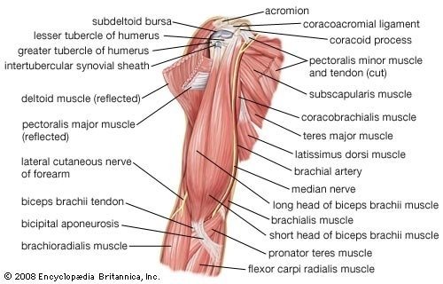
Pectoralis Major – The pectoralis major is a fan-shaped muscle covering the chest and comprised of clavicular and sternocostal regions.
- Attachments: The clavicular region originates from the clavicle and the sternocostal region originates from the sternum and the fascia of the oblique muscles of the abdomen. Both attach to the humerus.
- Actions: Adducts and rotates the upper arm.
Pectoralis Minor – The smaller pectoralis minor muscle lies beneath the pectoralis major.
- Attachments: The pectoralis minor originates from the third to fifth ribs and attaches to the scapula.
- Actions: Supports and depresses the scapula.
Serratus Anterior – The serratus anterior is located in the lateral wall of the chest.
- Attachments: The muscle is formed of several strips originating from the second to eighth ribs, each of which attaches to the scapula.
- Actions: Supports the scapula, allowing for elevation of the upper arm.
Intercostal Muscles
Intercostal muscles of the anterior trunk – Deep muscles of the chest and front of the arm, with the boundaries of the axilla. The intercostals are muscles between the ribs that form the chest cavity wall. Lying below the pectoral muscles, the intercostal muscles from the chest wall and play a key role in respiration. All intercostal muscles originate on the lower border of a rib and attach to the upper border of the rib below.
External Intercostals – The external intercostals are the most superficial of the intercostal muscles. They are continuous with the external oblique muscle of the abdomen.
- Actions: Elevate the ribs.
Internal Intercostals – Lying below the external intercostals, the internal intercostals are continuous with the internal oblique muscle of the abdomen.
- Actions: Elevate or depress the ribs.
Innermost Intercostals – The deepest lying of the intercostals, these muscles are similar in structure to the internal intercostals.
- Actions: Elevate or depress the ribs.
Other Muscles
Diaphragm – The diaphragm is a large, flat, sheet-like muscle that extends horizontally underneath the rib cage.
Functionally, the diaphragm separates the thoracic cavity, containing the lungs and heart and enclosed by the rib cage from the abdominal cavity, which contains the digestive organs. The diaphragm’s position allows it to aid in respiration. When it contracts, it physically moves the lungs and deforms the volume of the thoracic cavity.
- Attachments: The diaphragm has several points of origin along the sternum, the lower ribs, and the lower vertebrae. The muscle fibers combine into a central tendon, which ascends and attaches to the surface of the pericardium.
- Actions: Contracts, flattening and increasing the volume of the thoracic cavity. Relaxes and returns to the original shape, reducing the volume of the thoracic cavity.
Muscles of the Abdomen
The skeletal muscles of the abdomen form part of the abdominal wall, which holds and protects the gastrointestinal system. Five muscles form the abdominal wall, divided into vertical and flat groups. The flat muscles act to flex, laterally flex, and rotate the trunk. The fibers run in different directions and cross each other, strengthening the abdominal wall. The vertical muscles aid in compressing the abdominal cavity, stabilizing the pelvis, and depressing the ribs when a person is walking. Toward the midline, the muscles form aponeuroses, which merge into the linea alba.
Location of the external obliques: Highlighted in orange, the external obliques lie inferior to the pectoral muscles
External Oblique – The external oblique is the largest and most superficial of the flat muscles.
- Attachments: Originates from the lower ribs and attaches to the pelvis, forming an aponeurosis toward the midline and linea alba.
Internal Oblique – Lying deep to the external oblique, the internal oblique is smaller and thinner. Its fibers run perpendicular to the external oblique, improving the strength of the abdominal wall.
- Attachments: Originates from the pelvis and thoracolumbar fascia, running through the back. Attaches to the lower ribs and forms an aponeurosis toward the midline and linea alba.
Transversus Abdominis – The deepest of the flat muscles, the transversus abdominis consists of transversely-running fibers.
- Attachments: Originates from the lower ribs, thoracolumbar fascia, and pelvis, forming an aponeurosis toward the midline and linea alba.
Rectus Abdominis – A long vertical muscle that covers the abdomen, lying below the flat muscles. It is split through the midline by the linea alba formed from the aponeuroses of the abdominal muscles and separated by horizontal tendinous intersections which give rise to the six-pack.
- Attachments: Originates from the pubis and attaches to the lower edge of the rib cage and sternum.
Pyramidalis – Lying superficial to the rectus abdominal,s the pyramidalis is a small, triangular vertical muscle.
- Attachments: Originates from the pubis and attaches to the linea alba.
Posterior Muscles
Muscles of the posterior portion of the trunk include muscles of the back, suboccipital region, and perineum region.
Superficial Posterior Muscles
Location of the latissimus dorsi muscle – Highlighted in orange, the latissimus dorsi is a muscle of the posterior torso. The superficial posterior muscles are associated with the movement of the shoulder. As the name suggests, they are the most superficially located of the muscles covering the intermediate and intrinsic layers.
Trapezius – The trapezius is the most superficial muscle of the back and forms a broad flat triangle.
- Attachments: The trapezius originates from the skull and spine of the upper back and neck. It attaches to the clavicle and scapula.
- Actions: The superior region supports the arm and elevates and rotates the scapula, the intermediate region retracts the scapula, and the inferior region rotates and depresses the scapula.
Latissimus Dorsi – The latissimus dorsi originates from the lower back and covers a wide area.
- Attachments: The latissimus dorsi originates from the lower spine and ribs and the upper pelvis and fascia of the deep trunk muscles. The muscle converges into a tendon attaching to the humerus.
- Actions: Extends, adducts, and medially rotates the upper arm.
Levator Scapulae – A small, strap-like muscle that joins the neck to the scapula.
- Attachments: Originates from the side of the spine in the neck and attaches to the scapula.
- Actions: Elevates the scapula.
Rhomboid Major – Sits inferiorly to the levator scapulae.
- Attachments: Originates from the spine in the upper back and attaches to the scapula inferior to the levator scapulae attachment.
- Actions: Retracts and rotates the scapula.
Rhomboid Minor – Sits between the levator scapulae and rhomboid major, with which it is paired in action and function, this retracts and rotates the scapula.
Intermediate Posterior Muscles
The intermediate muscles of the posterior contribute to the movements of the ribcage during respiration.
Serratus Posterior Superior – The serratus posterior superior is a thin, rectangular-shaped muscle lying below the rhomboid muscles.
- Attachments: Originates from the lower spine and attaches to ribs 2 through 5.
- Actions: Elevates ribs 2 through 5.
Serratus Posterior Inferior – The serratus posterior inferior is a broad muscle lying beneath the latissimus dorsi.
- Attachments: Originates from the spine and attaches to ribs 9 through 12.
- Actions: Depresses ribs 9 through 12.
Intrinsic Posterior Muscles

The intrinsic muscles of the posterior are responsible for maintaining posture and facilitating the movement of the head and neck. They are divided into three layers.
Superficial Layer
Location of the splenius muscle – The splenius capitis is highlighted in orange, with the splenius cervicis directly below. Two muscles in the superficial layer are responsible for the rotation of the head.
Splenius Capitis – This thick rectangular muscle is the most superior of the next muscles.
- Attachments: Originates from the upper spine and attaches to the skull.
- Actions: Rotates and extends the head and neck.
Splenius cervicis – A small triangular-shaped muscle located immediately below the splenius capitis.
- Attachments: Originates from the spine and attaches several vertebrae higher.
- Actions: Rotates and extends the head and neck.
Intermediate Layer
Three columnar muscles in the intermediate layer are responsible for flexing and extending the neck as well as maintaining posture. All three originate from a common tendon associated with the pelvis.
Iliocostalis – The most laterally located of the three intermediate muscles.
- Attachments: Originates from the common tendon and attaches to the ribs and lower neck.
- Actions: Extends and controls abduction and adduction of the spine and neck.
Longissimus – Located between the iliocostalis and spinal muscles, it is the largest of the intermediate layer muscles.
- Attachments: Originates from the common tendon and attaches to the lower ribs, spine, and skull.
- Actions: Extends and controls abduction and adduction of the spine and neck.
Spinalis – The most medially-located and smallest of the three intermediate layer muscles.
- Attachments: Originates from the common tendon and attaches to the upper spine and skull.
- Actions: Extends, flexes, and controls abduction and adduction of the spine and neck.
Deep Layer
Two muscles in the deep layer are responsible for the maintenance of posture and rotation of the neck.
Semispinalis – The semispinalis is the most superficial of the deep muscles.
- Attachments: A broad origin on the upper regions of the spine, with each origin attaching several vertebrae higher or to the skull.
- Actions: Extends and rotates the head and maintains posture.
Multifidus – The multifidus is located underneath the semispinalis muscle, and is key in maintaining posture.
- Attachments: A broad origin up the length of the spine, with each origin attaching several vertebrae higher.
- Actions: Maintains posture through the spine.
Location of the multifidus muscle – Highlighted in orange, the multifidus muscle is a muscle of the posterior trunk and lies interior to a majority of muscles.
Key Terms
- Diaphragm – The key muscle in the control of respiration.
- Abdominal wall – A layer of muscle and fascia that protects and encloses the abdominal cavity, allowing for its compression as well as torso movement.
- Linea alba – A tough, fibrous line running down the midline of the abdomen, formed from the aponeuroses of the abdominal muscles.
- Intercostal – Muscles forming the chest wall, which aids in respiration.
- The back is characterized by numerous muscle groups which allow movement of the shoulder, head, and neck, as well as aid in respiration and maintain posture and balance.
- The superficial muscles of the back are responsible for the movement of the shoulder.
- The intermediate muscles of the back assist in the movement of the rib cage during respiration.
- The intrinsic back muscles facilitate the movement of the head and neck and are fundamental in maintaining posture and balance.
- The posterior or back muscles perform a wide range of functions, including the movement of the shoulder, head, and neck and assisting in respiration, posture, and balance. Posterior muscles are split into three groups depending on their physiological location.
- The anterior muscles of the trunk (torso) are associated with the front of the body, include chest and abdominal muscles. Chest muscles function in respiration while abdominal muscles function in torso movement and in the maintenance of balance and posture.
- The intercostal muscles from the chest wall and function in respiration.
- The diaphragm is a sheet-like muscle that extends underneath the rib cage and aids in respiration by physically moving the lungs.
- The obliques are abdominal muscles that assist during bending and twisting of the torso.
- The rectus abdominis is the muscles often referred to as the “six-pack abs” and is involved in numerous aspects of trunk stabilization and bending.
Frequently Asking Muscle of Face and Neck
- Anterior scalene – a muscle anterior to the middle scalene
- Appendicular – of the arms and legs axial of the trunk and head
- Buccinator – the muscle that compresses the cheek
- Corrugator supercilii – prime mover of the eyebrows
- Deglutition – swallowing
- Digastric – a muscle that has anterior and posterior bellies and elevates the hyoid bone and larynx when one swallows; it also depresses the mandible
- Epicranial aponeurosis – (also, galea aponeurosis) flat broad tendon that connects the frontalis and occipitalis
- Erector spinae group – large muscle mass of the back; primary extensor of the vertebral column
- Extrinsic eye muscles – originate outside the eye and insert into the outer surface of the white of the eye, and create eyeball movement
- Frontalis – the front part of the occipitofrontalis muscle
- Genioglossus – the muscle that originates on the mandible and allows the tongue to move downward and forward
- Geniohyoid – the muscle that depresses the mandible, and raises and pulls the hyoid bone anteriorly
- Hyoglossus – a muscle that originates on the hyoid bone to move the tongue downward and flatten it
- Iliocostalis cervicis – the muscle of the iliocostalis group associated with the cervical region
- Iliocostalis group – laterally placed muscles of the erector spinae
- Iliocostalis lumborum – the muscle of the iliocostalis group associated with the lumbar region
- Iliocostalis thoracic – the muscle of the iliocostalis group associated with the thoracic region
- Infrahyoid muscles – anterior neck muscles that are attached to, and inferior to the hyoid bone
- Lateral pterygoid – the muscle that moves the mandible from side to side
- Longissimus capitis – the muscle of the longissimus group associated with the head region
- Longissimus cervicis – the muscle of the longissimus group associated with the cervical region
- Longissimus group – intermediately placed muscles of the erector spinae
- Longissimus thoracis – the muscle of the longissimus group associated with the thoracic region
- Masseter – main muscle for chewing that elevates the mandible to close the mouth
- Mastication – chewing
- Medial pterygoid – the muscle that moves the mandible from side to side
- Middle scalene – longest scalene muscle, located between the anterior and posterior scalenes
- Multifidus – the muscle of the lumbar region that helps extend and laterally flex the vertebral column
- Mylohyoid – the muscle that lifts the hyoid bone and helps press the tongue to the top of the mouth
- Occipitalis – posterior part of the occipitofrontalis muscle
- Occipitofrontalis – the muscle that makes up the scalp with a frontal belly and an occipital belly
- Omohyoid – a muscle that has superior and inferior bellies and depresses the hyoid bone
- Orbicularis oculi – a circular muscle that closes the eye
- Orbicularis oris – a circular muscle that moves the lips
- Palatoglossus – the muscle that originates on the soft palate to elevate the back of the tongue
- Posterior scalene – smallest scalene muscle, located posterior to the middle scalene
- Scalene muscles – flex, laterally flex, and rotate the head; contribute to deep inhalation
- Segmental muscle group – interspinal and intertransversarii muscles that bring together the spinous and transverse processes of each consecutive vertebra
- Semispinalis capitis – transversospinales muscle associated with the head region
- Semispinalis services – transversospinales muscle associated with the cervical region
- Semispinalis thoracic – transversospinales muscle associated with the thoracic region
- Spinalis capitis – the muscle of the spinal group associated with the head region
- Spinalis cervicis – the muscle of the spinal group associated with the cervical region
- Spinalis group – medially placed muscles of the erector spinae
- Spinalis thoracis – the muscle of the spinal group associated with the thoracic region
- Splenius – posterior neck muscles; includes the splenius capitis and splenius cervicis
- Splenius capitis – neck muscle that inserts into the head region
- Splenius cervicis – neck muscle that inserts into the cervical region
- Sternocleidomastoid – the major muscle that laterally flexes and rotates the head
- Sternohyoid – a muscle that depresses the hyoid bone
- Sternothyroid – a muscle that depresses the larynx’s thyroid cartilage
- Styloglossus – a muscle that originates on the styloid bone, and allows upward and backward motion of the tongue
- Stylohyoid – a muscle that elevates the hyoid bone posteriorly
- Suprahyoid muscles – neck muscles that are superior to the hyoid bone
- Temporalis – a muscle that retracts the mandible
- Thyrohyoid – a muscle that depresses the hyoid bone and elevates the larynx’s thyroid cartilage
- Transversospinales – muscles that originate at the transverse processes and insert at the spinous processes of the vertebrae.
References