Pectoralis Major Muscle/Pectoralis major is the superior most and largest muscle of the anterior chest wall. It is a thick, fan-shaped muscle that lies underneath the breast tissue and forms the anterior wall of the axilla. Its origin lies anterior surface of the medial half of the clavicle, the anterior surface of the sternum, the first 7 costal cartilages, the sternal end of the sixth rib, and the aponeurosis of the external oblique of the anterior abdominal wall. The insertion of the pectoralis major is at the lateral lip of the intertubercular sulcus of the humerus. There are 2 heads of the pectoralis major, the clavicular and the sternocostal, which reference their area of origin[rx][rx]. The sternocostal head is described as having between 2 to 7 distinct segments.
Structure and Function of Pectoralis Major
The function of the pectoralis major is 3-fold and dependent on which heads of muscles are involved.[rx][rx]
Flexion, adduction and medial rotation of the arm at the glenohumeral joint
Clavicular head causes flexion of the extended arm
Sternoclavicular head causes extension of the flexed arm
The pectoralis major shows variation in muscle fiber length, differing from the majority of muscle fibers in the human body, which usually show uniform length. This configuration of the muscle fibers potentially allows for more power production through differing muscle shortening velocities.[rx]
Nerves of Pectoralis Major
The 2 heads of the pectoralis major have different nervous supplies. The clavicular head derives its nerve supply from the lateral pectoral nerve. The medial pectoral nerve innervates the sternocostal head. The lateral pectoral nerve arises directly from the lateral cord of the brachial plexus, and the medial pectoral nerve arises from the medial cord.[rx][rx]
Anatomy of the Pectoralis Major
Origin – Clavicular head, anterior sternum, costal cartilages 1 to 7, the sternal end rib 6, aponeurosis of the external oblique
Insertion – Lateral lip intertubercular sulcus of the humerus
Nervous innervation – Medial and lateral pectoral nerves (clavicular head C5, sternocostal head C6/7/8, T1)
Function – Flexion, adduction, and medial rotation of the arm at the glenohumeral joint; clavicular head causes flexion of the extended arm; sternoclavicular head causes extension of the flexed arm
Arterial supply – Pectoral artery (thoracoacromial trunk, the second branch of the axillary artery)
Venous drainage – Pectoral vein (drains into the subclavian vein)
Pectoralis major is the superior most and largest muscle of the anterior chest wall. It is a thick, fan-shaped muscle that lies underneath the breast tissue and forms the anterior wall of the axilla. Its origin lies anterior surface of the medial half of the clavicle, the anterior surface of the sternum, the first 7 costal cartilages, the sternal end of the sixth rib, and the aponeurosis of the external oblique of the anterior abdominal wall. The insertion of the pectoralis major is at the lateral lip of the intertubercular sulcus of the humerus. There are 2 heads of the pectoralis major, the clavicular and the sternocostal, which reference their area of origin[rx][rx]. The sternocostal head is described as having between 2 to 7 distinct segments.
Structure and Function of Pectoralis Major
The function of the pectoralis major is 3-fold and dependent on which heads of muscles are involved.[rx][rx]
Flexion, adduction and medial rotation of the arm at the glenohumeral joint
Clavicular head causes flexion of the extended arm
Sternoclavicular head causes extension of the flexed arm
The pectoralis major shows variation in muscle fiber length, differing from the majority of muscle fibers in the human body, which usually show uniform length. This configuration of the muscle fibers potentially allows for more power production through differing muscle shortening velocities.[rx]
Nerves of Pectoralis Major
The 2 heads of the pectoralis major have different nervous supplies. The clavicular head derives its nerve supply from the lateral pectoral nerve. The medial pectoral nerve innervates the sternocostal head. The lateral pectoral nerve arises directly from the lateral cord of the brachial plexus, and the medial pectoral nerve arises from the medial cord.[rx][rx]
Anatomy of the Pectoralis Major
Origin – Clavicular head, anterior sternum, costal cartilages 1 to 7, the sternal end rib 6, aponeurosis of the external oblique
Insertion – Lateral lip intertubercular sulcus of the humerus
Nervous innervation – Medial and lateral pectoral nerves (clavicular head C5, sternocostal head C6/7/8, T1)
Function – Flexion, adduction, and medial rotation of the arm at the glenohumeral joint; clavicular head causes flexion of the extended arm; sternoclavicular head causes extension of the flexed arm
Arterial supply – Pectoral artery (thoracoacromial trunk, the second branch of the axillary artery)
Venous drainage – Pectoral vein (drains into the subclavian vein)
What Is Patellar Tendinopathy/Patellar Tendinopathy is a painful condition of the knee caused by small tears in the patellar tendon that mainly occurs in sports requiring strenuous jumping. The tears are typically caused by accumulated stress on the patellar or quadriceps tendon. As the name implies, the condition is common in athletes from jumping sports such as volleyball, track (long and high jump), and basketball. The condition has a male predominance. Contrary to traditional belief, the jumper’s knee does not involve inflammation of the knee extensor tendons.
Jumper’s knee, or patellar tendinopathy (PT), is a chronic overuse injury of the patellar tendon. The prevalence is particularly high in jump sports athletes, such as in elite basketball players and volleyball players, respectively 32% and 45 %. However, also the prevalence among non-elite athletes is substantial and varies between 14.4% and 2.5% for different sports. Athletes with PT are often forced to reduce their training and competition levels because of patellar tendon pain. In some cases, athletes even have to quit their sporting activities. It is without a doubt that this condition can have an enormous impact on sports participation.
Stages of Patellar Tendinopathy
Depending on the duration of symptoms, Patellar Tendinopathy can be classified into 1 of 4 stages
Stage 1 – Pain only after activity, without functional impairment
Stage 2 – Pain during and after activity, although the patient is still able to perform satisfactorily in his or her sport
Stage 3 – Prolonged pain during and after activity, with increasing difficulty in performing at a satisfactory level
It is an overuse injury from repetitive overloading of the extensor mechanism of the knee. The microtears exceed the body’s healing mechanism unless the activity is stopped.
Running – Jumping or bounding are more common overall than Running
Athletes in jumping sports – High jump, Basketball, Football, Gymnastics
Pain after Exercise, especially prolonged Exercise and with knee flexion
Knee local corticosteroid injections and repetitive trauma to the knee extensor tendon
Leg Length Discrepancy
Pes Cavus
Insidious overall onset
Later – During Exercise and while at rest
Among the risk factors for patellar tendonitis are low ankle dorsiflexion, weak gluteal muscles, and muscle tightness, particularly in the calves, quadriceps muscle, and hamstrings.[rx]
Symptoms of Patellar Tendinopathy
Tenderness on palpation of the tendon, together with a characteristic history
Pain and tenderness around your patellar tendon.
Swelling.
Pain with jumping, running or walking.
Pain when bending or straightening your leg.
Tenderness behind the lower part of your kneecap.
pain below the kneecap, especially during sports, climbing stairs and bending the knee
A swollen knee joint
Knee stiffness
Leg or calf weakness
Pain when bending the knee
Strength – Knee extension weakness and Predisposing findings
Ankle dorsiflexion weakness
Hamstring tightness
Heel cord tightness
Quadriceps muscle tightness
Pain and decreased depth on single leg decline squat (LR+ 4 and LR- 0.5)
Extend unaffected knee
Squat with the affected leg
Diagnosis of Patellar Tendinopathy
Physical exam
Tenderness to palpation of the patellar tendon, just inferior to the patella, is the hallmark of the diagnosis. A clinical pearl is to palpate the tendon with the knee in extension as opposed to flexion. Palpation in flexion may mask a subtle jumper’s knee.
The patient may also have swelling of the tendon, and crepitus of the tendon with motion. Patients will also have pain with resisted knee extension.
A thorough knee exam including palpation of the joint lines, ligamentous and patellar stability and range of motion should also be performed to rule out other pathology.
Differential Diagnosis
Patellar tendon rupture
It can occur as an acute injury. Patients will have sudden, severe pain in the front of their knee and their knee will buckle. A defect can usually be appreciated in the patellar tendon, though sometimes this is difficult to assess if severe swelling is present. The patient will not be able to perform a straight leg raise, and they will have an extensor lag (lack of full active extension in the setting of full passive extension).
X-rays will show patella Alta (or high riding patella), and MRI will show the patellar tendon tear. Patients with patellar tendon ruptures should be placed in a knee immobilizer and referred to an orthopedic surgeon for urgent repair.
Patella fracture
It can occur as an acute injury. The patient will have pain over the patella itself. Diagnosis is usually made on an x-ray. Place in a knee immobilizer and refer to an orthopedic surgeon.
Patella chondromalacia (patellofemoral syndrome)
Presents with anterior knee pain. Pain is particularly bad with going up and downstairs. The pain with this condition is more proximal than with the jumper’s knee, and patients usually have no tenderness to palpation of the patellar tendon on the exam. Most often this is a diagnosis of exclusion.
Meniscus tears
Will have pain along the joint line. The pain is usually more lateral or medial than the jumper’s knee, but on occasion, the pain can be in the midline. Patients usually complain of clicking or popping in their knees. An MRI is diagnostic.
Fat pad syndrome
Inflammation of the fat pad that lies deep to the patellar tendon. Symptoms can be similar to jumper’s knee, but pain is around the tendon, and not on it. This may represent a spectrum of jumper’s knee and not a distinct entity. Regardless, the initial treatment is the same as that for the jumper’s knee.
Bone lesions
Tumors or infections are rare causes of anterior knee pain.
Radiograph
X-rays are usually negative for patients with jumper’s knee. On occasion, the x-ray can show shadows consistent with soft tissue swelling around the patellar tendon. In chronic cases, the x-ray may show calcifications in the patellar tendon. X-rays are most useful for ruling out concomitant pathology.
Advanced imaging
An MRI is usually not necessary in the early stages of the disease when the diagnosis is obvious on clinical exam. For more severe or chronic cases, an MRI can show if there are tears in the patella tendon. MRI’s are also most useful for ruling out concomitant pathology. For patients that cannot obtain an MRI, an ultrasound can also be diagnostic. However, an ultrasound will give limited information on intra-articular pathology.
Treatment of Patellar Tendinopathy
Treatment of Jumper’s Knee
Treatment for jumper’s knee includes
Rest and take a break from sports
Ice
Taping or wearing a knee support or strap just under the patella
Sitting with the leg raised
Massage therapy
Strengthening and stretching muscles through physical therapy or an at-home exercise program
Medication
Topical Medications – These prescription-strength creams, gels, ointments, patches, and sprays help relieve pain and inflammation through the skin.
Calcium & vitamin D3 – To improve bone health and healing tendon. As a general rule, men and women age 50 and older should consume 1,200 milligrams of calcium a day, and 600 international units of vitamin D a day.
Glucosamine & Diacerein, Chondroitin sulfate – can be used to tightening the loose tendon, cartilage, ligament, and cartilage, ligament regenerates cartilage or inhabits the further degeneration of cartilage, ligament.
Jumper’s Knee Surgery
If your injury is severe and other treatments have failed, you may be required to have surgery. The procedure consists of the doctor making a longitudinal or transverse incision over the patella tendon and then removing the abnormal tissue. After the surgery, it could take anywhere from 6 to 12 months to fully recover and begin training again. You should check with your doctor before beginning rehab and strengthening exercises after surgery.
Physical Therapy for Patellar Tendinopathy
Most patients respond to a conservative management program such as the one suggested below.
Activity modification – Decrease activities that increase kneecap and upper leg pressure (for example, jumping or squatting). Certain “loading” exercises may be prescribed.
Cryotherapy – Apply ice for 20 to 30 minutes, 4 to 6 times per day, especially after activity.
Joint motion and kinematics assessment – Hip, knee, and ankle joint range of motion are evaluated.
Strengthening – Specific exercises are often prescribed.
Other sport-specific joint, muscle, and tendon therapies may be prescribed.
Ultrasound or phonophoresis (ultrasound delivered medication) – may decrease pain symptoms. A special brace with a cutout for the kneecap and lateral stabilizer or taping may improve patellar tracking and provide stability. Sometimes arch supports or orthotics are used to improve foot and leg stability, which can reduce symptoms and help prevent future injury.
The treatment of jumper’s knee is often specific to the degree of involvement.
Stage 1
Stage I, which is characterized by pain only after activity and no undue functional impairment, is often treated with cryotherapy. The patient should use ice packs or ice massage after terminating the activity that exacerbates the pain and later again that evening. If aching persists, a course of regularly prescribed anti-inflammatory medications should be administered for 10 to14 days.
Stage II
In stage II, the patient has pain both during and after activity but is still able to participate in the sport satisfactorily. The pain may interfere with sleep. At this point, activities that cause increased loading of the patellar tendon (for example, running or jumping) should be avoided.
A comprehensive physical therapy program, as discussed above, should be implemented. For pain relief, the knee should be protected by avoiding high loads to the patellar tendon, and cryotherapy should continue. The athlete should be instructed in alternative conditioning to avoid injury to the affected area.
Once the pain improves, therapy should focus on knee, ankle, and hip joint range of motion, flexibility, and strengthening. If the pain becomes increasingly intense and if the athlete becomes more concerned about his or her performance, a local corticosteroid injection may be considered. The doctor will explain the pros and cons of these injections.
Stage III
In stage III, the patient’s pain is sustained, and performance and sport participation are adversely affected. Though discomfort increases, therapeutic measures similar to those described above should be continued along with not participating in activities that may worsen or prevent recovery from the injury. Relative rest for an extended period (for instance 3 to 6 weeks) may be necessary for stage III. Often, the athlete will be encouraged to continue an alternative cardiovascular and strength-training program.
If the condition does not improve with treatment, surgery may be considered. Some athletes will not be able to continue to participate in activities that worsen or prevent recovery from the problem.
Stage IV
Tendon rupture requires surgical repair.
Medical Issues and Complications
Knee immobilization is not recommended because it results in stiffness and may lead to other muscle or joint problems, further prolonging an athlete’s return to activity.
Consultations
Consultation with a physical medicine and rehabilitation specialist or an orthopedic specialist is recommended, particularly for Stage I cases that do not respond to conservative treatment and more severe cases (Stages II, III, and IV). Primary care sports medicine physicians can also be consulted.
Recovery Phase
Physical Therapy
An in-depth, stage-specific description of a conservative therapy program is described above. In brief, in the recovery phase, the athlete and therapist should work to restore pain-free joint range of motion and muscle flexibility, symmetric strength in the lower extremities, and joint sensation. Sport-specific training, including high-level sport-specific exercises, should then be initiated.
Consultations
Consultation with a physical medicine and rehabilitation specialist or an orthopedic specialist is recommended, particularly for Stage I cases that do not respond to conservative treatment or more severe cases (Stages II, III, IV).
Surgical Intervention
Surgical intervention is indicated for stage IV, and refractory stage III tendinopathy as noted above.
Maintenance Phase
Physical Therapy
An in-depth, stage-specific description of a conservative therapy program is described above. Briefly, once in the maintenance phase, the athlete should complete a sport-specific training program before returning to competition. The physician and physical therapist can assist the athlete in determining when to return to competition based on the patient’s symptoms, current physical examination findings, and functional test results. Once the athlete returns to play, he or she must work to maintain gains in flexibility and strength.
Rehabilitation Exercises
Stretching – Stretch
(1) flexors of the hip and knee (hamstrings, gastrocnemius, iliopsoas, rectus femoris, adductors),
(2) extensors of the hip and knee (quadriceps, gluteals),
(3) the iliotibial band (a large tendon on the outside of the hip and upper leg), and
(4) the surrounding tissues and structures of the kneecap.
Stretching Exercises
Test Flexibility
Lay on a flat surface, like a bench or couch.
Pull your knee up to your chest with your leg bent at the knee and your hands gripped under the knee.
Starting with this will prepare you for other stretches.
Quadriceps Stretch
In a standing position bring your leg up behind you to hold your toes in your hand.
Try to keep your knees together and to pull your leg up straight behind you, not to the side.
You will feel a stretch at the front of the leg.
Try to hold this position for 10 seconds when you first begin rehab and work up to 30 seconds when inflammation has gone down.
Strengthening Exercises
Thera-Band Knee Flexion (Prone)
Tie the TheraBand Resistance Band into a loop and secure one end close to the floor.
Lay on your stomach and place the other end of the loop around your ankle.
Begin with your knee straight and bend your knee against the band.
Hold and slowly return.
Thera Band Lunge
Start in a standing position with one leg in front of the other.
Hold the ends of the TheraBand Resistance Band in each hand while standing in the middle of the band with the front foot.
Bend the front knee, so the thigh is horizontal while the back knee goes towards the floor.
Try not to rest your back knee on the floor, instead of hover over it.
Hold and return to the starting position.
Prevention of Jumper’s Knee
It’s important to warm up before and cool down after exercising to prevent patellar tendonitis
Wear shoes that fit well and support your arch
Gradually increase the intensity of your workouts to reduce your risk of injury.
Anatomy of Latissimus Dorsi Muscle/Latissimus Dorsi Muscle is a broad, flat muscle that occupies the majority of the lower posterior thorax. The muscle’s primary function is of the upper extremity but is also considered to be a respiratory accessory muscle. Due to this muscle’s broad attachment on the spinous processes, ongoing research is looking at what, if any, role the muscle plays in the trunk movement. Currently, the evidence is mixed on how much influence this muscle has on spine extension, lateral flexion, or rotation. Even though the muscle has a broad attachment to the trunk and strong actions on the humerus, the use of this muscle for surgical transposition appears to have a limited effect on or restriction of normal function.[rx][rx][rx][rx]
The latissimus dorsi muscle is a broad, flat muscle that occupies the majority of the lower posterior thorax. The muscle’s primary function is of the upper extremity but is also considered to be a respiratory accessory muscle. Due to this muscle’s broad attachment on the spinous processes, ongoing research is looking at what, if any, role the muscle plays in the trunk movement. Currently, the evidence is mixed on how much influence this muscle has on spine extension, lateral flexion, or rotation. Even though the muscle has a broad attachment to the trunk and strong actions on the humerus, the use of this muscle for surgical transposition appears to have a limited effect on or restriction of normal function.
Actions of Latissimus Dorsi Muscle
Depression, adducts, extends and internally rotates the arm at the shoulder[rx]
Assists with deep inspiration and forced expiration
Nerve supply of Latissimus Dorsi Muscle
The latissimus dorsi is innervated by the sixth, seventh, and eighth cervical nerves through the thoracodorsal (long scapular) nerve. Electromyography suggests that it consists of six groups of muscle fibers that can be independently coordinated by the central nervous system.[rx]
Blood Supply of Latissimus Dorsi Muscle
The thoracodorsal nerve runs with the thoracodorsal artery and vein to supply the latissimus dorsi muscle. The nerve is close to the lymphatic vessels of the axilla, which are relevant during axillary lymph node dissection procedures. The subscapular lymph nodes are located in the posterior axillary fold, receiving lymph from the posterior thoracic wall and scapular region. Injury to the thoracodorsal nerve during axillary lymph node dissection is less frequent than other complications, including lymphedema and seroma formation.[rx]
What are the Symptoms of Latissimus Dorsi Pain?
The latissimus dorsi muscle covers the width of the middle and lower back and is more commonly known as the lats.
It may be difficult to tell whether the pain is located in the latissimus dorsi or other muscles in the shoulders or back. When the latissimus dorsi is injured, a person may feel pain in several places, including
These could be symptoms of a more severe condition.
Causes
The most common causes of pain result from overuse of the muscle and poor technique when working out.
The latissimus dorsi is used in everyday activities, including:
expanding the chest for breathing
pushing against armrests of a chair to stand
weightlifting with the upper body
rowing
throwing
performing bench-presses
overuse of the muscle
poor technique
exercising without warming up
have poor posture
continually reach overhead
chop wood
frequently shovel
golf
play baseball
row
ski
swim
play tennis
do exercises such as pull-ups or lat pulldowns
It is possible to tear the latissimus dorsi, and athletes are at a particular risk. Some athletes most likely to injure this muscle include:
water skiers
golfers
pitchers
gymnasts
Treatment
Treating for latissimus dorsi pain usually involves rest and physical therapy. While you rest, your doctor may recommend something called the RICE protocol:
R: resting your back and shoulders from, and cutting back on, physical activities
I: icing the painful area with an ice pack or cold compress
C: using compression by applying an elastic bandage
E: elevating the area by sitting upright or placing pillows behind your upper back or shoulder
You can also take nonsteroidal anti-inflammatory drugs, such as aspirin or ibuprofen, to help with the pain. If you have severe pain, your doctor may prescribe something stronger. Alternative treatments, such as cryotherapy or acupuncture, may also help.
Exercises for relief
Certain exercises can alleviate pain associated with the latissimus dorsi and strengthen the muscle to prevent further injury.
It is essential that a person consult an expert, such as a doctor or personal trainer, to ensure that the exercises are right for them and that they are using correct form.
The following two exercises can reduce latissimus dorsi pain. A doctor can recommend how often a person should perform these exercises. Never continue an exercise that is painful or too uncomfortable:
Back bow
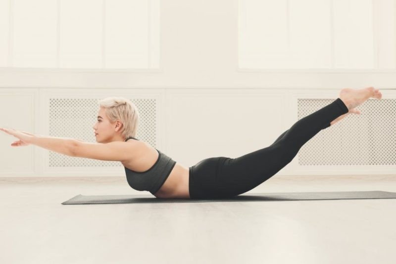
The back bow yoga pose can help reduce pain and strengthen the muscle. This pose is often referred to as “the Superman,” because it resembles how he flies.
To perform back bow:
Lay facedown on a yoga mat.
Extend straightened legs and arms away from the body, so that the arms are in front of the head.
Use the back to raise the shoulders and extended limbs toward the ceiling.
Hold the position for 10 seconds before lowering.
Pelvic raise or lift
To perform this exercise, a person should:
Lay flat on the back with the arms at the sides.
Bend the legs so that the heels are closer to the buttocks.
Lift the pelvis toward the ceiling.
Slowly lower it to the floor, keeping the hands and feet in place.
Prevention
A person can make certain lifestyle changes to prevent latissimus dorsi pain. These include:
using proper form during sports and exercise
avoiding overuse of the muscle
applying a heating pad to the area before exercising
warming up and cooling down before and after a workout
stretching gently after warming up and before cooling down
Latissimus Dorsi Muscle Pain/Latissimus Dorsi Muscle is a broad, flat muscle that occupies the majority of the lower posterior thorax. The muscle’s primary function is of the upper extremity but is also considered to be a respiratory accessory muscle. Due to this muscle’s broad attachment on the spinous processes, ongoing research is looking at what, if any, role the muscle plays in the trunk movement. Currently, the evidence is mixed on how much influence this muscle has on spine extension, lateral flexion, or rotation. Even though the muscle has a broad attachment to the trunk and strong actions on the humerus, the use of this muscle for surgical transposition appears to have a limited effect on or restriction of normal function.[rx][rx][rx][rx]
The latissimus dorsi muscle is a broad, flat muscle that occupies the majority of the lower posterior thorax. The muscle’s primary function is of the upper extremity but is also considered to be a respiratory accessory muscle. Due to this muscle’s broad attachment on the spinous processes, ongoing research is looking at what, if any, role the muscle plays in the trunk movement. Currently, the evidence is mixed on how much influence this muscle has on spine extension, lateral flexion, or rotation. Even though the muscle has a broad attachment to the trunk and strong actions on the humerus, the use of this muscle for surgical transposition appears to have a limited effect on or restriction of normal function.
Actions of Latissimus Dorsi Muscle
Depression, adducts, extends and internally rotates the arm at the shoulder[rx]
Assists with deep inspiration and forced expiration
Nerve supply of Latissimus Dorsi Muscle
The latissimus dorsi is innervated by the sixth, seventh, and eighth cervical nerves through the thoracodorsal (long scapular) nerve. Electromyography suggests that it consists of six groups of muscle fibers that can be independently coordinated by the central nervous system.[rx]
Blood Supply of Latissimus Dorsi Muscle
The thoracodorsal nerve runs with the thoracodorsal artery and vein to supply the latissimus dorsi muscle. The nerve is close to the lymphatic vessels of the axilla, which are relevant during axillary lymph node dissection procedures. The subscapular lymph nodes are located in the posterior axillary fold, receiving lymph from the posterior thoracic wall and scapular region. Injury to the thoracodorsal nerve during axillary lymph node dissection is less frequent than other complications, including lymphedema and seroma formation.[rx]
What are the Symptoms of Latissimus Dorsi Pain?
The latissimus dorsi muscle covers the width of the middle and lower back and is more commonly known as the lats.
It may be difficult to tell whether the pain is located in the latissimus dorsi or other muscles in the shoulders or back. When the latissimus dorsi is injured, a person may feel pain in several places, including
These could be symptoms of a more severe condition.
Causes of Latissimus Dorsi Muscle Pain
The most common causes of pain result from overuse of the muscle and poor technique when working out.
The latissimus dorsi is used in everyday activities, including:
expanding the chest for breathing
pushing against armrests of a chair to stand
weightlifting with the upper body
rowing
throwing
performing bench-presses
overuse of the muscle
poor technique
exercising without warming up
have poor posture
continually reach overhead
chop wood
frequently shovel
golf
play baseball
row
ski
swim
play tennis
do exercises such as pull-ups or lat pulldowns
It is possible to tear the latissimus dorsi, and athletes are at a particular risk. Some athletes most likely to injure this muscle include:
water skiers
golfers
pitchers
gymnasts
Treatment of Latissimus Dorsi Muscle Pain
Treating for latissimus dorsi pain usually involves rest and physical therapy. While you rest, your doctor may recommend something called the RICE protocol:
R: resting your back and shoulders from, and cutting back on, physical activities
I: icing the painful area with an ice pack or cold compress
C: using compression by applying an elastic bandage
E: elevating the area by sitting upright or placing pillows behind your upper back or shoulder
You can also take nonsteroidal anti-inflammatory drugs, such as aspirin or ibuprofen, to help with the pain. If you have severe pain, your doctor may prescribe something stronger. Alternative treatments, such as cryotherapy or acupuncture, may also help.
Exercises For Relief of Latissimus Dorsi Muscle Pain
Make a complete back workout by including exercises for your upper back and lower back.
One Arm Row on One Leg
Lat Pulls With Bands.
Barbell Rows
Dumbbell Pullovers
Renegade Row.
Pull-Ups
Dumbbell Rows.
One Arm Row.
Certain exercises can alleviate pain associated with the latissimus dorsi and strengthen the muscle to prevent further injury.
It is essential that a person consult an expert, such as a doctor or personal trainer, to ensure that the exercises are right for them and that they are using correct form.
The following two exercises can reduce latissimus dorsi pain. A doctor can recommend how often a person should perform these exercises. Never continue an exercise that is painful or too uncomfortable:
Back bow
The back bow yoga pose can help reduce pain and strengthen the muscle. This pose is often referred to as “the Superman,” because it resembles how he flies.
To perform back bow:
Lay facedown on a yoga mat.
Extend straightened legs and arms away from the body, so that the arms are in front of the head.
Use the back to raise the shoulders and extended limbs toward the ceiling.
Hold the position for 10 seconds before lowering.
Pelvic raise or lift
To perform this exercise, a person should:
Lay flat on the back with the arms at the sides.
Bend the legs so that the heels are closer to the buttocks.
Lift the pelvis toward the ceiling.
Slowly lower it to the floor, keeping the hands and feet in place.
Prevention
A person can make certain lifestyle changes to prevent latissimus dorsi pain. These include:
using proper form during sports and exercise
avoiding overuse of the muscle
applying a heating pad to the area before exercising
warming up and cooling down before and after a workout
stretching gently after warming up and before cooling down
What Is Latissimus Dorsi Muscle? Functions/Latissimus Dorsi Muscle is a broad, flat muscle that occupies the majority of the lower posterior thorax. The muscle’s primary function is of the upper extremity but is also considered to be a respiratory accessory muscle. Due to this muscle’s broad attachment on the spinous processes, ongoing research is looking at what, if any, role the muscle plays in the trunk movement. Currently, the evidence is mixed on how much influence this muscle has on spine extension, lateral flexion, or rotation. Even though the muscle has a broad attachment to the trunk and strong actions on the humerus, the use of this muscle for surgical transposition appears to have a limited effect on or restriction of normal function.[rx][rx][rx][rx]
The latissimus dorsi muscle is a broad, flat muscle that occupies the majority of the lower posterior thorax. The muscle’s primary function is of the upper extremity but is also considered to be a respiratory accessory muscle. Due to this muscle’s broad attachment on the spinous processes, ongoing research is looking at what, if any, role the muscle plays in the trunk movement. Currently, the evidence is mixed on how much influence this muscle has on spine extension, lateral flexion, or rotation. Even though the muscle has a broad attachment to the trunk and strong actions on the humerus, the use of this muscle for surgical transposition appears to have a limited effect on or restriction of normal function.
Actions of Latissimus Dorsi Muscle
Depression, adducts, extends and internally rotates the arm at the shoulder[rx]
Assists with deep inspiration and forced expiration
Nerve supply of Latissimus Dorsi Muscle
The latissimus dorsi is innervated by the sixth, seventh, and eighth cervical nerves through the thoracodorsal (long scapular) nerve. Electromyography suggests that it consists of six groups of muscle fibers that can be independently coordinated by the central nervous system.[rx]
Blood Supply of Latissimus Dorsi Muscle
The thoracodorsal nerve runs with the thoracodorsal artery and vein to supply the latissimus dorsi muscle. The nerve is close to the lymphatic vessels of the axilla, which are relevant during axillary lymph node dissection procedures. The subscapular lymph nodes are located in the posterior axillary fold, receiving lymph from the posterior thoracic wall and scapular region. Injury to the thoracodorsal nerve during axillary lymph node dissection is less frequent than other complications, including lymphedema and seroma formation.[rx]
What are the Symptoms of Latissimus Dorsi Pain?
The latissimus dorsi muscle covers the width of the middle and lower back and is more commonly known as the lats.
It may be difficult to tell whether the pain is located in the latissimus dorsi or other muscles in the shoulders or back. When the latissimus dorsi is injured, a person may feel pain in several places, including
These could be symptoms of a more severe condition.
Causes
The most common causes of pain result from overuse of the muscle and poor technique when working out.
The latissimus dorsi is used in everyday activities, including:
expanding the chest for breathing
pushing against armrests of a chair to stand
weightlifting with the upper body
rowing
throwing
performing bench-presses
overuse of the muscle
poor technique
exercising without warming up
have poor posture
continually reach overhead
chop wood
frequently shovel
golf
play baseball
row
ski
swim
play tennis
do exercises such as pull-ups or lat pulldowns
It is possible to tear the latissimus dorsi, and athletes are at a particular risk. Some athletes most likely to injure this muscle include:
water skiers
golfers
pitchers
gymnasts
Treatment
Treating for latissimus dorsi pain usually involves rest and physical therapy. While you rest, your doctor may recommend something called the RICE protocol:
R: resting your back and shoulders from, and cutting back on, physical activities
I: icing the painful area with an ice pack or cold compress
C: using compression by applying an elastic bandage
E: elevating the area by sitting upright or placing pillows behind your upper back or shoulder
You can also take nonsteroidal anti-inflammatory drugs, such as aspirin or ibuprofen, to help with the pain. If you have severe pain, your doctor may prescribe something stronger. Alternative treatments, such as cryotherapy or acupuncture, may also help.
Exercises For Relief
Make a complete back workout by including exercises for your upper back and lower back.
One Arm Row on One Leg
Lat Pulls With Bands.
Barbell Rows
Dumbbell Pullovers
Renegade Row.
Pull-Ups
Dumbbell Rows.
One Arm Row.
Certain exercises can alleviate pain associated with the latissimus dorsi and strengthen the muscle to prevent further injury.
It is essential that a person consult an expert, such as a doctor or personal trainer, to ensure that the exercises are right for them and that they are using correct form.
The following two exercises can reduce latissimus dorsi pain. A doctor can recommend how often a person should perform these exercises. Never continue an exercise that is painful or too uncomfortable:
Back bow
The back bow yoga pose can help reduce pain and strengthen the muscle. This pose is often referred to as “the Superman,” because it resembles how he flies.
To perform back bow:
Lay facedown on a yoga mat.
Extend straightened legs and arms away from the body, so that the arms are in front of the head.
Use the back to raise the shoulders and extended limbs toward the ceiling.
Hold the position for 10 seconds before lowering.
Pelvic raise or lift
To perform this exercise, a person should:
Lay flat on the back with the arms at the sides.
Bend the legs so that the heels are closer to the buttocks.
Lift the pelvis toward the ceiling.
Slowly lower it to the floor, keeping the hands and feet in place.
Prevention
A person can make certain lifestyle changes to prevent latissimus dorsi pain. These include:
using proper form during sports and exercise
avoiding overuse of the muscle
applying a heating pad to the area before exercising
warming up and cooling down before and after a workout
stretching gently after warming up and before cooling down
Origin Insertion Function of Latissimus Dorsi Muscle/Latissimus Dorsi Muscle is a broad, flat muscle that occupies the majority of the lower posterior thorax. The muscle’s primary function is of the upper extremity but is also considered to be a respiratory accessory muscle. Due to this muscle’s broad attachment on the spinous processes, ongoing research is looking at what, if any, role the muscle plays in the trunk movement. Currently, the evidence is mixed on how much influence this muscle has on spine extension, lateral flexion, or rotation. Even though the muscle has a broad attachment to the trunk and strong actions on the humerus, the use of this muscle for surgical transposition appears to have a limited effect on or restriction of normal function.[rx][rx][rx][rx]
The latissimus dorsi muscle is a broad, flat muscle that occupies the majority of the lower posterior thorax. The muscle’s primary function is of the upper extremity but is also considered to be a respiratory accessory muscle. Due to this muscle’s broad attachment on the spinous processes, ongoing research is looking at what, if any, role the muscle plays in the trunk movement. Currently, the evidence is mixed on how much influence this muscle has on spine extension, lateral flexion, or rotation. Even though the muscle has a broad attachment to the trunk and strong actions on the humerus, the use of this muscle for surgical transposition appears to have a limited effect on or restriction of normal function.
Actions of Latissimus Dorsi Muscle
Depression, adducts, extends and internally rotates the arm at the shoulder[rx]
Assists with deep inspiration and forced expiration
Nerve supply of Latissimus Dorsi Muscle
The latissimus dorsi is innervated by the sixth, seventh, and eighth cervical nerves through the thoracodorsal (long scapular) nerve. Electromyography suggests that it consists of six groups of muscle fibers that can be independently coordinated by the central nervous system.[rx]
Blood Supply of Latissimus Dorsi Muscle
The thoracodorsal nerve runs with the thoracodorsal artery and vein to supply the latissimus dorsi muscle. The nerve is close to the lymphatic vessels of the axilla, which are relevant during axillary lymph node dissection procedures. The subscapular lymph nodes are located in the posterior axillary fold, receiving lymph from the posterior thoracic wall and scapular region. Injury to the thoracodorsal nerve during axillary lymph node dissection is less frequent than other complications, including lymphedema and seroma formation.[rx]
What are the Symptoms of Latissimus Dorsi Pain?
The latissimus dorsi muscle covers the width of the middle and lower back and is more commonly known as the lats.
It may be difficult to tell whether the pain is located in the latissimus dorsi or other muscles in the shoulders or back. When the latissimus dorsi is injured, a person may feel pain in several places, including
These could be symptoms of a more severe condition.
Causes
The most common causes of pain result from overuse of the muscle and poor technique when working out.
The latissimus dorsi is used in everyday activities, including:
expanding the chest for breathing
pushing against armrests of a chair to stand
weightlifting with the upper body
rowing
throwing
performing bench-presses
overuse of the muscle
poor technique
exercising without warming up
have poor posture
continually reach overhead
chop wood
frequently shovel
golf
play baseball
row
ski
swim
play tennis
do exercises such as pull-ups or lat pulldowns
It is possible to tear the latissimus dorsi, and athletes are at a particular risk. Some athletes most likely to injure this muscle include:
water skiers
golfers
pitchers
gymnasts
Treatment
Treating for latissimus dorsi pain usually involves rest and physical therapy. While you rest, your doctor may recommend something called the RICE protocol:
R: resting your back and shoulders from, and cutting back on, physical activities
I: icing the painful area with an ice pack or cold compress
C: using compression by applying an elastic bandage
E: elevating the area by sitting upright or placing pillows behind your upper back or shoulder
You can also take nonsteroidal anti-inflammatory drugs, such as aspirin or ibuprofen, to help with the pain. If you have severe pain, your doctor may prescribe something stronger. Alternative treatments, such as cryotherapy or acupuncture, may also help.
Exercises For Relief
Make a complete back workout by including exercises for your upper back and lower back.
One Arm Row on One Leg
Lat Pulls With Bands.
Barbell Rows
Dumbbell Pullovers
Renegade Row.
Pull-Ups
Dumbbell Rows.
One Arm Row.
Certain exercises can alleviate pain associated with the latissimus dorsi and strengthen the muscle to prevent further injury.
It is essential that a person consult an expert, such as a doctor or personal trainer, to ensure that the exercises are right for them and that they are using correct form.
The following two exercises can reduce latissimus dorsi pain. A doctor can recommend how often a person should perform these exercises. Never continue an exercise that is painful or too uncomfortable:
Back bow
The back bow yoga pose can help reduce pain and strengthen the muscle. This pose is often referred to as “the Superman,” because it resembles how he flies.
To perform back bow:
Lay facedown on a yoga mat.
Extend straightened legs and arms away from the body, so that the arms are in front of the head.
Use the back to raise the shoulders and extended limbs toward the ceiling.
Hold the position for 10 seconds before lowering.
Pelvic raise or lift
To perform this exercise, a person should:
Lay flat on the back with the arms at the sides.
Bend the legs so that the heels are closer to the buttocks.
Lift the pelvis toward the ceiling.
Slowly lower it to the floor, keeping the hands and feet in place.
Prevention
A person can make certain lifestyle changes to prevent latissimus dorsi pain. These include:
using proper form during sports and exercise
avoiding overuse of the muscle
applying a heating pad to the area before exercising
warming up and cooling down before and after a workout
stretching gently after warming up and before cooling down
How do you stretch your latissimus dorsi?/Latissimus Dorsi Muscle is a broad, flat muscle that occupies the majority of the lower posterior thorax. The muscle’s primary function is of the upper extremity but is also considered to be a respiratory accessory muscle. Due to this muscle’s broad attachment on the spinous processes, ongoing research is looking at what, if any, role the muscle plays in the trunk movement. Currently, the evidence is mixed on how much influence this muscle has on spine extension, lateral flexion, or rotation. Even though the muscle has a broad attachment to the trunk and strong actions on the humerus, the use of this muscle for surgical transposition appears to have a limited effect on or restriction of normal function.[rx][rx][rx][rx]
The latissimus dorsi muscle is a broad, flat muscle that occupies the majority of the lower posterior thorax. The muscle’s primary function is of the upper extremity but is also considered to be a respiratory accessory muscle. Due to this muscle’s broad attachment on the spinous processes, ongoing research is looking at what, if any, role the muscle plays in the trunk movement. Currently, the evidence is mixed on how much influence this muscle has on spine extension, lateral flexion, or rotation. Even though the muscle has a broad attachment to the trunk and strong actions on the humerus, the use of this muscle for surgical transposition appears to have a limited effect on or restriction of normal function.
Actions of Latissimus Dorsi Muscle
Depression, adducts, extends and internally rotates the arm at the shoulder[rx]
Assists with deep inspiration and forced expiration
Nerve supply of Latissimus Dorsi Muscle
The latissimus dorsi is innervated by the sixth, seventh, and eighth cervical nerves through the thoracodorsal (long scapular) nerve. Electromyography suggests that it consists of six groups of muscle fibers that can be independently coordinated by the central nervous system.[rx]
Blood Supply of Latissimus Dorsi Muscle
The thoracodorsal nerve runs with the thoracodorsal artery and vein to supply the latissimus dorsi muscle. The nerve is close to the lymphatic vessels of the axilla, which are relevant during axillary lymph node dissection procedures. The subscapular lymph nodes are located in the posterior axillary fold, receiving lymph from the posterior thoracic wall and scapular region. Injury to the thoracodorsal nerve during axillary lymph node dissection is less frequent than other complications, including lymphedema and seroma formation.[rx]
What are the Symptoms of Latissimus Dorsi Pain?
The latissimus dorsi muscle covers the width of the middle and lower back and is more commonly known as the lats.
It may be difficult to tell whether the pain is located in the latissimus dorsi or other muscles in the shoulders or back. When the latissimus dorsi is injured, a person may feel pain in several places, including
These could be symptoms of a more severe condition.
Causes
The most common causes of pain result from overuse of the muscle and poor technique when working out.
The latissimus dorsi is used in everyday activities, including:
expanding the chest for breathing
pushing against armrests of a chair to stand
weightlifting with the upper body
rowing
throwing
performing bench-presses
overuse of the muscle
poor technique
exercising without warming up
have poor posture
continually reach overhead
chop wood
frequently shovel
golf
play baseball
row
ski
swim
play tennis
do exercises such as pull-ups or lat pulldowns
It is possible to tear the latissimus dorsi, and athletes are at a particular risk. Some athletes most likely to injure this muscle include:
water skiers
golfers
pitchers
gymnasts
Treatment
Treating for latissimus dorsi pain usually involves rest and physical therapy. While you rest, your doctor may recommend something called the RICE protocol:
R: resting your back and shoulders from, and cutting back on, physical activities
I: icing the painful area with an ice pack or cold compress
C: using compression by applying an elastic bandage
E: elevating the area by sitting upright or placing pillows behind your upper back or shoulder
You can also take nonsteroidal anti-inflammatory drugs, such as aspirin or ibuprofen, to help with the pain. If you have severe pain, your doctor may prescribe something stronger. Alternative treatments, such as cryotherapy or acupuncture, may also help.
Exercises for relief
Certain exercises can alleviate pain associated with the latissimus dorsi and strengthen the muscle to prevent further injury.
It is essential that a person consult an expert, such as a doctor or personal trainer, to ensure that the exercises are right for them and that they are using correct form.
The following two exercises can reduce latissimus dorsi pain. A doctor can recommend how often a person should perform these exercises. Never continue an exercise that is painful or too uncomfortable:
Back bow
The back bow yoga pose can help reduce pain and strengthen the muscle. This pose is often referred to as “the Superman,” because it resembles how he flies.
To perform back bow:
Lay facedown on a yoga mat.
Extend straightened legs and arms away from the body, so that the arms are in front of the head.
Use the back to raise the shoulders and extended limbs toward the ceiling.
Hold the position for 10 seconds before lowering.
Pelvic raise or lift
To perform this exercise, a person should:
Lay flat on the back with the arms at the sides.
Bend the legs so that the heels are closer to the buttocks.
Lift the pelvis toward the ceiling.
Slowly lower it to the floor, keeping the hands and feet in place.
Prevention
A person can make certain lifestyle changes to prevent latissimus dorsi pain. These include:
using proper form during sports and exercise
avoiding overuse of the muscle
applying a heating pad to the area before exercising
warming up and cooling down before and after a workout
stretching gently after warming up and before cooling down
What does latissimus dorsi muscle do?/Latissimus Dorsi Muscle is a broad, flat muscle that occupies the majority of the lower posterior thorax. The muscle’s primary function is of the upper extremity but is also considered to be a respiratory accessory muscle. Due to this muscle’s broad attachment on the spinous processes, ongoing research is looking at what, if any, role the muscle plays in the trunk movement. Currently, the evidence is mixed on how much influence this muscle has on spine extension, lateral flexion, or rotation. Even though the muscle has a broad attachment to the trunk and strong actions on the humerus, the use of this muscle for surgical transposition appears to have a limited effect on or restriction of normal function.[rx][rx][rx][rx]
The latissimus dorsi muscle is a broad, flat muscle that occupies the majority of the lower posterior thorax. The muscle’s primary function is of the upper extremity but is also considered to be a respiratory accessory muscle. Due to this muscle’s broad attachment on the spinous processes, ongoing research is looking at what, if any, role the muscle plays in the trunk movement. Currently, the evidence is mixed on how much influence this muscle has on spine extension, lateral flexion, or rotation. Even though the muscle has a broad attachment to the trunk and strong actions on the humerus, the use of this muscle for surgical transposition appears to have a limited effect on or restriction of normal function.
Actions of Latissimus Dorsi Muscle
Depression, adducts, extends and internally rotates the arm at the shoulder[rx]
Assists with deep inspiration and forced expiration
Nerve supply of Latissimus Dorsi Muscle
The latissimus dorsi is innervated by the sixth, seventh, and eighth cervical nerves through the thoracodorsal (long scapular) nerve. Electromyography suggests that it consists of six groups of muscle fibers that can be independently coordinated by the central nervous system.[rx]
Blood Supply of Latissimus Dorsi Muscle
The thoracodorsal nerve runs with the thoracodorsal artery and vein to supply the latissimus dorsi muscle. The nerve is close to the lymphatic vessels of the axilla, which are relevant during axillary lymph node dissection procedures. The subscapular lymph nodes are located in the posterior axillary fold, receiving lymph from the posterior thoracic wall and scapular region. Injury to the thoracodorsal nerve during axillary lymph node dissection is less frequent than other complications, including lymphedema and seroma formation.[rx]
What are the Symptoms of Latissimus Dorsi Pain?
The latissimus dorsi muscle covers the width of the middle and lower back and is more commonly known as the lats.
It may be difficult to tell whether the pain is located in the latissimus dorsi or other muscles in the shoulders or back. When the latissimus dorsi is injured, a person may feel pain in several places, including
These could be symptoms of a more severe condition.
Causes
The most common causes of pain result from overuse of the muscle and poor technique when working out.
The latissimus dorsi is used in everyday activities, including:
expanding the chest for breathing
pushing against armrests of a chair to stand
weightlifting with the upper body
rowing
throwing
performing bench-presses
overuse of the muscle
poor technique
exercising without warming up
have poor posture
continually reach overhead
chop wood
frequently shovel
golf
play baseball
row
ski
swim
play tennis
do exercises such as pull-ups or lat pulldowns
It is possible to tear the latissimus dorsi, and athletes are at a particular risk. Some athletes most likely to injure this muscle include:
water skiers
golfers
pitchers
gymnasts
Treatment
Treating for latissimus dorsi pain usually involves rest and physical therapy. While you rest, your doctor may recommend something called the RICE protocol:
R: resting your back and shoulders from, and cutting back on, physical activities
I: icing the painful area with an ice pack or cold compress
C: using compression by applying an elastic bandage
E: elevating the area by sitting upright or placing pillows behind your upper back or shoulder
You can also take nonsteroidal anti-inflammatory drugs, such as aspirin or ibuprofen, to help with the pain. If you have severe pain, your doctor may prescribe something stronger. Alternative treatments, such as cryotherapy or acupuncture, may also help.
Exercises for relief
Certain exercises can alleviate pain associated with the latissimus dorsi and strengthen the muscle to prevent further injury.
It is essential that a person consult an expert, such as a doctor or personal trainer, to ensure that the exercises are right for them and that they are using correct form.
The following two exercises can reduce latissimus dorsi pain. A doctor can recommend how often a person should perform these exercises. Never continue an exercise that is painful or too uncomfortable:
Back bow
The back bow yoga pose can help reduce pain and strengthen the muscle. This pose is often referred to as “the Superman,” because it resembles how he flies.
To perform back bow:
Lay facedown on a yoga mat.
Extend straightened legs and arms away from the body, so that the arms are in front of the head.
Use the back to raise the shoulders and extended limbs toward the ceiling.
Hold the position for 10 seconds before lowering.
Pelvic raise or lift
To perform this exercise, a person should:
Lay flat on the back with the arms at the sides.
Bend the legs so that the heels are closer to the buttocks.
Lift the pelvis toward the ceiling.
Slowly lower it to the floor, keeping the hands and feet in place.
Prevention
A person can make certain lifestyle changes to prevent latissimus dorsi pain. These include:
using proper form during sports and exercise
avoiding overuse of the muscle
applying a heating pad to the area before exercising
warming up and cooling down before and after a workout
stretching gently after warming up and before cooling down
What exercises work the latissimus dorsi?/Latissimus Dorsi Muscle is a broad, flat muscle that occupies the majority of the lower posterior thorax. The muscle’s primary function is of the upper extremity but is also considered to be a respiratory accessory muscle. Due to this muscle’s broad attachment on the spinous processes, ongoing research is looking at what, if any, role the muscle plays in the trunk movement. Currently, the evidence is mixed on how much influence this muscle has on spine extension, lateral flexion, or rotation. Even though the muscle has a broad attachment to the trunk and strong actions on the humerus, the use of this muscle for surgical transposition appears to have a limited effect on or restriction of normal function.[rx][rx][rx][rx]
The latissimus dorsi muscle is a broad, flat muscle that occupies the majority of the lower posterior thorax. The muscle’s primary function is of the upper extremity but is also considered to be a respiratory accessory muscle. Due to this muscle’s broad attachment on the spinous processes, ongoing research is looking at what, if any, role the muscle plays in the trunk movement. Currently, the evidence is mixed on how much influence this muscle has on spine extension, lateral flexion, or rotation. Even though the muscle has a broad attachment to the trunk and strong actions on the humerus, the use of this muscle for surgical transposition appears to have a limited effect on or restriction of normal function.
Actions of Latissimus Dorsi Muscle
Depression, adducts, extends and internally rotates the arm at the shoulder[rx]
Assists with deep inspiration and forced expiration
Nerve supply of Latissimus Dorsi Muscle
The latissimus dorsi is innervated by the sixth, seventh, and eighth cervical nerves through the thoracodorsal (long scapular) nerve. Electromyography suggests that it consists of six groups of muscle fibers that can be independently coordinated by the central nervous system.[rx]
Blood Supply of Latissimus Dorsi Muscle
The thoracodorsal nerve runs with the thoracodorsal artery and vein to supply the latissimus dorsi muscle. The nerve is close to the lymphatic vessels of the axilla, which are relevant during axillary lymph node dissection procedures. The subscapular lymph nodes are located in the posterior axillary fold, receiving lymph from the posterior thoracic wall and scapular region. Injury to the thoracodorsal nerve during axillary lymph node dissection is less frequent than other complications, including lymphedema and seroma formation.[rx]
What are the Symptoms of Latissimus Dorsi Pain?
The latissimus dorsi muscle covers the width of the middle and lower back and is more commonly known as the lats.
It may be difficult to tell whether the pain is located in the latissimus dorsi or other muscles in the shoulders or back. When the latissimus dorsi is injured, a person may feel pain in several places, including
These could be symptoms of a more severe condition.
Causes
The most common causes of pain result from overuse of the muscle and poor technique when working out.
The latissimus dorsi is used in everyday activities, including:
expanding the chest for breathing
pushing against armrests of a chair to stand
weightlifting with the upper body
rowing
throwing
performing bench-presses
overuse of the muscle
poor technique
exercising without warming up
have poor posture
continually reach overhead
chop wood
frequently shovel
golf
play baseball
row
ski
swim
play tennis
do exercises such as pull-ups or lat pulldowns
It is possible to tear the latissimus dorsi, and athletes are at a particular risk. Some athletes most likely to injure this muscle include:
water skiers
golfers
pitchers
gymnasts
Treatment
Treating for latissimus dorsi pain usually involves rest and physical therapy. While you rest, your doctor may recommend something called the RICE protocol:
R: resting your back and shoulders from, and cutting back on, physical activities
I: icing the painful area with an ice pack or cold compress
C: using compression by applying an elastic bandage
E: elevating the area by sitting upright or placing pillows behind your upper back or shoulder
You can also take nonsteroidal anti-inflammatory drugs, such as aspirin or ibuprofen, to help with the pain. If you have severe pain, your doctor may prescribe something stronger. Alternative treatments, such as cryotherapy or acupuncture, may also help.
Exercises For Relief
Make a complete back workout by including exercises for your upper back and lower back.
One Arm Row on One Leg
Lat Pulls With Bands.
Barbell Rows
Dumbbell Pullovers
Renegade Row.
Pull-Ups
Dumbbell Rows.
One Arm Row.
Certain exercises can alleviate pain associated with the latissimus dorsi and strengthen the muscle to prevent further injury.
It is essential that a person consult an expert, such as a doctor or personal trainer, to ensure that the exercises are right for them and that they are using correct form.
The following two exercises can reduce latissimus dorsi pain. A doctor can recommend how often a person should perform these exercises. Never continue an exercise that is painful or too uncomfortable:
Back bow
The back bow yoga pose can help reduce pain and strengthen the muscle. This pose is often referred to as “the Superman,” because it resembles how he flies.
To perform back bow:
Lay facedown on a yoga mat.
Extend straightened legs and arms away from the body, so that the arms are in front of the head.
Use the back to raise the shoulders and extended limbs toward the ceiling.
Hold the position for 10 seconds before lowering.
Pelvic raise or lift
To perform this exercise, a person should:
Lay flat on the back with the arms at the sides.
Bend the legs so that the heels are closer to the buttocks.
Lift the pelvis toward the ceiling.
Slowly lower it to the floor, keeping the hands and feet in place.
Prevention
A person can make certain lifestyle changes to prevent latissimus dorsi pain. These include:
using proper form during sports and exercise
avoiding overuse of the muscle
applying a heating pad to the area before exercising
warming up and cooling down before and after a workout
stretching gently after warming up and before cooling down
What type of muscle is the latissimus dorsi?/Latissimus Dorsi Muscle is a broad, flat muscle that occupies the majority of the lower posterior thorax. The muscle’s primary function is of the upper extremity but is also considered to be a respiratory accessory muscle. Due to this muscle’s broad attachment on the spinous processes, ongoing research is looking at what, if any, role the muscle plays in the trunk movement. Currently, the evidence is mixed on how much influence this muscle has on spine extension, lateral flexion, or rotation. Even though the muscle has a broad attachment to the trunk and strong actions on the humerus, the use of this muscle for surgical transposition appears to have a limited effect on or restriction of normal function.[rx][rx][rx][rx]
The latissimus dorsi muscle is a broad, flat muscle that occupies the majority of the lower posterior thorax. The muscle’s primary function is of the upper extremity but is also considered to be a respiratory accessory muscle. Due to this muscle’s broad attachment on the spinous processes, ongoing research is looking at what, if any, role the muscle plays in the trunk movement. Currently, the evidence is mixed on how much influence this muscle has on spine extension, lateral flexion, or rotation. Even though the muscle has a broad attachment to the trunk and strong actions on the humerus, the use of this muscle for surgical transposition appears to have a limited effect on or restriction of normal function.
Actions of Latissimus Dorsi Muscle
Depression, adducts, extends and internally rotates the arm at the shoulder[rx]
Assists with deep inspiration and forced expiration
Nerve supply of Latissimus Dorsi Muscle
The latissimus dorsi is innervated by the sixth, seventh, and eighth cervical nerves through the thoracodorsal (long scapular) nerve. Electromyography suggests that it consists of six groups of muscle fibers that can be independently coordinated by the central nervous system.[rx]
Blood Supply of Latissimus Dorsi Muscle
The thoracodorsal nerve runs with the thoracodorsal artery and vein to supply the latissimus dorsi muscle. The nerve is close to the lymphatic vessels of the axilla, which are relevant during axillary lymph node dissection procedures. The subscapular lymph nodes are located in the posterior axillary fold, receiving lymph from the posterior thoracic wall and scapular region. Injury to the thoracodorsal nerve during axillary lymph node dissection is less frequent than other complications, including lymphedema and seroma formation.[rx]
What are the Symptoms of Latissimus Dorsi Pain?
The latissimus dorsi muscle covers the width of the middle and lower back and is more commonly known as the lats.
It may be difficult to tell whether the pain is located in the latissimus dorsi or other muscles in the shoulders or back. When the latissimus dorsi is injured, a person may feel pain in several places, including
These could be symptoms of a more severe condition.
Causes
The most common causes of pain result from overuse of the muscle and poor technique when working out.
The latissimus dorsi is used in everyday activities, including:
expanding the chest for breathing
pushing against armrests of a chair to stand
weightlifting with the upper body
rowing
throwing
performing bench-presses
overuse of the muscle
poor technique
exercising without warming up
have poor posture
continually reach overhead
chop wood
frequently shovel
golf
play baseball
row
ski
swim
play tennis
do exercises such as pull-ups or lat pulldowns
It is possible to tear the latissimus dorsi, and athletes are at a particular risk. Some athletes most likely to injure this muscle include:
water skiers
golfers
pitchers
gymnasts
Treatment
Treating for latissimus dorsi pain usually involves rest and physical therapy. While you rest, your doctor may recommend something called the RICE protocol:
R: resting your back and shoulders from, and cutting back on, physical activities
I: icing the painful area with an ice pack or cold compress
C: using compression by applying an elastic bandage
E: elevating the area by sitting upright or placing pillows behind your upper back or shoulder
You can also take nonsteroidal anti-inflammatory drugs, such as aspirin or ibuprofen, to help with the pain. If you have severe pain, your doctor may prescribe something stronger. Alternative treatments, such as cryotherapy or acupuncture, may also help.
Exercises for relief
Certain exercises can alleviate pain associated with the latissimus dorsi and strengthen the muscle to prevent further injury.
It is essential that a person consult an expert, such as a doctor or personal trainer, to ensure that the exercises are right for them and that they are using correct form.
The following two exercises can reduce latissimus dorsi pain. A doctor can recommend how often a person should perform these exercises. Never continue an exercise that is painful or too uncomfortable:
Back bow
The back bow yoga pose can help reduce pain and strengthen the muscle. This pose is often referred to as “the Superman,” because it resembles how he flies.
To perform back bow:
Lay facedown on a yoga mat.
Extend straightened legs and arms away from the body, so that the arms are in front of the head.
Use the back to raise the shoulders and extended limbs toward the ceiling.
Hold the position for 10 seconds before lowering.
Pelvic raise or lift
To perform this exercise, a person should:
Lay flat on the back with the arms at the sides.
Bend the legs so that the heels are closer to the buttocks.
Lift the pelvis toward the ceiling.
Slowly lower it to the floor, keeping the hands and feet in place.
Prevention
A person can make certain lifestyle changes to prevent latissimus dorsi pain. These include:
using proper form during sports and exercise
avoiding overuse of the muscle
applying a heating pad to the area before exercising
warming up and cooling down before and after a workout
stretching gently after warming up and before cooling down
Latissimus Dorsi Muscle is a broad, flat muscle that occupies the majority of the lower posterior thorax. The muscle’s primary function is of the upper extremity but is also considered to be a respiratory accessory muscle. Due to this muscle’s broad attachment on the spinous processes, ongoing research is looking at what, if any, role the muscle plays in the trunk movement. Currently, the evidence is mixed on how much influence this muscle has on spine extension, lateral flexion, or rotation. Even though the muscle has a broad attachment to the trunk and strong actions on the humerus, the use of this muscle for surgical transposition appears to have a limited effect on or restriction of normal function.[rx][rx][rx][rx]
The latissimus dorsi muscle is a broad, flat muscle that occupies the majority of the lower posterior thorax. The muscle’s primary function is of the upper extremity but is also considered to be a respiratory accessory muscle. Due to this muscle’s broad attachment on the spinous processes, ongoing research is looking at what, if any, role the muscle plays in the trunk movement. Currently, the evidence is mixed on how much influence this muscle has on spine extension, lateral flexion, or rotation. Even though the muscle has a broad attachment to the trunk and strong actions on the humerus, the use of this muscle for surgical transposition appears to have a limited effect on or restriction of normal function.
Actions of Latissimus Dorsi Muscle
Depression, adducts, extends and internally rotates the arm at the shoulder[rx]
Assists with deep inspiration and forced expiration
Nerve supply of Latissimus Dorsi Muscle
The latissimus dorsi is innervated by the sixth, seventh, and eighth cervical nerves through the thoracodorsal (long scapular) nerve. Electromyography suggests that it consists of six groups of muscle fibers that can be independently coordinated by the central nervous system.[rx]
Blood Supply of Latissimus Dorsi Muscle
The thoracodorsal nerve runs with the thoracodorsal artery and vein to supply the latissimus dorsi muscle. The nerve is close to the lymphatic vessels of the axilla, which are relevant during axillary lymph node dissection procedures. The subscapular lymph nodes are located in the posterior axillary fold, receiving lymph from the posterior thoracic wall and scapular region. Injury to the thoracodorsal nerve during axillary lymph node dissection is less frequent than other complications, including lymphedema and seroma formation.[rx]
What are the Symptoms of Latissimus Dorsi Pain?
The latissimus dorsi muscle covers the width of the middle and lower back and is more commonly known as the lats.
It may be difficult to tell whether the pain is located in the latissimus dorsi or other muscles in the shoulders or back. When the latissimus dorsi is injured, a person may feel pain in several places, including
These could be symptoms of a more severe condition.
Causes
The most common causes of pain result from overuse of the muscle and poor technique when working out.
The latissimus dorsi is used in everyday activities, including:
expanding the chest for breathing
pushing against armrests of a chair to stand
weightlifting with the upper body
rowing
throwing
performing bench-presses
overuse of the muscle
poor technique
exercising without warming up
have poor posture
continually reach overhead
chop wood
frequently shovel
golf
play baseball
row
ski
swim
play tennis
do exercises such as pull-ups or lat pulldowns
It is possible to tear the latissimus dorsi, and athletes are at a particular risk. Some athletes most likely to injure this muscle include:
water skiers
golfers
pitchers
gymnasts
Treatment
Treating for latissimus dorsi pain usually involves rest and physical therapy. While you rest, your doctor may recommend something called the RICE protocol:
R: resting your back and shoulders from, and cutting back on, physical activities
I: icing the painful area with an ice pack or cold compress
C: using compression by applying an elastic bandage
E: elevating the area by sitting upright or placing pillows behind your upper back or shoulder
You can also take nonsteroidal anti-inflammatory drugs, such as aspirin or ibuprofen, to help with the pain. If you have severe pain, your doctor may prescribe something stronger. Alternative treatments, such as cryotherapy or acupuncture, may also help.
Exercises for relief
Certain exercises can alleviate pain associated with the latissimus dorsi and strengthen the muscle to prevent further injury.
It is essential that a person consult an expert, such as a doctor or personal trainer, to ensure that the exercises are right for them and that they are using correct form.
The following two exercises can reduce latissimus dorsi pain. A doctor can recommend how often a person should perform these exercises. Never continue an exercise that is painful or too uncomfortable:
Back bow
The back bow yoga pose can help reduce pain and strengthen the muscle. This pose is often referred to as “the Superman,” because it resembles how he flies.
To perform back bow:
Lay facedown on a yoga mat.
Extend straightened legs and arms away from the body, so that the arms are in front of the head.
Use the back to raise the shoulders and extended limbs toward the ceiling.
Hold the position for 10 seconds before lowering.
Pelvic raise or lift
To perform this exercise, a person should:
Lay flat on the back with the arms at the sides.
Bend the legs so that the heels are closer to the buttocks.
Lift the pelvis toward the ceiling.
Slowly lower it to the floor, keeping the hands and feet in place.
Prevention
A person can make certain lifestyle changes to prevent latissimus dorsi pain. These include:
using proper form during sports and exercise
avoiding overuse of the muscle
applying a heating pad to the area before exercising
warming up and cooling down before and after a workout
stretching gently after warming up and before cooling down