Category Archive Anatomy A – Z

ByRx Harun

Teres Minor; Origin, Nerve Supply, Functions

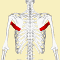
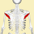
Teres Minor is a narrow elongated muscle of the rotator cuff. The muscle originates from the lateral border and adjacent posterior surface of the corresponding right or left scapula and inserts at both the greater tubercle of the humerus and the posterior surface of the joint capsule.[rx]

The primary function of the teres minor is to modulate the action of the deltoid, preventing the humeral head from sliding upward as the arm is abducted. It also functions to rotate the humerus laterally. The teres minor is innervated by the axillary nerve.[rx]

At a Glance of Teres minor

  • Function – Lateral rotation of the arm, stabilize glenohumeral joint
  • Origin – Lateral/axillary border and adjacent posterior aspect of the scapula
  • Insertion – Inferior aspect of the greater tubercle on the humerus
  • Innervation – Axillary nerve (C5, C6)

[stextbox id=’custom’ defcaption=”true”]

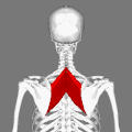
Muscles on the dorsum of the left scapula, and the Triceps brachii muscle:
Details
Origin the lateral border of the scapula
Insertion inferior facet of the greater tubercle of the humerus
Artery posterior circumflex humeral artery and the circumflex scapular artery
Nerve axillary nerve (C5-C6)
Actions laterally rotates the arm, stabilizes the humerus
Identifiers
Latin musculus teres minor
TA A04.6.02.010
FMA 32550
Anatomical terms of muscle


[/stextbox]

Teres Minor

Nerve Supply of Teres Minor

The muscle is innervated by the posterior branch of the axillary nerve where it forms a pseudoganglion.[rx] A pseudoganglion has no nerve cells but nerve fibers are present. Damage to the fibers innervating the teres minor is clinically significant. Sometimes a group of muscle fibers from teres minor may be fused with infraspinatus.

Functions of Teres Minor

The infraspinatus and teres minor attach to the head of the humerus; as part of the rotator cuff, they help hold the humeral head in the glenoid cavity of the scapula. They work in tandem with the posterior deltoid to externally (laterally) rotate the humerus, as well as adduction. Teres Minor can produce only very small scapular plane adduction during maximal contraction with an adductor moment arm of approximately 0.2 cm at 45° of shoulder internal rotation and approximately 0.1 cm at 45° of shoulder external rotation.


References

Teres Minor


ByRx Harun

Teres Major; Origin, Functions, Nerve Supply

Teres Major /Teres Major Muscle is a muscle of the upper limb. It attaches to the scapula and the humerus and is one of the seven scapulohumeral muscles. It is a thick but somewhat flattened muscle. The teres major muscle (from Latin teres, meaning “rounded”) is positioned above the latissimus dorsi muscle and assists in the extension and medial rotation of the humerus. This muscle is commonly confused as a rotator cuff muscle, but it is not because it does not attach to the capsule of the shoulder joint, unlike the teres minor muscle for example.

The primary function of the teres minor is to modulate the action of the deltoid, preventing the humeral head from sliding upward as the arm is abducted. It also functions to rotate the humerus laterally. The teres minor is innervated by the axillary nerve.[rx]

At a Glance of Teres major

  • Function: Adduction and medial rotation of the arm
  • Origin: Posterior surface of the scapula at its inferior angle
  • Insertion: Intertubercular groove on its medial aspect
  • Innervation: Lower scapular nerve (C5, C6)

[stextbox id=’custom’ defcaption=”true”]

Teres major muscle (in red) seen from the back (posterior to anterior perspective).
Details
Origin Posterior aspect of the inferior angle of the scapula
Insertion The medial lip of the intertubercular sulcus of the humerus
Artery Subscapular and circumflex scapular arteries
Nerve Lower subscapular nerve (segmental levels C5 and C6)
Actions adduct the humerus, Internal rotation (medial rotation) of the humerus, extend the humerus from flexed position, Depress shoulder
Identifiers
Latin Musculus teres major
TA A04.6.02.011
FMA 32549
Anatomical terms of muscle


[/stextbox]

What Is Teres Major Muscle?

Nerve Supply of Teres Major Muscle

Teres major is supplied primarily by the lower subscapular nerve and additionally by the thoracodorsal nerve (middle subscapular nerve). These are distal to the upper subscapular nerve. These three nerves branch off the posterior cord of the brachial plexus. The nerves that innervate teres major consist of fibers from spinal nerves C5-C8.

Functions of Teres Major Muscle

The teres major is a medial rotator and adductor of the humerus and assists the latissimus dorsi in drawing the previously raised humerus downwards and backward (extension, but not hyperextension). It also helps stabilize the humeral head in the glenoid cavity.


References

Teres Major


ByRx Harun

Functions Nerve Supply of Teres Major Muscle

Functions Nerve Supply of Teres Major Muscle/Teres Major Muscle is a muscle of the upper limb. It attaches to the scapula and the humerus and is one of the seven scapulohumeral muscles. It is a thick but somewhat flattened muscle. The teres major muscle (from Latin teres, meaning “rounded”) is positioned above the latissimus dorsi muscle and assists in the extension and medial rotation of the humerus. This muscle is commonly confused as a rotator cuff muscle, but it is not because it does not attach to the capsule of the shoulder joint, unlike the teres minor muscle for example.

The primary function of the teres minor is to modulate the action of the deltoid, preventing the humeral head from sliding upward as the arm is abducted. It also functions to rotate the humerus laterally. The teres minor is innervated by the axillary nerve.[rx]

At a Glance of Teres major

  • Function: Adduction and medial rotation of the arm
  • Origin: Posterior surface of the scapula at its inferior angle
  • Insertion: Intertubercular groove on its medial aspect
  • Innervation: Lower scapular nerve (C5, C6)

[stextbox id=’custom’ defcaption=”true”]

Teres major muscle (in red) seen from the back (posterior to anterior perspective).
Details
Origin Posterior aspect of the inferior angle of the scapula
Insertion The medial lip of the intertubercular sulcus of the humerus
Artery Subscapular and circumflex scapular arteries
Nerve Lower subscapular nerve (segmental levels C5 and C6)
Actions adduct the humerus, Internal rotation (medial rotation) of the humerus, extend the humerus from a flexed position, Depress shoulder
Identifiers
Latin Musculus teres major
TA A04.6.02.011
FMA 32549
Anatomical terms of muscle


[/stextbox]

Teres Major Muscle

Nerve Supply of Teres Major Muscle

Teres major is supplied primarily by the lower subscapular nerve and additionally by the thoracodorsal nerve (middle subscapular nerve). These are distal to the upper subscapular nerve. These three nerves branch off the posterior cord of the brachial plexus. The nerves that innervate teres major consist of fibers from spinal nerves C5-C8.

Functions of Teres Major Muscle

The teres major is a medial rotator and adductor of the humerus and assists the latissimus dorsi in drawing the previously raised humerus downwards and backward (extension, but not hyperextension). It also helps stabilize the humeral head in the glenoid cavity.


References

Functions Nerve Supply of Teres Major Muscle


ByRx Harun

What Is Teres Major Muscle? Functions

What Is Teres Major Muscle?/Teres Major Muscle is a muscle of the upper limb. It attaches to the scapula and the humerus and is one of the seven scapulohumeral muscles. It is a thick but somewhat flattened muscle. The teres major muscle (from Latin teres, meaning “rounded”) is positioned above the latissimus dorsi muscle and assists in the extension and medial rotation of the humerus. This muscle is commonly confused as a rotator cuff muscle, but it is not because it does not attach to the capsule of the shoulder joint, unlike the teres minor muscle for example.

The primary function of the teres minor is to modulate the action of the deltoid, preventing the humeral head from sliding upward as the arm is abducted. It also functions to rotate the humerus laterally. The teres minor is innervated by the axillary nerve.[rx]

At a Glance of Teres major

  • Function: Adduction and medial rotation of the arm
  • Origin: Posterior surface of the scapula at its inferior angle
  • Insertion: Intertubercular groove on its medial aspect
  • Innervation: Lower scapular nerve (C5, C6)

[stextbox id=’custom’ defcaption=”true”]

Teres major muscle (in red) seen from the back (posterior to anterior perspective).
Details
Origin Posterior aspect of the inferior angle of the scapula
Insertion The medial lip of the intertubercular sulcus of the humerus
Artery Subscapular and circumflex scapular arteries
Nerve Lower subscapular nerve (segmental levels C5 and C6)
Actions adduct the humerus, Internal rotation (medial rotation) of the humerus, extend the humerus from flexed position, Depress shoulder
Identifiers
Latin Musculus teres major
TA A04.6.02.011
FMA 32549
Anatomical terms of muscle


[/stextbox]

What Is Teres Major Muscle?

Nerve Supply of Teres Major Muscle

Teres major is supplied primarily by the lower subscapular nerve and additionally by the thoracodorsal nerve (middle subscapular nerve). These are distal to the upper subscapular nerve. These three nerves branch off the posterior cord of the brachial plexus. The nerves that innervate teres major consist of fibers from spinal nerves C5-C8.

Functions of Teres Major Muscle

The teres major is a medial rotator and adductor of the humerus and assists the latissimus dorsi in drawing the previously raised humerus downwards and backward (extension, but not hyperextension). It also helps stabilize the humeral head in the glenoid cavity.


References

What Is Teres Major Muscle?


ByRx Harun

Functions of Rhomboid Minor Muscle

Functions of Rhomboid Minor Muscle/Rhomboid Minor Muscle is a small skeletal muscle on the back that connects the scapula with the vertebrae of the spinal column. Located inferior to levator scapulae and superior to rhomboid major, it acts together with the latter to keep the scapula pressed against the thoracic wall. It lies deep to trapezius but superficial to the long spinal muscles.[rx]

Rhomboid Major Muscles often simply called the rhomboids, are rhombus-shaped muscles associated with the scapula. There are two rhomboid muscles on each side of the upper back. The large rhombus-shaped muscle, located under the trapezius muscle in the upper part of the thoracic region of the back, and also the small muscle, in the same way, participate in the movement of the scapula.

  • Rhomboid major muscle
  • Rhomboid minor muscle

At a Glance of Rhomboid minor

  • Function – Retracts and rotates the scapula
  • Origin – Spinous processes of C7 to T1 vertebrae
  • Insertion – Medial border of the scapula
  • Innervation – Dorsal scapular nerve (C5)

Anatomy of Rhomboid Minor Muscles

Rhomboid Minor

Origin and Insertion

  • The rhomboid minor arises from the inferior border of the nuchal ligament, from the spinous processes of the seventh cervical and first thoracic vertebrae, and from the intervening supraspinous ligaments. It is inserted into a small area of the medial border of the scapula at the level of the scapular spine.[rx]

Innervation and Blood supply

  • The nerve supply comes from the dorsal scapular nerve, with most of its fibers derived from the C5 nerve root and an only minor contribution from C4 or C6. [rx] The rhomboid minor gets its arterial blood supply from the dorsal scapular artery.

Functions of Rhomboid Major Muscles

  • Together with the rhomboid major, the rhomboid minor retracts the scapula when trapezius is contracted. Acting as a synergist to the trapezius, the rhomboid major and minor elevate the medial border of the scapula medially and upward, working in tandem with the levator scapulae muscle to rotate the scapulae downward. While other shoulder muscles are active, the rhomboid major and minor stabilize the scapula. [rx]
  • The rhomboid major helps to hold the scapula (and thus the upper limb) onto the ribcage. Other muscles that perform this function include the serratus anterior and pectoralis minor.
  • Both rhomboids (major and minor) also act to retract the scapula, pulling it towards the vertebral column.
  • The rhomboids work collectively with the levator scapulae muscles to elevate the medial border of the scapula, downwardly rotating the scapula with respect to the glenohumeral joint. Antagonists to this function (upward rotators of the scapulae) are the serratus anterior and lower fibers of the trapezius. If the lower fibers are inactive, the serratus anterior and upper trapezius work in tandem with rhomboids and elevators to elevate the entire scapula.


References

Functions of Rhomboid Minor Muscle


ByRx Harun

Origin Nerve Supply Of Rhomboid Minor Muscle

Origin Nerve Supply Of Rhomboid Minor Muscle/Rhomboid Minor Muscle is a small skeletal muscle on the back that connects the scapula with the vertebrae of the spinal column. Located inferior to levator scapulae and superior to rhomboid major, it acts together with the latter to keep the scapula pressed against the thoracic wall. It lies deep to trapezius but superficial to the long spinal muscles.[rx]

Rhomboid Major Muscles often simply called the rhomboids, are rhombus-shaped muscles associated with the scapula. There are two rhomboid muscles on each side of the upper back. The large rhombus-shaped muscle, located under the trapezius muscle in the upper part of the thoracic region of the back, and also the small muscle, in the same way, participate in the movement of the scapula.

  • Rhomboid major muscle
  • Rhomboid minor muscle

At a Glance of Rhomboid minor

  • Function – Retracts and rotates the scapula
  • Origin – Spinous processes of C7 to T1 vertebrae
  • Insertion – Medial border of the scapula
  • Innervation – Dorsal scapular nerve (C5)

Anatomy of Rhomboid Minor Muscles

Rhomboid Minor

Origin and Insertion

  • The rhomboid minor arises from the inferior border of the nuchal ligament, from the spinous processes of the seventh cervical and first thoracic vertebrae, and from the intervening supraspinous ligaments. It is inserted into a small area of the medial border of the scapula at the level of the scapular spine.[rx]

Innervation and Blood supply

  • The nerve supply comes from the dorsal scapular nerve, with most of its fibers derived from the C5 nerve root and an only minor contribution from C4 or C6. [rx] The rhomboid minor gets its arterial blood supply from the dorsal scapular artery.

Functions of Rhomboid Major Muscles

  • Together with the rhomboid major, the rhomboid minor retracts the scapula when trapezius is contracted. Acting as a synergist to the trapezius, the rhomboid major and minor elevate the medial border of the scapula medially and upward, working in tandem with the levator scapulae muscle to rotate the scapulae downward. While other shoulder muscles are active, the rhomboid major and minor stabilize the scapula. [rx]
  • The rhomboid major helps to hold the scapula (and thus the upper limb) onto the ribcage. Other muscles that perform this function include the serratus anterior and pectoralis minor.
  • Both rhomboids (major and minor) also act to retract the scapula, pulling it towards the vertebral column.
  • The rhomboids work collectively with the levator scapulae muscles to elevate the medial border of the scapula, downwardly rotating the scapula with respect to the glenohumeral joint. Antagonists to this function (upward rotators of the scapulae) are the serratus anterior and lower fibers of the trapezius. If the lower fibers are inactive, the serratus anterior and upper trapezius work in tandem with rhomboids and elevators to elevate the entire scapula.


References

Origin Nerve Supply Of Rhomboid Minor Muscle


ByRx Harun

Rhomboid Minor; Origin, Nerve Supply, Functions

Rhomboid Minor Muscle is a small skeletal muscle on the back that connects the scapula with the vertebrae of the spinal column. Located inferior to levator scapulae and superior to rhomboid major, it acts together with the latter to keep the scapula pressed against the thoracic wall. It lies deep to trapezius but superficial to the long spinal muscles.[rx]

Rhomboid Major Muscles often simply called the rhomboids, are rhombus-shaped muscles associated with the scapula. There are two rhomboid muscles on each side of the upper back. The large rhombus-shaped muscle, located under the trapezius muscle in the upper part of the thoracic region of the back, and also the small muscle, in the same way, participate in the movement of the scapula.

  • Rhomboid major muscle
  • Rhomboid minor muscle

At a Glance of Rhomboid minor

  • Function – Retracts and rotates the scapula
  • Origin – Spinous processes of C7 to T1 vertebrae
  • Insertion – Medial border of the scapula
  • Innervation – Dorsal scapular nerve (C5)

Anatomy of Rhomboid Minor Muscles

Rhomboid Minor

Origin and Insertion

  • The rhomboid minor arises from the inferior border of the nuchal ligament, from the spinous processes of the seventh cervical and first thoracic vertebrae, and from the intervening supraspinous ligaments. It is inserted into a small area of the medial border of the scapula at the level of the scapular spine.[rx]

Innervation and Blood supply

  • The nerve supply comes from the dorsal scapular nerve, with most of its fibers derived from the C5 nerve root and an only minor contribution from C4 or C6. [rx] The rhomboid minor gets its arterial blood supply from the dorsal scapular artery.

Functions of Rhomboid Major Muscles

  • Together with the rhomboid major, the rhomboid minor retracts the scapula when trapezius is contracted. Acting as a synergist to the trapezius, the rhomboid major and minor elevate the medial border of the scapula medially and upward, working in tandem with the levator scapulae muscle to rotate the scapulae downward. While other shoulder muscles are active, the rhomboid major and minor stabilize the scapula. [rx]
  • The rhomboid major helps to hold the scapula (and thus the upper limb) onto the ribcage. Other muscles that perform this function include the serratus anterior and pectoralis minor.
  • Both rhomboids (major and minor) also act to retract the scapula, pulling it towards the vertebral column.
  • The rhomboids work collectively with the levator scapulae muscles to elevate the medial border of the scapula, downwardly rotating the scapula with respect to the glenohumeral joint. Antagonists to this function (upward rotators of the scapulae) are the serratus anterior and lower fibers of the trapezius. If the lower fibers are inactive, the serratus anterior and upper trapezius work in tandem with rhomboids and elevators to elevate the entire scapula.


References

Rhomboid Minor


ByRx Harun

What Is Rhomboid Minor Muscle, Functions

What Is Rhomboid Minor Muscle/Rhomboid Minor Muscle is a small skeletal muscle on the back that connects the scapula with the vertebrae of the spinal column. Located inferior to levator scapulae and superior to rhomboid major, it acts together with the latter to keep the scapula pressed against the thoracic wall. It lies deep to trapezius but superficial to the long spinal muscles.[rx]

Rhomboid Major Muscles often simply called the rhomboids, are rhombus-shaped muscles associated with the scapula. There are two rhomboid muscles on each side of the upper back. The large rhombus-shaped muscle, located under the trapezius muscle in the upper part of the thoracic region of the back, and also the small muscle, in the same way, participate in the movement of the scapula.

  • Rhomboid major muscle
  • Rhomboid minor muscle

At a Glance of Rhomboid minor

  • Function – Retracts and rotates the scapula
  • Origin – Spinous processes of C7 to T1 vertebrae
  • Insertion – Medial border of the scapula
  • Innervation – Dorsal scapular nerve (C5)

Anatomy of Rhomboid Minor Muscles

What Is Rhomboid Minor Muscle

Origin and Insertion

  • The rhomboid minor arises from the inferior border of the nuchal ligament, from the spinous processes of the seventh cervical and first thoracic vertebrae, and from the intervening supraspinous ligaments. It is inserted into a small area of the medial border of the scapula at the level of the scapular spine.[rx]

Innervation and Blood supply

  • The nerve supply comes from the dorsal scapular nerve, with most of its fibers derived from the C5 nerve root and an only minor contribution from C4 or C6. [rx] The rhomboid minor gets its arterial blood supply from the dorsal scapular artery.

Functions of Rhomboid Major Muscles

  • Together with the rhomboid major, the rhomboid minor retracts the scapula when trapezius is contracted. Acting as a synergist to the trapezius, the rhomboid major and minor elevate the medial border of the scapula medially and upward, working in tandem with the levator scapulae muscle to rotate the scapulae downward. While other shoulder muscles are active, the rhomboid major and minor stabilize the scapula. [rx]
  • The rhomboid major helps to hold the scapula (and thus the upper limb) onto the ribcage. Other muscles that perform this function include the serratus anterior and pectoralis minor.
  • Both rhomboids (major and minor) also act to retract the scapula, pulling it towards the vertebral column.
  • The rhomboids work collectively with the levator scapulae muscles to elevate the medial border of the scapula, downwardly rotating the scapula with respect to the glenohumeral joint. Antagonists to this function (upward rotators of the scapulae) are the serratus anterior and lower fibers of the trapezius. If the lower fibers are inactive, the serratus anterior and upper trapezius work in tandem with rhomboids and elevators to elevate the entire scapula.


References

What Is Rhomboid Minor Muscle


ByRx Harun

Rhomboid Major; Origin, Nerve Supply, Functions

Rhomboid Major Muscles often simply called the rhomboids, are rhombus-shaped muscles associated with the scapula. There are two rhomboid muscles on each side of the upper back. The large rhombus-shaped muscle, located under the trapezius muscle in the upper part of the thoracic region of the back, and also the small muscle, in the same way, participate in the movement of the scapula.

  • Rhomboid major muscle
  • Rhomboid minor muscle

At a Glance of Rhomboid Major

  • Function – Retracts and rotates the scapula
  • Origin – Spinous processes of T2 to T5 vertebrae
  • Insertion – Inferomedial border of the scapula
  • Innervation – Dorsal scapular nerve (C5)

Anatomy of Rhomboid Major 

Rhomboid Major Muscles

The rhomboid major muscle originates from the spinous processes of vertebrae T2-T5, while it inserts to the medial border of the scapula. The rhomboid minor muscle originates from the nuchal ligament and spinous processes of vertebrae C7-T1. Both muscles are innervated by the dorsal scapular nerve, a branch of the brachial plexus. Their functions are the following:[rx][rx][rx]

  • Drawing scapula superomedially
  • Supporting scapula
  • Rotating glenoid cavity inferiorly

The rhomboid major arises from the spinous processes of the thoracic vertebrae T2 to T5 as well as the supraspinous ligament. It inserts on the medial border of the scapula, from about the level of the scapular spine to the scapula’s inferior angle. [rx]

The rhomboid major is considered a superficial back muscle. It is deep to the trapezius and is located directly inferior to the rhomboid minor. As the word rhomboid suggests, the rhomboid major is diamond-shaped. The major in its name indicates that it is the larger of the two rhomboids. The two rhomboids are sometimes fused into a single muscle. [rx]

Nerve Supply of Rhomboid Major

The scapular and circumflex arteries. Rhomboid major labeled at lower left. The rhomboid major, like the rhomboid minor, is innervated by the ventral primary ramus via the dorsal scapular nerve (C5).

Blood supply

Both rhomboid muscles also derive their arterial blood supply from the dorsal scapular artery.

Functions of Rhomboid Major

  • The rhomboid major helps to hold the scapula (and thus the upper limb) onto the ribcage. Other muscles that perform this function include the serratus anterior and pectoralis minor.
  • Both rhomboids (major and minor) also act to retract the scapula, pulling it towards the vertebral column.
  • The rhomboids work collectively with the levator scapulae muscles to elevate the medial border of the scapula, downwardly rotating the scapula with respect to the glenohumeral joint. Antagonists to this function (upward rotators of the scapulae) are the serratus anterior and lower fibers of the trapezius. If the lower fibers are inactive, the serratus anterior and upper trapezius work in tandem with rhomboids and elevators to elevate the entire scapula.


References

Rhomboid Major


ByRx Harun

Rhomboid Major Muscles; Origin, Nerve Supply, Functions

Rhomboid Major Muscles often simply called the rhomboids, are rhombus-shaped muscles associated with the scapula. There are two rhomboid muscles on each side of the upper back. The large rhombus-shaped muscle, located under the trapezius muscle in the upper part of the thoracic region of the back, and also the small muscle, in the same way, participate in the movement of the scapula.

  • Rhomboid major muscle
  • Rhomboid minor muscle

At a Glance of Rhomboid Major

  • Function – Retracts and rotates the scapula
  • Origin – Spinous processes of T2 to T5 vertebrae
  • Insertion – Inferomedial border of the scapula
  • Innervation – Dorsal scapular nerve (C5)

Anatomy of Rhomboid Major Muscles

Rhomboid Major Muscles

The rhomboid major muscle originates from the spinous processes of vertebrae T2-T5, while it inserts to the medial border of the scapula. The rhomboid minor muscle originates from the nuchal ligament and spinous processes of vertebrae C7-T1. Both muscles are innervated by the dorsal scapular nerve, a branch of the brachial plexus. Their functions are the following:[rx][rx][rx]

  • Drawing scapula superomedially
  • Supporting scapula
  • Rotating glenoid cavity inferiorly

The rhomboid major arises from the spinous processes of the thoracic vertebrae T2 to T5 as well as the supraspinous ligament. It inserts on the medial border of the scapula, from about the level of the scapular spine to the scapula’s inferior angle. [rx]

The rhomboid major is considered a superficial back muscle. It is deep to the trapezius and is located directly inferior to the rhomboid minor. As the word rhomboid suggests, the rhomboid major is diamond-shaped. The major in its name indicates that it is the larger of the two rhomboids. The two rhomboids are sometimes fused into a single muscle. [rx]

Nerve Supply of Rhomboid Major Muscles

The scapular and circumflex arteries. Rhomboid major labeled at lower left. The rhomboid major, like the rhomboid minor, is innervated by the ventral primary ramus via the dorsal scapular nerve (C5).

Blood supply

Both rhomboid muscles also derive their arterial blood supply from the dorsal scapular artery.

Functions of Rhomboid Major Muscles

  • The rhomboid major helps to hold the scapula (and thus the upper limb) onto the ribcage. Other muscles that perform this function include the serratus anterior and pectoralis minor.
  • Both rhomboids (major and minor) also act to retract the scapula, pulling it towards the vertebral column.
  • The rhomboids work collectively with the levator scapulae muscles to elevate the medial border of the scapula, downwardly rotating the scapula with respect to the glenohumeral joint. Antagonists to this function (upward rotators of the scapulae) are the serratus anterior and lower fibers of the trapezius. If the lower fibers are inactive, the serratus anterior and upper trapezius work in tandem with rhomboids and elevators to elevate the entire scapula.


References

Rhomboid Major Muscles


ByRx Harun

What Is Levator scapulae Muscle? Functions

What Is Levator scapulae Muscle? /Levator scapulae is a skeletal muscle situated at the back and side of the neck. As the Latin name suggests, its main function is to lift the scapula. The levator scapulae originate from the posterior tubercle of the transverse process of cervical vertebrae one to four. The muscle is inserted into the medial border of the scapula extending from a superior angle to junction of the spine and medial border of the scapula.[rx]

The levator scapulae may lie deep to the Sternocleidomastoid at its origin, deep or adjacent to the splenius capitis at its origin and mid-portion, and deep to the trapezius in its lower portion.

At a Glance 0f Levator Scapulae

  • Function – Elevates the scapula.
  • Origin – Transverse processes of the C1 to C4 vertebrae
  • Insertion – Medial border of the scapula
  • Innervation – C3, C4, and the Dorsal scapular nerve (C5)

Levator scapulae

Relations

One of the muscles within the floor of the posterior triangle of the neck, the superior part of levator scapulae is covered by sternocleidomastoid and its inferior part by the trapezius. It is bounded in front by the scalenus medius and behind by splenius cervicis. The spinal accessory nerve crosses laterally in the middle part of the muscle and the dorsal scapular nerve may lie deep to or pass through it.[rx]

Origin of Levator Scapulae

Levator scapulae is a thin muscle that descends from the neck to the scapula. It originates from the transverse processes of C1 (atlas), C2 (axis), C3, and C4. The fibers run diagonally from these points to the medial scapular border just above where the spine of the scapula connects. Origination points of this muscle can vary in the neck and posterior skull.

Nerve supply

The levator scapulae are supplied by two or three branches of the fourth and fifth cervical nerves,[rx] and frequently by a branch from the dorsal scapular nerve.[rx]

Blood supply

The levator scapulae are supplied by the dorsal scapular artery. Normally, this artery has a small branch which passes laterally to the supraspinatus fossa of the scapula, and in a third of cases, this branch supplies the muscle. If the dorsal scapular artery comes off the transverse cervical artery, the parent transverse cervical artery splits, the dorsal scapular artery passes medially, while the transverse cervical artery passes laterally.[rx]

The Functions of Levator Scapulae

  • Function: Elevates the scapula. When the spine is fixed, levator scapulae elevates the scapula and rotates its inferior angle medially.[rx] It often works in combination with other muscles like the rhomboids and pectoralis minor to rotate down.
  • Elevating or rotating one shoulder at a time would require muscles to stabilize the cervical spine and keep it immobile so it does not flex or rotate. Elevating both at once with equal amounts of pull on both sides of cervical spinal origins would counteract these forces. The downward rotation would be prevented by the co-contraction of other muscles that elevate the spine, the upper fibers of the trapezius, which is an upward rotator.
  • When the shoulder is fixed, levator scapulae rotates to the same side and flexes the cervical spine laterally.[rx] When both shoulders are fixed, a simultaneous co-contraction of both levator scapulae muscles in equal amounts would not produce lateral flexion or rotation and may produce straight flexion or extension of the cervical spine.


References

Levator scapulae


ByRx Harun

Levator scapulae Muscle, Functions, Nerve Supply

Levator scapulae Muscle/Levator scapulae is a skeletal muscle situated at the back and side of the neck. As the Latin name suggests, its main function is to lift the scapula. The levator scapulae originate from the posterior tubercle of the transverse process of cervical vertebrae one to four. The muscle is inserted into the medial border of the scapula extending from a superior angle to junction of the spine and medial border of the scapula.[rx]

The levator scapulae may lie deep to the Sternocleidomastoid at its origin, deep or adjacent to the splenius capitis at its origin and mid-portion, and deep to the trapezius in its lower portion.

At a Glance 0f Levator Scapulae

  • Function – Elevates the scapula.
  • Origin – Transverse processes of the C1 to C4 vertebrae
  • Insertion – Medial border of the scapula
  • Innervation – C3, C4, and the Dorsal scapular nerve (C5)

Levator scapulae

Relations

One of the muscles within the floor of the posterior triangle of the neck, the superior part of levator scapulae is covered by sternocleidomastoid and its inferior part by the trapezius. It is bounded in front by the scalenus medius and behind by splenius cervicis. The spinal accessory nerve crosses laterally in the middle part of the muscle and the dorsal scapular nerve may lie deep to or pass through it.[rx]

Origin of Levator Scapulae

Levator scapulae is a thin muscle that descends from the neck to the scapula. It originates from the transverse processes of C1 (atlas), C2 (axis), C3, and C4. The fibers run diagonally from these points to the medial scapular border just above where the spine of the scapula connects. Origination points of this muscle can vary in the neck and posterior skull.

Nerve supply

The levator scapulae are supplied by two or three branches of the fourth and fifth cervical nerves,[rx] and frequently by a branch from the dorsal scapular nerve.[rx]

Blood supply

The levator scapulae are supplied by the dorsal scapular artery. Normally, this artery has a small branch which passes laterally to the supraspinatus fossa of the scapula, and in a third of cases, this branch supplies the muscle. If the dorsal scapular artery comes off the transverse cervical artery, the parent transverse cervical artery splits, the dorsal scapular artery passes medially, while the transverse cervical artery passes laterally.[rx]

The Functions of Levator Scapulae

  • Function: Elevates the scapula. When the spine is fixed, levator scapulae elevates the scapula and rotates its inferior angle medially.[rx] It often works in combination with other muscles like the rhomboids and pectoralis minor to rotate down.
  • Elevating or rotating one shoulder at a time would require muscles to stabilize the cervical spine and keep it immobile so it does not flex or rotate. Elevating both at once with equal amounts of pull on both sides of cervical spinal origins would counteract these forces. The downward rotation would be prevented by the co-contraction of other muscles that elevate the spine, the upper fibers of the trapezius, which is an upward rotator.
  • When the shoulder is fixed, levator scapulae rotates to the same side and flexes the cervical spine laterally.[rx] When both shoulders are fixed, a simultaneous co-contraction of both levator scapulae muscles in equal amounts would not produce lateral flexion or rotation and may produce straight flexion or extension of the cervical spine.


References

Levator scapulae


Translate »