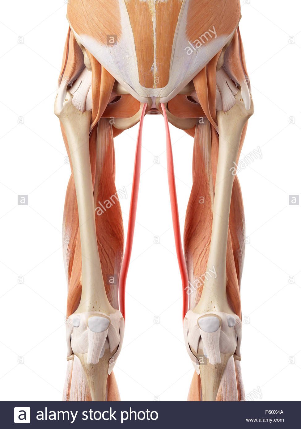
Gracilis is a long thin muscle located in the medial compartment of the thigh. It originates on the medial aspect of the ischiopubic ramus and joins together with the sartorius and semitendinosus muscle tendons to form the pes anserine, which inserts on the superior medial tibia, medial to the tibial tuberosity. The gracilis has a spiral arrangement of muscle fiber bundles and the muscle fibers insert onto the anterior aspect of the gracilis tendon obliquely, making it a spiral unipennate muscle. There are 5 to 7 muscle fiber bundle compartments in the gracilis muscle, with nerve branches coursing along with each compartment, which may indicate independent neuromuscular compartment functioning. Crossing both the hip and knee joints, the gracilis muscle performs hip adduction, hip flexion, hip internal rotation, knee flexion, and knee internal rotation.[rx][rx][rx][rx][rx]

Origin and Insertion
Gracilis is a thin, flat, long muscle that attaches to the coxal bone and tibia. It starts out broad and then tapers off as it approaches its insertion point. The muscle originates through a thin aponeurosis from three sites located on the ischium and pubis
- medial margins of the lower half of the anterior body of pubis
- the entire surface of the inferior pubic ramus
- a small portion of the ramus of ischium close to its adjoining point with the inferior pubic ramus
The muscle fibers travel inferiorly and eventually blend into a round tendon which courses posterior to the sartorius tendon and passes the medial condyle of the femur. At the level of the proximal part of the tibia, the gracilis tendon curves and then fans out around the medial condyle of the tibia. Here, it joins the pes anserinus, which represents a conjoined tendon comprising the tendons of three different muscles; gracilis, sartorius, and semitendinosus.
At its insertion, the tendon is situated immediately above that of the semitendinosus muscle, and its upper edge is overlapped by the tendon of the sartorius muscle, which it joins to form the pes anserinus. The pes anserinus is separated from the medial collateral ligament of the knee-joint by a bursa.
Blood Supply
The gracilis obtains its vascular supply from the
- The medial circumflex femoral artery
- The superficial femoral artery
- The deep femoral artery
- Descending genicular artery
- Anterior branch of the obturator artery.
- The anterior division of the internal iliac artery
- Inferior epigastric artery
- The external iliac artery can give off the obturator artery
- The external iliac artery turns into the common femoral artery as it passes the inguinal ligament.
- The common femoral artery splits into the superficial and deep femoral artery.
- The deep femoral artery then gives rise to the medial and lateral circumflex femoral arteries, while the superficial femoral artery gives rise to the descending genicular artery near its terminal end.[rx][r][rx][rx]
Nerves Supply
- The obturator nerve originates from the L2 to L4 spinal nerve roots, and it traverses along the iliopectineal line, enters the obturator canal, and exits from the obturator forearm and splits into the anterior and posterior branches. The gracilis muscle receives innervation from the anterior branch of the obturator nerve.
Function
Gracilis acts on the hip and knee joints, resulting in several movements
- Strong leg flexion and medial (internal) rotation around the knee joint when the knee is in a semi flexed position.
- Weak thigh flexion and adduction around the hip joint, simply aiding the other, more powerful thigh adductors.
- The gracilis is a long, thin muscle located in the medial compartment of the thigh. There are 5 to 7 muscle fiber bundle compartments in the gracilis muscle, with nerve branches coursing along with each compartment, which may indicate independent neuromuscular compartment functioning.
- Crossing both the hip and knee joints, the gracilis muscle performs hip adduction, hip flexion, hip internal rotation, knee flexion, and knee internal rotation.[rx][rx][rx][rx][rx]